The Ulutas Medical Journal · ISSN 2149-0430 (print), 2149-388X (online) · https://www.ulutasmedicaljournal.com/?mno=151284
Original Article · Open access
Evaluation of the Results of Patients Who Have Retrograde Intramedullary Nailing Surgery for Femur Supracondylar Fractures
Also available in: العربية · Türkçe · 中文
- Department of Orthopedics and Traumatology, Acıbadem University, Istanbul, Turkey ROR
- Department of Orthopedics and Traumatology, Selahhadin Eyyubi Hospital, Diyarbakır, Turkey ROR
- Department of Orthopedics and Traumatology, Adana Metro Hospital, Adana, Turkey ROR
- Department of Orthopedics and Traumatology, Fırat University, Elazığ, Turkey ROR
Abstract
- Introduction
- Supracondylar femur fractures are usually seen after high-energy trauma. Our study evaluated the surgical results of patients who underwent retrograde intramedullary nailing surgery.
- Materials and Methods
- Thirty patients were treated with retrograde intramedullary nailing for femoral supracondylar fractures in a single clinic between January 2015 and September 2018. The clinical, functional, and radiological findings were analyzed retrospectively. Demographic data of the patients in our study, length of hospital stay, time until surgery, presence of additional trauma, complication rates, type of trauma, HSS and NEER scoring criteria for functional evaluation, and AO classification were used to determine fracture types.
- Results
- Eighteen (60%) patients had high-energy trauma as the etiological cause, while 12 (40%) had fractures due to low-energy trauma. There were four open fractures and 26 closed fractures in the patients. The patients were evaluated according to the HSS and NEER evaluation systems. According to the HSS scoring system, 8 (26.7%) had excellent, 9 (30%) good, 12 (40%) moderate, and 1 (3%) poor results. According to the NEER scoring system, 7 (23.3%) excellent, 10 (33.3%) good, and 13 (43.4%) intermediate results were obtained.
- Conclusion
- In supracondylar femur fractures, retrograde intramedullary nailing is a method that should be preferred in appropriate cases because of the low amount of bleeding, rapid mobilization, good union since the fracture hematoma is not evacuated, and good postoperative functional results.
Keywords
Full text
Introduction
Distal femur fractures are among the most challenging fracture types to be treated by many surgeons from the past to the present (1). The strong muscles attached to this area create profound fracture displacements (2). As a result, of these displacements (3), severe damage to neurovascular structures by fracture fragments and extremity losses increases the importance of fractures in this region (4). In addition, due to the ever-developing industry, personal armament, and intensifying traffic, work and traffic accidents and firearm injuries have increased significantly. Parallel to these, there is an increase in distal femur fractures (5).
Although distal femur fractures in adults are not as common as some other fractures, they are more likely to be associated with additional injuries since they often develop due to high-energy trauma (7). Therefore, even only femoral shaft fractures have a high mortality rate. In the past, distal femur fractures were mainly treated with conservative methods. However, surgical treatment increased in conservative treatment due to insufficient anatomical reduction, joint gradation, loss of knee motion, and delayed mobilization. In addition, innovations, osteo-synthesis techniques, quality in implants, extensive surgical experience and development of rehabilitation techniques have contributed(1). Different views have been put forward in treating distal femur fractures, and many surgeons have tried to treat these fractures with various implants. Retrograde nailing is one of the techniques applied in distal femur fractures. Being minimally invasive, protecting the fracture hematoma, and early mobilization are the advantages of this surgical technique.
Our study compared the surgical results of our patients who underwent retrograde intramedullary nailing in supracondylar femur fractures with the literature.
Materials and Methods
Ethical Statement
The study received ethics committee approval (approval no: 2018/14/12). Because the study was designed retrospectively, no written informed consent form was obtained from patients. Patients diagnosed with supracondylar femur fracture who applied to the emergency department between January 2015 and September 2018 were evaluated retrospectively using the hospital information management system. Patients who did not accept surgical treatment or who could not undergo surgical treatment for any reason, patients who died during hospitalization after surgery, patients whose postoperative follow-ups could not be reached, patients with incomplete or inconsistent data, and patients who were treated with a different surgical treatment other than retrograde intramedullary nailing was excluded. We had 61 patients diagnosed with a supracondylar femur fracture. A total of 37 patients underwent retrograde intramedullary nail surgery. Seven patients were excluded because they did not meet inclusion criteria. Our study is single-center. The same team performed the surgeries. While examining the follow-up data looked at clinical pain, limitation of movement, and presence of complaints such as limping; In patients who were thought to have length differences, the distance between the SIAS and the medial malleolus was measured, and the length differences were noted. In addition, the patients were evaluated according to age, gender, side, etiology, open or closed fracture, fracture type according to AO fracture classification, presence of concomitant trauma, time until the operation, surgical time, length of hospital stay, type of anesthesia, duration of union, and follow-up period. All this information was transferred to forms created. Clinical improvement in patients; It was accepted that the patients could fully load their extremities, not feel pain, and do their daily work without any problems.
Radiological improvement was accepted as the appearance of the callus bridge in the AP and lateral planes, where the patient can give total weight, and the disappearance of the fracture line on X-ray films. Neer et al. (2) and Leung et al. (1) Knee-Rating Scale Hospital for Special Surgery evaluation criteria were used to evaluate the results. In the Neer evaluation system, patients are evaluated in 2 parts, functional and anatomical, over 100 points. According to the scores obtained in the Neer evaluation system, the results are assessed as follows: Perfect: >85, Good: 55-70, Medium: 70-85, Bad:<55. Leung et al. In the modified Knee-Rating Scale of the HSS system, the results are evaluated as follows: Perfect:>85, Good: 70-84, Medium: 60-69, Bad:<60 (1).
Surgical Method
All patients were placed on the operating table in the supine position. The knee was flexed to 30 degrees. An arthrotomy was performed with a 5 cm incision over the patellar tendon. With the help of a guide wire, access was provided from the appropriate place. After reduction, the intramedullary guide wire was sent proximally. Reamerization was done. The retrograde nail was placed. Proximal and distal locking screws were sent. Knee and ankle movements were started in early postoperative period. Weight-bearing was started at an average of 4-6 weeks.
The SPSS (Statistical Packages for the Social Sciences) v21 package program was used for statistical analysis. Categorical measurements were summarized as number and percentage, and continuous measurements as mean± standard deviation or median. The comparison of union time with the type of fracture (open/closed), gender, and type of anesthesia was made with the Mann-Whitney U test. The comparison of union time with fracture classification (AO fracture classification), ROM of the joints, operation time, comparison of ROM of the joints, and patients' age was made by the Kruskal Wallis test. Chi-square test statistics were used to compare categorical measures between groups. A p-value of <0.05 was considered significant in all tests.
Results
When the patients treated with the diagnosis of supracondylar femur fracture by applying retrograde intramedullary nailing between the dates of January 2015 and September 2018 were examined by the exclusion criteria, the data of 30 patients were obtained. When the demographic characteristics of the patients were examined, 12 (40%) of 30 patients were female, and 18 (60%) were male.
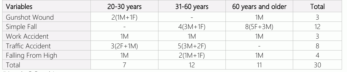
The youngest of the patients in our study was male and 24; the oldest was female and 88 years old. The mean age of the cases was 45.7 years. In our research, 7 (23.4%) patients were between 20-30 years old, 12 (40%) patients were between 31-60 years old, and 11 (36.6%) patients were over 60 years old. Fractures were on the right side in 21 (70%) patients, on the left side in 9 (30%) patients, and the dominant side was the right side. The distribution of etiological causes according to age groups in the patients is shown in Table 1. Looking at the distribution of distal femur fractures in 30 patients according to AO classification, 17 cases are Type A1 fractures, 7 are Type A2 fractures, and 6 are Type A3 fractures.
Since 18 cases were caused by high-energy trauma, they had additional pathologies. For example, there were isolated distal femur fractures in 25 of 30 patients and other pathologies and distal femur fractures in 5. In addition, there were humerus shaft fractures in 1 case, tibia diaphyseal fractures in 1 case, femoral neck fractures in 1 case, distal radius fracture in 1 case, and contralateral femur intertrochanteric fracture in 1 patient.
While general anesthesia was applied in 21 patients, spinal anesthesia was used in 9. Patients were operated on an average of 2.1 (1–6) days after trauma. While 10 of our patients were operated on the first day, 12 on the second day, and six on the third day, the operation was allowed on the fifth and sixth days for 2 of our patients by the intensive care physicians. The shortest follow-up period of patients with retrograde nails is 20 weeks, and the most prolonged follow-up is 52 weeks. The average follow-up time is 30 weeks. The mean time to union was 26 (18-48) weeks. The mean hospital stay was 8.3 (4-30) days.
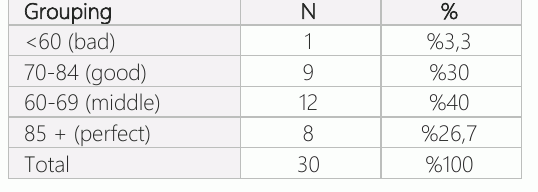
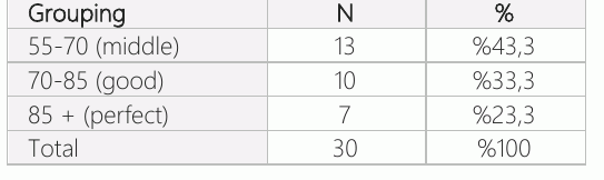
According to the HSS knee scoring system, excellent results were obtained in 8 (26.7%) patients, good results in 9 (30%), moderate in 12 (40%) patients, and poor outcomes in 1 (3%) patient (table 2). According to the Neer grading system, 7 (23.3%) patients had excellent, 10 (33.3%) good results, and 13 (43.3%) intermediate results. None of the patients had a bad outcome (table 3). Thirteen of our patients had pain when they got tired, 6 had pain with air exchange, 2 had limitation of movement and function, and 9 had no complaints of pain. When we evaluated the knee motion function, 17 patients had a range of motion between 120 and 135 degrees, seven patients had a range of 11-120 degrees, and six patients had a range of motion between 80-110 degrees. Five of the patients had thigh atrophy. On the other hand, it was observed that there was no restriction in walking in 8 patients like before the accident, there was a mild restriction in walking for a long time in 21 patients, and in 1 patient, he sometimes used a cane due to pain.
Considering the X-ray findings, the post-op alignment was anatomical in 24 patients. Four patients had 5-10 degrees of varus angulation in the AP plane. Shortness of 1 cm was observed in one patient and 2 cm in one patient.
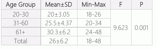
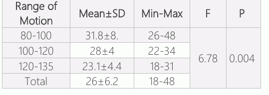
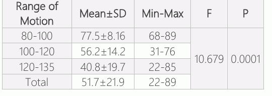
Patients were classified as 20-30, 31-60, and over 61 years old according to their age groups. When these groups were compared according to their union time, it was seen that the age groups were significant according to the union time, and the union time increased as the age increased (p<0.05) (Table 4). When the union times of the patients were compared according to the AO fracture classification, there was no significant difference in union time according to AO fracture classification (p>0.05). When the patients were compared in terms of the relationship between open or closed fractures and union times, it was seen that there was no significant relationship between open or closed fractures and union times (p>0.05). When the union time was compared according to the male and female genders, it was observed that there was no significant difference between union times according to gender (p>0.05). When the patients were compared in terms of the relationship between the joint range of motion and union time, it was seen that there was a significant relationship between the joint range of motion and union time (p<0.05) (table 5). When the union time was evaluated according to the anesthesia applied, no significant correlation was found between the time of union and the anesthesia used (p>0.05). When union time was evaluated according to operation time, no correlation was found between union time and operation time. When age groups are evaluated to etiological reason, there was a difference between these groups and etiological causes. It was observed that high-energy trauma was more common in the young age group (p<0.05).
When the etiological causes were compared according to gender, the etiological causes were not significant according to male and female gender (p>0.05). When the relationship between the joint range of motion and age was compared, it was seen that there was a significant difference between the degree of joint motion and age. Furthermore, young patients' joint movements were better (p<0.05).
Discussion
Since the femur is the largest and strongest bone in the body and has strong muscle groups, fractures in this region usually occur due to high-energy trauma, supracondylar femur fractures tend to be comminuted, and open fractures due to severe trauma. Additional pathologies such as patella fracture, tibia fracture and ligament injury accompany (3). Due to the increase in the elderly population, the risk of developing osteoporosis in women, and the fact that women live longer than men, the frequency of distal femur fractures due to low-energy traumas such as simple falls is increasing. In our study, injury type and gender distribution were compatible with the literature. Anatomical features such as the wide medullary canal of the distal femur, thin cortex, and high cancellous bone density and accompanying additional pathologies affect the treatment selection method. Purpose of treatment selection; It is to reduce the fracture by providing the average length and axis of the bone with the least invasive procedure, mobilize the patient as soon as possible, and ensure the fracture's union.
There is no definite consensus on the boundaries of the distal femoral diaphysis region. Muller; Fractures involving the distal 1/3 of the femur are roughly called distal femoral diaphyseal fractures. In our study, fractures matching Müller's definition were included (4). As a result of developments in medicine and other fields of science, the average life expectancy is increasing worldwide, and the elderly population is increasing. It is a fact that the incidence of distal femoral fractures will increase with older adults in our country. In our study, the mean age of distal femur fractures was 40.9 in men, 52.5 in women, and 45.7 in all our patients. We believe that the higher average age of the patients in our study group compared to the literature is the gradual increase in the elderly population since men are slightly more active in social and business life than women and distal femoral fractures are seen slightly higher in the male population. Distal femur fractures are more common in young and elderly patient populations. While these fractures occur primarily due to high-energy trauma in young people, they occur primarily due to low-energy trauma in older people. Since distal femur fractures are usually caused by high-energy trauma, additional pathologies often accompany. In the literature, in our patient group, another orthopedic trauma accompanied by distal femoral diaphysis fracture in 5 patients, and our additional trauma rate was found to be 16.6%, which is lower than the literature. However, accompanying additional trauma could not be shown to have a significant effect on clinical scores (p>0.05). Since distal femur fractures are usually caused by severe trauma, their incidence is high with open fractures. In the literature, the incidence of open fractures varies between 8-38% (5).
In our study, 4 (13.4%) of the cases were open fractures, 1 was type 1, and 3 were typed 3A open fractures according to Gustillo Anderson Classification. No complication related to open fracture was detected in any of these patients. We think this is because we quickly started debridement, irrigation, and appropriate antibiotic treatment for these open fractures and operated as soon as possible.
No consensus exists on whether the surgery will be performed early or late. In severe traumas that cause this type of fracture, life-threatening injuries should be treated, and then fracture detection should be addressed. In studies conducted on patients with multi-traumatic femur fractures in the literature, it has been reported that if the patient is hemodynamically stable, not hypothermic or coagulopathic, stable femoral fracture fixation should be performed in the first 24 hours. Thus, the patient will be mobilized early, with pulmonary complications and systemic infections, thus reducing mortality (6).
However, problems such as irreversible pulmonary embolism, acute respiratory distress syndrome (ARDS), and multiorgan failure can result from early surgical intervention in admission to the hospital. Such complications that would cause high morbidity or mortality did not develop in our patients. We believe that supracondylar femur fractures should be operated on immediately if the patient is hemodynamically stable.
The basic principle in surgical treatment is the anatomical fixation of the joint surfaces, providing the axis and length of the femoral body and rigid fixation that can allow early movement. Many techniques and implants are used in the surgery of distal femur fractures. Requires extensive soft tissue scraping in a plate and screw fixation, increasing the infection rate; The risk of nonunion increases due to impaired periosteal blood circulation and emptying of the fracture hematoma. In addition, the central load distribution in intramedullary nails shifts laterally in plate-screw fixation, and the transfer of most of the load on the plate increases the risk of implant failure. However, it can be preferred in cases where plate-screw osteo-synthesis does not technically require much equipment, and there is not enough technical opportunity to apply intramedullary nails. Advantages of retrograde intramedullary nails; not draining the fracture hematoma, not scraping the periosteum, and allowing the better union of the fracture (8).
The retrograde intramedullary nail technique is advantageous because it has less blood loss, can be made with a small incision of 5 cm, and has a short operation time. In addition, since it provides the length and a more rigid fixation opportunity, it allows the patient to be mobilized by loading more quickly (8). There is no consensus on using the intramedullary nail patients with impaired hemodynamics. technique with or without engraving. As a result Emphasized that the risk of ARDS, pulmonary embolism, multiple organ failure, pneumonia, and death did not increase after early fracture detection in patients with chest injuries (7). In our study, we performed surgery in an average of 2.1 days (1-6 days) from our patient's of the developments in nail design, thinner but stronger nails have started to be produced, which has further increased the discussions on the carving process. Clinical studies have reported that carving has some drawbacks, especially in patients with multi-trauma and lung damage (9). We used the carved technique in all our cases. No pathology in lung functions was encountered in any patients. Union in femur fractures, lack of tenderness at the fracture line can be said to be the time when solid callus tissue is seen on the radiographs. Fractures with an average union of up to 6 months are called delayed union fractures of 6 to 9 months, and fractures with more than nine months and no callus tissue on the radiographs are called nonunion (10). We attribute the higher rate of the union of the patients in our study group compared to the literature to the high mean age of our cases.
Neer and HSS scoring systems are primarily used to evaluate distal femur fractures. In this study, we used the Neer and HSS scoring system. We used both scoring systems because one scoring system was insufficient. In our study, the mean was 74.3 (good) according to the Neer scoring system and 74.6 (good) according to the HSS scoring system.
Analyzing the fracture type well is necessary to choose the appropriate surgical technique and implant before the operation. According to the AO-ASIF classification, retrograde intramedullary nailing can be applied in A1, A2, A3, C1, and C2 fractures. In this study, 17 cases were identified as A1, 7 as A2, and 6 as A3, according to the AO-ASIF classification. There is no consensus regarding post-surgical mobilization and weight-bearing (11).
Most surgeons have agreed to mobilize without weight bearing if the fixation is stable. Rehabilitation should be started early after surgery to prevent joint movement limitation and increase and shape cartilage nutrition, especially in intra-articular fractures (12). We start our patients with quadriceps exercises on the first postoperative day and raise them to their feet with double crutches. A statistically significant correlation was found between the patients with a good range of motion and the union duration (p<0.05). This finding was attributed to the fact that early and good rehabilitation after surgery affected the union positively. Again, in this study, it was observed that the older age group had more limitations in joint movement. We attribute this to the decrease in physical capacity of people in the advanced age group, systemic diseases, and low adaptation to rehabilitation.
Many complications may occur during or after retrograde intramedullary nailing. The most important of these is infection. Rates between 0-7% are reported in the literature (13). Papadokosdakis et al. (14), in a meta-analysis study, retrograde İ.M., reported the incidence of septic arthritis as 0.18% in nailing patients. Predisposing factors for infection; include open fractures, high-energy traumas, extensive surgical dissection, prolonged operative time, and inadequate fixation (15). No severe postoperative infection was observed in the patients included in our study.
After distal femur fractures, patients frequently complain of a limited range of motion and knee pain. Besides the surgical technique, these injuries occur in the quadriceps muscle and other soft tissues. The limitation of joint movement can be kept to a minimum by placing the nail with the correct technique and post-op rehabilitation (16). In this study, we found the knee joint range of motion of our patients to be 118 degrees on average, which was similar to the literature (17).
In the studies, retrograde İ.M. knee pain has been reported in patients after nailing. It has been stated that this is due to articular cartilage damage in the intercondylar notch (18). All 5 of our cases had muscle atrophy in the thigh. The post-op alignment was anatomical in 24 patients considering the X-ray findings. 4 patients had 5-10 degrees of varus angulation in the AP plane. Shortness of 1 cm was observed in one patient and 2 cm in one patient.
There are some limitations of our study. First, our most important limitations are that it is a retrospective study, the number of patients is limited to 30, and the follow-up period is short. Prospective studies are needed inlarger groups.
Conclusion
Retrograde intramedullary nailing provides a significant advantage in multi-trauma patients due to the short operation time and low bleeding. In addition, we think that one of the most important advantages is that it positively affects the joint’s range of motion since the union is in the early period.
Declarations
- Author contributions (CRediT)
- All authors contributed equally to all 14 CRediT roles (Conceptualization, Data curation, Formal analysis, Funding acquisition, Investigation, Methodology, Project administration, Resources, Software, Supervision, Validation, Visualization, Writing – original draft, Writing – review & editing).
- Funding
-
Funding information was not reported in the published version of this article.
- Conflicts of interest
- The authors declare no conflict of interest.
- Ethics approval
- The study received ethics committee approval (approval no: 2018/14/12). Because the study was designed retrospectively, no written informed consent form was obtained from patients.
- Data availability
- A data availability statement was not reported in the published version of this article.
References (18)
14 of 18 references are linked to their DOI.
- Leung KS, Shen WY, So WS, Mui LT, Grosse A. Interlocking intramedullary nailing for supracondylar and intercondylar fractures of the distal part of the femur. J Bone Joint Surg 1991; 73: 332-340 DOI Crossref PubMed Google Scholar
- Neer CS, Grantham AS, Shelton ML. Supracondylar fracture of the adult femur. J Bone Joint Surg 1967; 49: 591-613 DOI Crossref PubMed Google Scholar
- Paley D. Normal lower limb alignment and joint orientation. Principles of Deformity Correction 2002; 1-17 DOI Crossref PubMed Google Scholar
- Müller ME, Allgöver. Manual of internal fixation. Third Edition. Springer-Verlag 1991; 548-550 DOI Crossref PubMed Google Scholar
- Ali MA, Hussain SA, Khan MS. Evaluation of results of interlocking nails in femur fractures due to high velocity gunshot injuries. J Ayub Med Coll Abbottabad. 2008 Jan-Mar;20(1):16-9 PubMed Google Scholar
- Arazi M, Yel M, Oktar MN, Ogtin TC, Memik R. Erişkin femur cisim kırıklarının kilitli intramedüller çivileme ile tedavisi. Acta Orthop Trauma Turc 1999; 33: 126-130. PubMed Google Scholar
- Brundage SI, McGhan R, Jurkovich G, Mack CD, Maier RV. Timing of femur fracture fixation. Effect on outcome in patient with thoracic and head injuries. J Trauma 2002; 52(2): 299-307 DOI Crossref PubMed Google Scholar
- Neubauer T, Ritter E, Potschka T, Karlbauer A, Wagner M. Retrograde nailing of femoral fractures. Acta Chir Orthop Traumatol Cech. 2008 Jun;75(3):158-66 DOI Crossref PubMed Google Scholar
- Heiney JP, Battula S, O'Connor JA, Ebraheim N, Schoenfeld AJ, Vrabec G. Distal femoral fixation: a biomechanical comparison of retrograde nail, retrograde intramedullary nail, and prototype locking retrograde nail. Clin Biomech (Bristol, Avon). 2012 Aug;27(7):692-6 DOI Crossref PubMed Google Scholar
- Han QT, Tang HW. [Treament of distal fractures of femur by supracondylar intramedullary interlocking nail in 59 patients]. Zhongguo Gu Shang. 2009 May;22(5):388-9 PubMed Google Scholar
- Albareda-Albareda J, Gabarre- Raso S, Rosell-Pradas J, Puértolas-Broto S, Ibarz-Montaner E, Redondo-Trasobares B, Gómez-Vallejo J, Blanco-Rubio N, Sánchez-Gimeno M, Herrera-Rodríguez A, Gracia-Villa L. Biomechanical behavior of retrograde intramedullary nails in distal femoral fractures. Injury. 2021 Jul;52 Suppl 4:S76-S86 DOI Crossref PubMed Google Scholar
- Wall R, Syed F, Arastu M, Riemer B, Boutefnouchet T. Treatment of Supracondylar Periprosthetic Femoral Fractures with Retrograde Intramedullary Nailing Versus Distal Femoral Plating: A systematic review and meta-analysis of current evidence. Orthop Traumatol Surg Res. 2022 Nov 25:103489 DOI Crossref PubMed Google Scholar
- Netter FH. Musculoscletal system. The CIBA Cellection of Medical IIIustration. CIBA Geigy Corporation 2010; 76-97 PubMed Google Scholar
- Papadokostakis G, Papakostidis C, Dimitriou R, Giannoudis PV. The role and efficacy of retrograting nailing for the treatment of diaphyseal and distal femoral fractures: a systematic review of the literature. Injury 2005; 36(7):813-822 DOI Crossref PubMed Google Scholar
- Gill S, Mittal A, Raj M, Singh P, Singh J, Kumar S. Extra Articular Supracondylar Femur Fractures Managed with Locked Distal Femoral Plate or Supracondylar Nailing: A Comparative Outcome Study. J Clin Diagn Res. 2017 May;11(5):RC19-RC23 DOI Crossref PubMed Google Scholar
- Wu CC, Tai CL. Retrograde nailing of a femoral supracondyle. Orthopedics. 2012 Apr;35(4):e491-6 DOI Crossref PubMed Google Scholar
- Wu CC. Retrograde locked intramedullary nailing for aseptic supracondylar femoral nonunion following failed locked plating. J Orthop Surg (Hong Kong). 2015 Aug;23(2):155-9 DOI Crossref PubMed Google Scholar
- Du YR, Ma JX, Wang S, Sun L, Wang Y, Lu B, Bai HH, Hu YC. Comparison of Less Invasive Stabilization System Plate and Retrograde Intramedullary Nail in the Fixation of Femoral Supracondylar Fractures in the Elderly: A Biomechanical Study. Orthop Surg. 2019 Apr;11(2):311-317 DOI Crossref PubMed Google Scholar
Respond to this article
Comments on a published article are welcome as a Letter to the Editor.
Get an e-mail when new articles in your fields are published
Questions to the authors
Questions are moderated: an editor approves each question, the authors answer, and an editor approves the answer before both appear here.
No answered questions yet.
Ask the authors a question
Discussion
Comments are moderated: they appear after the writer confirms the e-mail address and an editor approves them. Each comment carries a conflict-of-interest statement.
No comments yet.
Write a comment
Tell me when this article is cited
Leave your e-mail address and confirm it. These notices are not being sent yet; your confirmed address is kept for them and used for nothing else.
Read the full text
Open the PDF here
Loading the PDF…
If the PDF does not show here, use the download button above.
How to cite this article
Yilmaz E, Ulusoy I, Kivrak A, Igrek S, Yilmaz E. Evaluation of the Results of Patients Who Have Retrograde Intramedullary Nailing Surgery for Femur Supracondylar Fractures. Ulutas Med J. 2023;9(2):66-73. doi:10.5455/umj.20230427065545
Yilmaz, E., Ulusoy, I., Kivrak, A., Igrek, S., & Yilmaz, E. (2023). Evaluation of the Results of Patients Who Have Retrograde Intramedullary Nailing Surgery for Femur Supracondylar Fractures. The Ulutas Medical Journal, 9(2), 66–73. https://doi.org/10.5455/umj.20230427065545
Yilmaz, E. et al. (2023) 'Evaluation of the Results of Patients Who Have Retrograde Intramedullary Nailing Surgery for Femur Supracondylar Fractures', The Ulutas Medical Journal, 9(2), pp. 66–73. doi: 10.5455/umj.20230427065545.
@article{umj151284,
  title = {Evaluation of the Results of Patients Who Have Retrograde Intramedullary Nailing Surgery for Femur Supracondylar Fractures},
  author = {Yilmaz, Edip and Ulusoy, Ibrahim and Kivrak, Aybars and Igrek, Servet and Yilmaz, Erhan},
  journal = {The Ulutas Medical Journal},
  year = {2023},
  volume = {9},
  number = {2},
  pages = {66--73},
  doi = {10.5455/umj.20230427065545},
  url = {https://www.ulutasmedicaljournal.com/?mno=151284},
  issn = {2149-388X}
}