The Ulutas Medical Journal · ISSN 2149-0430 (print), 2149-388X (online) · https://www.ulutasmedicaljournal.com/?mno=76905
Original Article · Open access
Utilization of Corticocancellous Bone Graft, Demineralized Bone Matrix and Synthetic Ceramic Bioglass for Defective Bone Fractures in Rats
- Department of Orthopedics and Traumatology, Tarsus State Hospital, Mersin, Turkey ROR
- Hatay State Hospital, Department of Orthopedics and Traumatology, Hatay, Turkey ROR
- Mustafa Kemal University, Department of Orthopedics and Traumatology, Hatay, Turkey ROR
Abstract
- Introduction
- Trauma, bone infections, and bone defects that emerge during the attempts as musculoskeletal tumor surgery can be treated with bone grafts. The aim of this study is to investigate the level of bone fracture healing that was treated using different kinds of grafts.
- Materials and Methods
- 40 male Wistar-Albino rats were used as 4 different groups with 10 rats each. The groups included the sham, the corticocancellous analog bone graft, the demineralized bone matrix, and the synthetic ceramic bioglass. All rat groups were evaluated biomechanically and histopathologically.
- Results
- The average scores of the histological evaluations were as follows: 5.5±1.64 in the sham, 5.62±2.38 in the group-1, 6.9±1.19 in the group-2, and 7.4±1.17 in the group-3. There was a significant difference among all groups in terms of the histopathological findings (p=0,041). The p-values for the group pairs were as follows: the sham and group-2, p=0.857; the sham and group-3, p=0.049; the sham and group-4, p=0.011; groups 2 and 3, p=0.22; groups 2 and 4, p=0.087; groups 3 and 4, p=0.37.
- Conclusion
- Histopathologic findings showed the presence of a supporting effect of allograft on the healing of defective bone fractures. However, biomechanical positive effects of allografts were seen by the demineralized bone matrix and bioactive glass-ceramic.
Keywords
Full text
Introduction
The skeletal system is a vital structure that provides the necessary support for the proper functioning of the organs and systems of the human body. This system is subjected to frequent injury with environmental factors and human factors (1). The skeletal system injuries can vary from a simple soft tissue injury to a severe condition, such as a fracture, depending on the severity of the trauma (2). The pathology that results from a fracture is not limited to bone tissue. The surrounding soft tissues and organ systems will be negatively affected (3, 4). The two major factors that influence the healing process of the fracture are the blood supply and the stability of the fracture (5). It is well known that several other factors also influence the healing process of a bone fracture, such as condition and configuration of the bone tissue, the periosteum, and the surrounding muscles/soft tissue. These factors' healthy function and their relationships with each other will affect the healing of the fracture (6). The studies on fracture healing focus on the fixation of the fracture, the systemic and local factors that influence the fracture, electrical and audio stimulation of the fracture, and hyperbaric oxygen applications. Many scientists are trying to develop new approaches to better understand the healing process of the bone, and ensure healthy healing of the skeletal system (5, 7).
The bone tissue has an extensive regeneration capacity; it can completely restore its original structure and function (8). However, in some situations such as trauma, infection, cysts, and tumors, the bone defects may not completely heal. In these situations, the bone defects may need to be replaced with bone grafts to initiate, accelerate or facilitate the healing process (9). In this study, the rats' right femurs were fractured to form a bone defect, followed by intramedullary fixation. In the sixth week, the fractures were treated with different grafts. The histopathological and biomechanical aspects of the different graft treatment were investigated.
Materials and Methods
Study Design
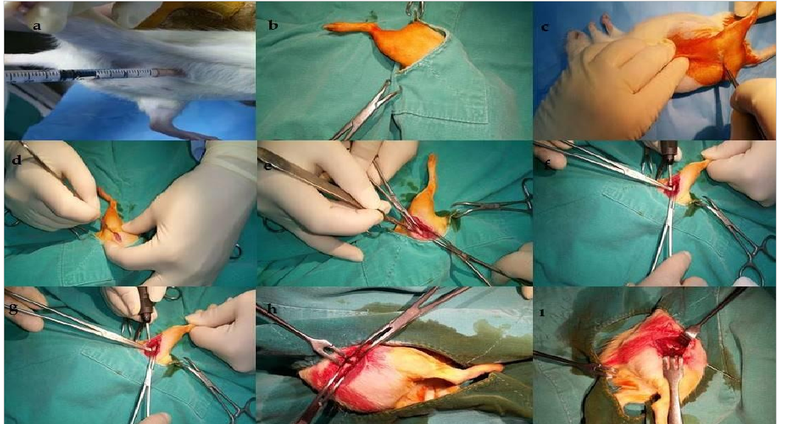
In this experimental study, we investigated the effects of different graft materials on the healing process of the defective rat femur fractures. The study was carried out at our Experimental Animal Research Laboratory with a total of 80 male Wistar-Albino rats, obtained from the Laboratory Animals and Research Laboratory of Institute. The study was approved by the Research and Examination Committee of the Mustafa Kemal University Medical Faculty Medicine regarding the use of experimental animals (25.12.2016 - 2014/11/1). Wistar Albino rats were all males and older than 12 weeks, and weighed between 280-320 grams (Figure-1).
Experimental Groups
The rats were randomly divided into 4 groups. The group-1 was treated with the cancellous allograft (TranZgraft®, TBI/Tissue International, USA), group-2 was treated with demineralized bone matrix (DBM) (SureFuse®, Hans Biomed Corp., Seoul, Korea), and the group-3 was treated with bioactive glass-ceramic (BonAlive putty®, BonAlive Biomaterials Ltd., Turku, Finland). The control (sham group) was not treated with any graft material. There were no limitations on the diets of the rats. The subjects were kept in the optimal rat shelter, at an average of 22oC temperature and a 12 hours light/12 hours dark cycle. The subjects were fed with dry rat food.
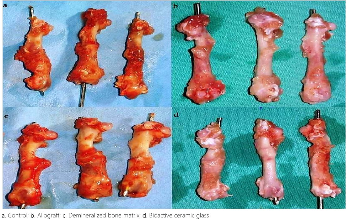
The care and nutrition of the subjects were provided by the designated personnel and was monitored by a veterinarian. After 6 weeks, all rats were sacrificed with overdose of ketamine (100mg/kg/ketamine) and cervical dislocation. The sacrificed subjects' femurs were removed from the previous incision site and excised (Figure-2). The femurs were kept in a 10% buffer formalin solution until the histopathological and the biomechanical examination.
Surgical Technique
The dose of the drug was individually determined through the weight of each subject. The anesthetic drug [combination of 50 mg/kg Ketamine HCl (Ketalar®, Pfizer, USA) and 10 mg/kg Xylazine (Rompun ®, Bayer, Turkey)] was applied from right groin intraperitoneally. After the administration of the anesthetic drug, the local area was sterilized with a Betadine scrub (10% povidoneiod, Merkez-Turkey). An antibiotherapy was applied before and after the operation (IV 50 mg/kgCefazolin sodium). The skin was dissected from the lateral of the shaft with a 3-cm longitudinal incision. The muscles were bluntly dissected to reveal the femoral shaft. The defect was created (2 times the size of the shaft) by cutting the femoral shaft from the middle with a powered saw. The femur was defectively fixated with a 2 mm Kirschner wire (retrograde intramedullary). The part of the wire that remained outside was cut off with a chisel. In the control group, the formed defective region was not filled with a graft. In Group-1, the defective fracture was treated with a cancellous (freeze-dried) allograft; in group-2, with demineralized bone matrix; and in group-3, with bioactive glass-ceramic. The incision area was washed with 10cc of the sterile saline solution and sutured with a basic suture with a 2/0 silk thread. The wound was wiped with povidone-iodine.
Biomechanical Assessments
In the first step, the muscle tissue was completely removed from the femurs, as this is necessary for an accurate measurement. The soft tissue can alter the refracture of the bone. The bones were tried and fractured from the initial fracture line. The values that resulted from the fracture were digitally recorded. Two rats were excluded from the study. One rat from the allograft group had osteomyelitis, and another one rat from the glass-ceramic group did not heal and developed an abscess. The bones that were obtained from the sacrificed rats were at once tested with the three-point bending method, at room temperature (23 °C). This test was conducted by adding two attachments to the ZWICK/Z100 drawbench (Zwick Roell AG, Ulm, Germany). The femurs were placed to the lower attachment to provide an anteroposterior bending force. The effective span was 20 mm. The pull force was applied symmetrically to the center of the rat femur, at the callus line. The initial pull force was 0 Newton and was increased at 20 mm/min until the femur was fractured. The ultimate load at the point of fracture was digitally recorded by a computer.
Histopathological Evaluation
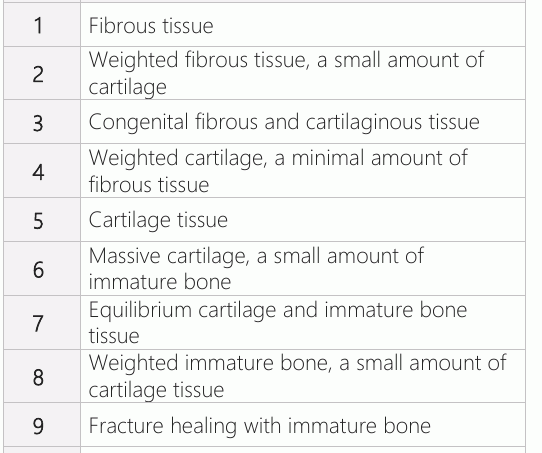
There were a total of 40 rats that were divided into four groups of 10 rats. The surrounding soft tissue was removed from each femur, and the femurs were kept in a 10% neutral formalin solution for 48 h. Subsequently, the specimens were decalcified with a 10% formic acid solution. After one week of decalcification, an auto-technicon was used to dehydrate, clear and embed the femurs in paraffin. The longitudinal sections (5-6 μm) were perpendicular to the fracture line and in close proximity to the callus. They were later stained with hematoxylin and eosin (HE). The prepared slides' micrographs were evaluated by a pathology specialist with a binocular research microscope. The slides were evaluated regarding fibrous tissue, cartilage, new bone,and mature bone content, according to the scale reported by Huo et al. (10)(Table-1).
Statistical Analysis
Means and standard deviation are used to report normally distributed data. It was agreed at the start of analysis that any variable with 5% or more missing data would be excluded. The obtained data were analyzed with the SPSS v21 Statistical Package (IBM Inc., Chicago, Illinois, USA), through the Kruskal Wallis analysis. The groups were compared in pairs with the Mann- Whitney U-test. In all the comparisons, a p-value of <0.05 was considered significant.
Results
Biomechanical Findings
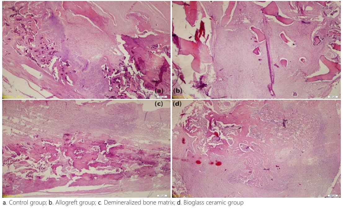
The femurs obtained from the rats that were sacrificed on week six were tested with the three-point bending test (11). After the removal of the intramedullary material, the number of samples with sufficient integrity for the test were as follows: 10 femurs from the control group, 9 femurs from group 1, 10 femurs from group 2 and 8 femurs from group 3. The data from the biomechanical test are presented in Figure-3. The callus formation in the control group mostly had a regular appearance. The samples of study groups had fibrous tissue formation, but in some of the samples, this appearance was replaced with cartilage and immature bone formation. The average scores of histological evaluations were as follows: 5.5±1.64 in control, 5.62±2.38 in the group-1, 6.9±1.19 in the group-2, and 7.4±1.17 in the group-3. The distribution of the histopathological scores is presented in Figure-4.
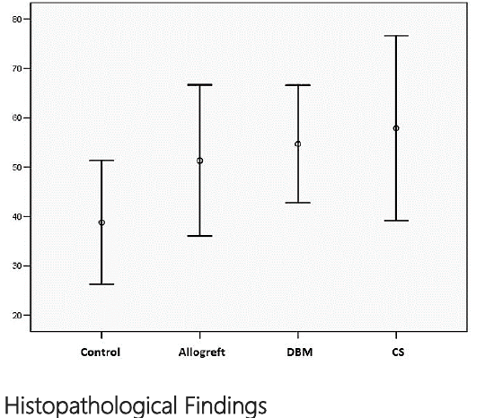
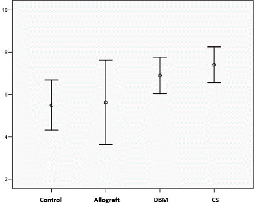
There was a significant difference between the histopathological findings (p=0,041). The p-values for the group pairs were as follows: the control group and group 1, 0.857; the control group and group 2, 0.049; the control group and group 3, 0.011; groups 1 and 2, 0.22; groups 1 and 3, 0.087; groups 2 and 3, 0.37. The analyses revealed that the difference was significant only between the control group and groups 2 and 3 (Figure-5).
Discussion
Various methods have been developed to improve the orthopedic healing process of bone fractures. The applications that were found to improve bone healing started to be commonly used in clinical practice (12). There are also graft applications that are developed to biologically improve the healing process (13). The applications serve 2 important purposes: the replacement of a lost bone, and the correction of bone deformities to restore integrity and function. In our study, we created a defective fracture in the femur and stabilized it with intramedullary fixation. The purpose was to compare the biomechanical and histopathological results of the treatments including different kinds of grafts. We believe that the findings will contribute to the routine graft practices performing by surgeons.
Until recently, orthopedic surgeons could only use autologous bone grafts and allografts to replace bones during treatment. The bone autograft is regarded as the gold standard for the treatment of bone defects due to the low immune response (14). However, developments in tissue engineering applications, it is now possible to choose among many different options; including ceramics, demineralized bone matrix, bone marrow, autologous growth factors, and composite grafts. The basis of bone tissue engineering is to create a response that will lead to healing in the desired anatomical area. The treatment aims to regenerate and repair the bone tissue through the stimulation of a combination of cells and inducing factors. Tissue engineering tries to imitate the body's natural tissue formation process to create functional living tissues (15). There are different factors that influence this process, such as the variety of the cell at the injury site, and the levels of different factors (16, 17). Closed fractures are regarded to be easier to heal than open fractures, as the hematoma has not drained, and there is less damage to the surrounding soft tissue. The experimental studies have also used closed fracture methods. In the open fracture model, the healing process is longer, and thus, it is easier to determine the rate of healing between different groups (16). In our study, we have chosen to experiment with osteotomy in order to be able to form a bone defect and apply the grafts to the fracture line. The experimental studies concerning bone healing use different animal models, such as dogs, rats, rabbits, sheep, and pigs. In general, it can be said that the large animal models are used to develop new devices, and the small animal models are used to better understand the molecular mechanisms and genetics of healing. The fact that most of the studies on fracture healing were performed on rats led us to make this decision (18). We decided to use isogenic mice that we obtained from the experimental animal research laboratory of our university. The studies on bone fracture healing that use rats mostly experiment on the tibia or the femur, due to the ease of manipulation, exposure, and preparation of the slides (19). Also, the morphology of the Wistar-type rats' femur is similar to that of the human (20). Therefore, Wistar type rats were preferred in fracture healing studies.
In the defective fracture studies, the purpose is to create a gap in the site of the fracture in order to investigate the activities of different modalities, such as grafts, growth factors, and hormones; and to subsequently fill this gap. The most commonly used model for the experimental studies is the rabbit and rat skull defects (21, 22). In our clinic, we mostly encounter defective fractures in the long bones, which are internally or externally fixated. In light of the literature, we chose to work with the femur and used the intramedullary nailing method which is accepted as the gold standard for femur fractures. After the formation of the defective fracture, the bone was fixated with the intramedullary application of the 2 mm K-wire. It was easy to apply the technique and to obtain the material, however, there were structures that were not rigid. We accepted this drawback to be an advantage, as it would provide more reliable data regarding the observation of secondary bone healing, and that there is a more prominent callus formation in non-rigid fixations. Due to the small size of the rat femur, most of the studies choose intramedullary fixation. After the intramedullary fixation, the femurs were not further fixated. The studies mostly examine the healing in the sixth week (23, 24). This is because, at six weeks, the callus is now entirely replaced by the bone tissue; and the mineralization can be observed. Thus, we have chosen to examine the fractures in the sixth week.
The literature presents several different methods of histologically examining the tissue. Allen et al. have given the tissues a score from 0 to 4, concerning the healing process of the callus tissue. Huo et al. have given a score from 1 to 10 to determine the cellular changes in the fracture site (10). This scale uses the fibrous, cartilage, mature and immature bone tissues in the healing site to determine the degree of healing. In our study, we have used a histologic evaluation scale developed by Huo et al.
There are several studies concerning the effects of different grafts on healing. Tiedeman et al. have used the demineralized matrix in the nonunion tibia fractures of dogs (25). Their histological examination was performed on the 12th week instead of the 6 week, and they observed lots of cartilage, isolated calcified cartilage regions, and new bone formation through endochondral ossification. Bernabe et al. have investigated the effects of bioactive glass and calcium sulfate on the healing of rat tibias (26). On the 10th postoperative day, there were no differences between the study groups. On the 30th postop. day, the bone formation was more prominent in the calcium sulfate barrier group. Rabie et al. have implanted endochondral autogenous graft, demineralized bone, and a combination of these two grafts to observe the healing process of the rabbit parietal bones (27). They reported that the bone healing was through endochondral ossification in all groups and that the healing had significantly improved in the group where the two materials were used together: the demineralized bone powder had increased the osteoinduction of both the receiving site and the bone graft.
There are many studies indicating that the demineralized bone matrix has similar results to autologous bone graft in the nonunion of long bones and the bone losses that occur due to fractures. Tiedeman et al. (28) have compared the bone marrow and DBM composite graft treatment (10 subjects) with the autograft from the iliac cristae (8 subjects). They reported that after a 12-month radiological follow-up, 9 out of 10 patients with the DBM treatment had fully recovered, whereas only 6 out of 8 patients had a complete recovery. DBM is a mixture of bone morphogenic proteins (BMP) and it is immunogenic. The BMP mixtures are obtained from purified bone extracts. Johnson et al. have used a human BMP-DBM composite to treat 25 patients with resistant nonunion with a partial or complete segmental defect (29). The average follow-up was 21 months (5-82 months). They obtained 14 perfect, five good and five moderate results. Amorosa et al. have compared the physiological load on the callus tissue following the application of allograft, autograft, and scaffolding to the rat femur segmental defective fracture (9). They have found that the biomechanical results were different between the groups. The rats were divided into four groups, and it was observed that the biomechanical results of the autograft and the allograft groups were better than those of the scaffold. In our study, we compared histopathological and biomechanical results of the different graft materials: freeze-dried cancellous allograft,demineralized bone matrix, and bioactive glass granules.
In the current study, the biomechanical data which were obtained from the three-point bending test were digitally measured and recorded. The control and study groups were not found to be significantly different. However, the callus tissue was macroscopically observed to be larger; the data were higher in the study group, which indicates the positive effect of graft use on the healing process of a defective bone. We believe a study with more extended healing periods may yield significant results. We saw that an immature bone tissue had developed in all groups except the control group. In the control group, cartilaginous and fibrous tissue were present in approximately equal parts. The comparison of the histological findings and scores revealed a significant difference between the control group and the graft groups. The different graft groups' histological scores also differed among themselves. This difference may be due to the different healing rates that arose from the non-rigid fixation. However, the significant difference between the control group and the graft groups indicate that these materials improve the healing process.
The limitation of this study is the lack of radiographic way. Similar studies radiologically evaluated parameters such as the bridging status, the prominence of the fracture line, and the width of the callus. As we believed that these parameters might lead to errors in measurement, we did not include in our study.
Conclusion
In our study, we have found that the presence of an allograft, demineralized bone matrix and bioactive glass-ceramic did not improve the biomechanical strength of the fractured bone; however, the histopathological findings indicate that these grafts improve the healing process. These findings indicate that the graft materials can replace autografts, when the autograft leads to morbidity in the donor site or where sufficient material cannot be obtained due to the massive bone loss. We hope that the longer-term follow-up results of these materials will provide more meaningful results.
Declarations
- Author contributions (CRediT)
- All authors contributed equally to all 14 CRediT roles (Conceptualization, Data curation, Formal analysis, Funding acquisition, Investigation, Methodology, Project administration, Resources, Software, Supervision, Validation, Visualization, Writing – original draft, Writing – review & editing).
- Funding
-
Funding information was not reported in the published version of this article.
- Conflicts of interest
- The authors have declared no conflict of interest for the present article.
- Ethics approval
- Ethics approval details were not reported in the published version of this article.
- Data availability
- A data availability statement was not reported in the published version of this article.
References (29)
26 of 29 references are linked to their DOI.
- Trotta DR, Gorny C, Jr., Zielak JC, Gonzaga CC, Giovanini AF, Deliberador TM. Bone repair of critical size defects treated with mussel powder associated or not with bovine bone graft: Histologic and histomorphometric study in rat calvaria. Journal of Cranio- Maxillo-Facial Surgery. 2014;42(6):738-43 DOI Crossref PubMed Google Scholar
- Ozturk R, Karakoc Y. Arthroscopic Treatment of Benign Tumors and Tumor-Like Lesions Located in and Around the Knee Joint. Ulutas Med J. 2018;4(1):19-24 DOI Crossref PubMed Google Scholar
- Adriani E, Di Paola B, Alfieri A, De Fenu E. Femoral Fixation With Curve Cross- Pin System in Arthroscopic Posterior Cruciate Ligament Reconstruction. Arthrosc Tech. 2018;7(4):e289-e98 DOI Crossref PubMed Google Scholar
- Sheikh Z, Zhang YL, Tamimi F, Barralet J. Effect of processing conditions of dicalcium phosphate cements on graft resorption and bone formation. Acta Biomater. 2017;53:526-35 DOI Crossref PubMed Google Scholar
- Yeoh JC, Taylor BA. Osseous Healing in Foot and Ankle Surgery with Autograft, Allograft, and Other Orthobiologics. Orthop Clin North Am. 2017;48(3):359-69 DOI Crossref PubMed Google Scholar
- Kong CH, Steffi C, Shi Z, Wang W. Development of mesoporous bioactive glass nanoparticles and its use in bone tissue engineering. J Biomed Mater Res B Appl Biomater. 2018 DOI Crossref PubMed Google Scholar
- Espregueira-Mendes J, Andrade R et al. Augmentation of Patellar Tendon Repair With Autologous Semitendinosus Graft-Porto Technique. Arthrosc Tech. 2017;6(6):e2271 -e6 DOI Crossref PubMed Google Scholar
- Paulo IJ, Miliane G, Gabriela KB, Conrado L, Edilson E, Mamie I. Bone repair of critical size defects treated with autogenic, allogenic, or xenogenic bone grafts alone or in combination with rhBMP‐2. Clinical Oral Implants Research. 2016;27(5):558- PubMed Google Scholar
- Amorosa LF, Lee CH, Aydemir AB, Nizami S, Hsu A, Patel NR, et al. Physiologic load-bearing characteristics of autografts, allografts, and polymer-based scaffolds in a critical sized segmental defect of long bone: an experimental study. International Journal of Nanomedicine. 2013;8:1637-43 DOI Crossref PubMed Google Scholar
- Huo MH, Troiano NW, Pelker RR, Gundberg CM, Friedlaender GE. The influence of ibuprofen on fracture repair: Biomechanical, biochemical, histologic, and histomorphometric parameters in rats. Journal of Orthopaedic Research. 1991; 9(3):383-90 DOI Crossref PubMed Google Scholar
- Adlakha VK, Srivastava N, Ch P, na, Grover J. Gingival Depigmentation: Esthetic Enhancement Made Simple and Efficient. Ulutas Med J. 2015;1(1):7-9 DOI Crossref PubMed Google Scholar
- Claes L, Willie B. The enhancement of bone regeneration by ultrasound. Progress in Biophysics and Molecular Biology. 2007;93(1-3):384-98 DOI Crossref PubMed Google Scholar
- Hart LE. Low-Intensity Pulsed Ultrasound Therapy for Fracture Healing: A Meta-Analysis. Clinical Journal of Sport Medicine. 2003;13(2):127 DOI Crossref PubMed Google Scholar
- Schultze-Mosgau S, Keweloh M, Wiltfang J, Kessler P, Neukam FW. Histomorphometric and densitometric changes in bone volume and structure after avascular bone grafting in the extremely atrophic maxilla. British Journal of Oral and Maxillofacial Surgery. 2001;39(6):439-47 DOI Crossref PubMed Google Scholar
- Hing KA. Bone repair in the twenty-first century: biology, chemistry or engineering? Philosophical Transactions of the Royal Society A: Mathematical, Physical and Engineering Sciences. 2004;362(1825):2821-50 DOI Crossref PubMed Google Scholar
- Jackson RA, McDonald MM, Nurcombe V, Little DG, Cool SM. The use of heparan sulfate to augment fracture repair in a rat fracture model. Journal of Orthopaedic Research. 2006;24 (4):636-44 DOI Crossref PubMed Google Scholar
- Zimmermann G, Henle P, Kusswetter M, Moghaddam A, Wentzensen A, Richter W, et al. TGF-β1 as a marker of delayed fracture healing. Bone. 2005;36(5):779-85 DOI Crossref PubMed Google Scholar
- Rasubala L, Yoshikawa H, Nagata K, Iijima T, Ohishi M. Platelet-derived growth factor and bone morphogenetic protein in the healing of mandibular fractures in rats. British Journal of Oral and Maxillofacial Surgery. 2003;41(3):173-8 DOI Crossref PubMed Google Scholar
- Cao Y, Mori S, Mashiba T, Kaji Y, Manabe T, Iwata K, et al. 1α,25-Dihydroxy- 2β (3-hydroxy propoxy) vitamin D3 (ED-71) suppressed callus remodeling but did not interfere with fracture healing in rat femora. Bone. 2007;40(1):132-9 DOI Crossref PubMed Google Scholar
- Jäger M, Sager M, Lensing-Höhn S, Krauspe R. The critical size bony defect in a small animal for bone healing studies (I): Comparative anatomical study on rat femur. Biomedizinische Technik/Biomedical Engineering. 2005;50(4): 107-10 PubMed Google Scholar
- An YH, Hill CM, Friedman RJ. Animal Models of Orthopaedic Implant Infection. Medical Implications of Biofilms: Cambridge University Press. p. 59-85 DOI Crossref PubMed Google Scholar
- Eckardt H, Christensen KS, Lind M, Hansen ES, Hall DWR, Hvid I. Recombinant human bone morphogenetic protein 2 enhances bone healing in an experimental model of fractures at risk of non-union. Injury. 2005;36(4):489-94 DOI Crossref PubMed Google Scholar
- Cebesoy O, Tutar E, Kose KC, Baltaci Y, Bagci C. Effect of strontium ranelate on fracture healing in rat tibia. Joint Bone Spine. 2007;74(6):590-3 DOI Crossref PubMed Google Scholar
- Einhorn TA. Enhancement of fracture-healing. The Journal of Bone & Joint Surgery. 1995;77(6):940-56 DOI Crossref PubMed Google Scholar
- Tiedeman JJ, Connolly JF, Strates BS, Lippiello L. Treatment of nonunion by percutaneous injection of bone marrow and demineralized bone matrix. An experimental study in dogs. Clin Orthop Relat Res. 1991(268):294-302 PubMed Google Scholar
- Bernabé PFE, Melo LGN, Cintra LTA, Gomes-Filho JE, Dezan Jr E, Nagata MJH. Bone healing in critical-size defects treated with either bone graft, membrane, or a combination of both materials: a histological and histometric study in rat tibiae. Clinical Oral Implants Research. 2011;23(3):384-8 DOI Crossref PubMed Google Scholar
- Rabie ABM, Wong RWK, Hägg U. Composite autogenous bone and demineralized bone matrices used to repair defects in the parietal bone of rabbits. British Journal of Oral and Maxillofacial Surgery. 2000;38(5):565-70 DOI Crossref PubMed Google Scholar
- Tiedeman JJ. The role of a composite, demineralized bone matrix and bone marrow in the treatment of osseous defects. Journal of Pediatric Orthopaedics. 1996;16(3):413 DOI Crossref PubMed Google Scholar
- Johnson EE, Urist MR, Finerman GA. Resistant nonunions and partial or complete segmental defects of long bones. Treatment with implants of a composite of human bone morphogenetic protein (BMP) and autolyzed, antigen-extracted, allogeneic (AAA) bone. Clin Orthop Relat Res. 1992(277):229-37 DOI Crossref PubMed Google Scholar
Read the full text
Open the PDF here
Loading the PDF…
If the PDF does not show here, use the download button above.
How to cite this article
CANSEVER SA, Aydogan Z, Ozden R. Utilization of Corticocancellous Bone Graft, Demineralized Bone Matrix and Synthetic Ceramic Bioglass for Defective Bone Fractures in Rats. Ulutas Med J. 2019;5(4):233-242. doi:10.5455/umj.20191206042123
CANSEVER, S. A., Aydogan, Z., & Ozden, R. (2019). Utilization of Corticocancellous Bone Graft, Demineralized Bone Matrix and Synthetic Ceramic Bioglass for Defective Bone Fractures in Rats. The Ulutas Medical Journal, 5(4), 233–242. https://doi.org/10.5455/umj.20191206042123
CANSEVER, S.A., Aydogan, Z. and Ozden, R. (2019) 'Utilization of Corticocancellous Bone Graft, Demineralized Bone Matrix and Synthetic Ceramic Bioglass for Defective Bone Fractures in Rats', The Ulutas Medical Journal, 5(4), pp. 233–242. doi: 10.5455/umj.20191206042123.