The Ulutas Medical Journal · ISSN 2149-0430 (print), 2149-388X (online) · https://www.ulutasmedicaljournal.com/?mno=152892
Original Article · Open access
The Effect of Barrier Gel and Free Fat Graft on the Formation of Epidural Fibrosis in the Laminectomy Model
- Department of Orthopedic Surgery, Doha Clinic Hospital, Doha, Qatar ROR
- Department of Orthopedic Surgery, Icon Medical Center, Doha, Qatar
- Department of Orthopedic Surgery, The View Hospital, Doha, Qatar
Abstract
- Introduction
- This study aims to compare the efficacy of ADCON-L and free fat graft used to prevent epidural fibrosis in the experimental laminectomy model in rabbits.
- Materials and Methods
- Thirty adult male New Zealand-type white rabbits weighing 2500-3000 grams were used. Three groups of ten were formed. New subjects and all subjects who replaced subjects who died during the study and follow-up period were sacrificed at the end of the 12th week. Group 1 was determined as the control group. Free fat grafts were placed into the laminectomy areas in group 2. In group 3, ADCON-L was injected into the surgical area. Subjects were sacrificed at week 12. After decalcification, laminectomy zones were taken into routine tissue follow-up for histopathological examination.
- Results
- When group-1 and group-3 were compared regarding fibroblastic proliferation, there was a statistically significant difference (p<0.001). When group-2 and group-3 were compared, it was determined that fibroblastic proliferation was statistically higher in group-2 (p<0.001). When group-1 and group-3 were compared in terms of collagen amount, the amount of collagen in group-3 was significantly less (p<0.001). When the groups were evaluated statistically regarding the adhesion of the scar tissue to the dura, there was a significant difference between group-1 and group-2 (p<0.01). Adhesion was significantly less in group-3 compared to group-2 (p<0.001).
- Conclusion
- We concluded that ADCON-L did not cause FBS syndrome. So, ADCON-L is more successful than free fat graft in the prevention of epidural fibrosis after laminectomy in spinal surgery.
Keywords
Full text
Introduction
Met Fibrotic tissue formation, a physiological part of wound healing after all surgical interventions, can become a problem when intense (1, 2). Undesirable density fibrotic tissue that develops in the epidural space, dura surface, and around the nerve root after laminotomy/laminectomy applied in patients such as narrow spinal canal (NSC), lumbar disc herniation (LDH) who do not benefit from conservative treatment and have surgical indications for Failed Back Surgery (FBS) syndrome (3, 4). In order to prevent epidural fibrosis or the resulting fibrosis from reaching the spinal dura mater, it is necessary to create an anatomical or physiological barrier between the dura mater and the m. erector spinae. For this purpose, various biological and non-biological materials have been used for many years. These include fat grafts as solid barriers, fascia grafts, kiel bone grafts, polyvinyl alcohol hydrogel membrane, polytetrafluoroethylene membrane, polylactic acid membrane, vicryl mesh, hyaluronate as the viscous solution, gel as fibrinolytic agent, recombinant plasminogen activator, urokinase gel, hemostat. Sponge, microfibrillar collagen, methylprednisolone, triamcinolone, prednisolone, ketoprofen as an anti-inflammatory agent, dexamethasone, CO2 laser, and Antiadhesion Barrier Gel (ADCON-L), a carbohydrate polymer developed in recent years for the prevention of epidural fibrosis, were used (5–10). However, there is no adopted standard treatment protocol to prevent or minimize occurrence of epidural fibrosis (11–13). It is well known that ADCON-L, a carbohydrate polymer developed in recent years, is highly effective in preventing epidural fibrosis, and this is proportional to clinical improvement (14). Frederickson showed in vitro that astrocytes respond to damage by forming a gliotic scar, that this gliotic scar creates a barrier against the spread of neuritis in the brain, and that proteoglycans, a component of this scar, can inhibit the spread of neuritis (15). Studies with proteoglycans and related molecules have shown that this substance can act as a barrier against fibroblasts; this formed the scientific logic in the production of ADCON-L (16, 17). It has been reported that with further tests on the proteoglycan molecule, it has been determined that the main responsible for the barrier effect is the carbohydrate content, and ADCON-L was developed with this basic logic (15, 18).
This study aims to compare the efficacy of ADCON-L and free fat graft used to prevent epidural fibrosis in experimental laminectomy model in rabbits.
Materials and Methods
Study Design
This study was carried out within the facilities of Erciyes University Faculty of Medicine (E.U.T.F) Hakan Çetinsaya Experimental and Clinical Research, Orthopedics and Traumatology and Pathology departments.
Ethical Statement
Ethics Committee approval was obtained from the Erciyes University, Faculty of Medicine, the experimental research ethics Committee (Approval No: 02.02.43, 2003). Our study used 30 adults (1-year-old) male New Zealand-type white rabbits weighing 2500-3000 grams. Three groups of ten were formed. New subjects and all subjects who replaced subjects who died during the study and follow-up period were sacrificed at the end of the 12th week.
Group I (n: 10) control group: The group that underwent laminectomy but no material was placed in the distance.
Group II (n:10) free fat graft group: The autologous free fat graft was placed in the distance after laminectomy.
Group III (n:10) ADCON-L group: The group injected with ADCON-L gel into the (Gliatech, Cleveland, OH,USA) laminectomy distance after laminectomy.
The subjects were taken into operation after fasting for 4 hours. Anesthesia was provided to the subjects by intramuscular injection of 40 mg/kg ketamine hydrochloride (Ketalar 50 mg/ml, 10ml vial, Eczacıbaşı İlaç Sanayi Istanbul, Turkey). Cefazolin sodium (Cefazol I.M. vial 1000 mg, Mustafa Nevzat) at 70 mg/kg was administered intramuscularly for prophylactic purposes. The lumbar region was shaved, and the hair was removed. Subjects were fixed in the prone position on the operating table prepared for this study. After determining the distance in the lumbar region for the operation area, a skin incision of approximately 4 cm was made over the spinous processes from the midline. The iliac wings were palpated, and the L5 level was determined. The lumbar fascia was opened from the midline, and the paravertebral muscles were stripped bilaterally by blunt dissection. After the spinous process was removed with a rongeur, a total laminectomy was performed at the L5 level with the help of a drill.
Bleeding was stopped by applying a compress and using Surgicell. Surgicell was removed after bleeding control. After the operation area was washed with physiological saline, the necrotic paravertebral muscles were deprived. No other action was taken in the operation area of Group I. Autologous free fat graft was placed in the laminectomy distance of Group II, covering the dura mater. The free fat graft was obtained by releasing the adipose tissue in the epidural space. 0.5cc of ADCON-L was injected into the laminectomy distance of Group III (19).
Subjects were sacrificed at week 12. After the skin was opened in all subjects, the prevertebral region was reached with a transverse incision approximately 2 cm distal to and 2 cm proximal to the L5 level. The vertebral column, which was cut with bone scissors, was removed as a whole and sliced at 5mm intervals. It was detected in 10% formaldehyde solution in the Department of Pathology for 48-72 hours. After fixation, they were decalcified in 10% EDTA solution. After decalcification, laminectomy zones were taken into routine tissue follow-up for histopathological examination. After decalcification, laminectomy zones were taken into routine tissue follow-up for histopathological exam. Sections of 3-4 micron thickness were obtained from the blocked tissues with a microtome and stained with Hematoxylin-Eosin (H&E) and Masson Trichrome (MT). The criteria of He et al. (20) were used in the histopathological evaluation of the obtained preparations by light microscopy (Table 1).
Statistical Analysis
Statistical Package for Social Sciences (SPSS) for Windows 16 program was used for statistical analysis. According to the Kolmogorov Smirnov test distribution analysis, the "Kruskal Wallis Variance Analysis Test" was used for the statistical evaluation of the data obtained in this study, and the "Man Whitney U Test" was used for the comparison of subgroups. The results were evaluated at the 95% confidence interval and the significance level of p<0.05.
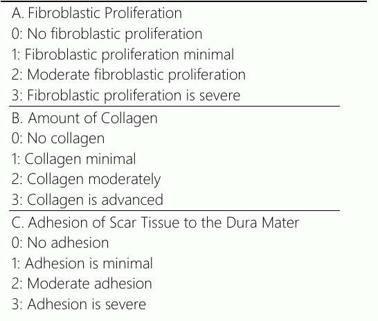
Results
Macroscopic
After sacrification, the entire vertebral column was separated from the surrounding soft tissues and sliced at 5mm intervals. Macroscopically, an increase in fibrous tissue was observed in the laminectomy areas. Large spaces were observed between the spinal cord, dura mater, and fibrous tissue formed macroscopically in the subjects using ADCON-L. The other two groups had no significant difference (Table 2).
Microscopic - Fibroblastic Proliferation
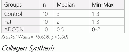
When the subjects sacrificed at the end of the 12th week were evaluated regarding fibroblastic proliferation, six advanced, three moderate, and one minimal fibroblastic proliferation were observed in group-1. In group-2, 3 had severe, 5 had moderate fibroblastic proliferation, and 2 had minimal fibroblastic proliferation.
In group-3, moderate fibroblastic proliferation was observed in one and minimal fibroblastic proliferation in 4, while fibroblastic proliferation was not detected in 5. In addition, we observed fibroblastic proliferation was more intense in the parts adjacent to paraspinal muscles (Table 2). When the groups were compared regarding fibroblastic proliferation, no difference was observed between group-1 and group-2. A difference was found when group-1 [3(1–3)] and group-3 were compared [0.5(1, 2)] (p<0.001). When group-2 and group-3 [0.5 (1, 2)] were compared, it was determined that fibroblastic proliferation was statistically higher in group-2 [2 (1–3)] (p<0.001) (Table 2).
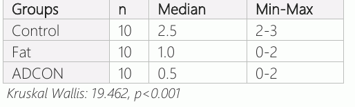
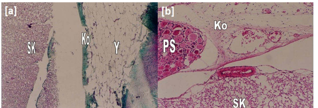
All the groups were evaluated regarding collagen synthesis. In the group-1, advanced collagen synthesis was observed in five and moderate collagen synthesis in five. In group-2 (Fat), moderate collagen synthesis was detected in five and minimal collagen synthesis in seven, while collagen synthesis was not detected in two (Figure 1a). In group-3 (ADCON), moderate collagen synthesis was detected in one, minimal collagen synthesis in four, and no collagen synthesis in five (Figure 1b). When the groups were compared in terms of collagen amount, no statistical difference was found between group-2 [1.0 (0-2)] and group-3 [0.5 (0-2)] (p>0.05). The amount of collagen was found to be more intense in group-1 [2.5 (2, 3)] than in group-2 [1.0 (0-2)] (p<0.001). When group-1 and group-3 were compared in terms of collagen amount, the amount of collagen in group-3 was less (p<0.001) (Table 3).
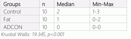
Adhesion of Scar Tissue to the Dura Mater When evaluated regarding the adhesion of the scar tissue to the dura mater, four severe, four moderate, and 2 minimal adhesions were observed in group-1. In group-2, moderate adhesion was observed in four, minimal adhesion was observed in two, and no adhesion was observed in four (Figure 2a). None of the group-3 had scar tissue adhesion to the dura mater (Figure 2b).
When the groups were evaluated statistically regarding the adhesion of the scar tissue to the dura, there was a significant difference between group-1 and group-2, and adhesion was less in group-2 (p<0.01). Adhesion was significantly less in group-3 compared to group-2 (p<0.001). When group-1 and group-3 were compared in terms of the adhesion of the scar tissue to the dura, the adhesion was significantly less in group-3 (p<0.001) (Table 4).
In the samples obtained from the control subjects, it was observed that there was intense scar tissue in the laminectomy area, and the scar tissue progressed toward the spinal canal and adhered to the dura mater. The scar tissue was an extension of the scar tissue under the paraspinal muscles rather than originating from the inside of the spinal canal. Adhesion was higher in the parts of the laminectomy defect close to the bone in the free fat graft group. In addition, in some samples, it was observed that the fat graft was not wholly absorbed and dispersed in the form of islets (Figure 1a, 2a). In cross-sections of the distances using ADCON- L, gaps were observed between the dura mater around the medulla spinalis and surrounding muscle layer and nerve roots (Figure 3). No residual ADCON-L was found in any subjects, and ADCON-L was resorbed. No foreign body reaction to ADCON-L was observed in any subjects (Figure 3). In some subjects, foreign body type inflammatory granulation tissue consisting of multinuclear giant cells, fibroblasts, capillary vessel proliferation, and a small number of PMNL (Polymorph Core Leukocyte) that phagocytized them was seen around suture material used in some subjects.
Discussion
In spinal surgery, epidural fibrosis, which develops on the dural sac and around the nerve root, especially after decompression and LDH surgeries, causes neurological symptoms with the effect of traction or compression, causes the patient's complaints to persist, and/or causes additional complaints, is one of the problems that still awaits a solution in spinal surgery. Research on the causes, prevention, and treatment of epidural fibrosis is ongoing, and opinions differ. Epidural fibrosis is one of the causes accepted to cause FBS syndrome, and it is reported to cause clinical complaints at a rate of 6-25% (4, 21, 22).
Histological examinations in experimental studies showed that after lumbar laminectomy and discectomy, a large hematoma appeared in the first two days, and fibrosis began in 2 weeks and became evident in 4 weeks (23, 24). In a study, he et al. examined rats by sacrificing them on the 8th, 15 , 30 , and 90 days (20). They reported that the laminectomy membrane started to appear on the 15th day, the laminectomy site was closed after 30 days, and after 90 days, massive scar tissue was located on the defect and adhered to the dura mater (20). Songer et al. reported that the adhesion maturity or strength was not different at the 12th and 26th weeks in their examination in dogs at the 2nd, 4th, 12th, and 26th weeks (8). Our study aimed to see the final state of scar tissue formation by sacrificing all subjects at the end of the 12th week.

Gerszten et al. found in their study on dogs that the laminectomy defect was covered with scar tissue originating from the paraspinal muscles and advancing toward the epidural region (25). Yang et al. (26) and Sobti et al. (27) found similar results in similar studies. Our study found that fibroblastic proliferation was more in the parts close to the paraspinal muscles. This finding can be interpreted as a substance that will be placed in the laminectomy or laminectomy area and will prevent the progression of the scar and may block the laminectomy membrane. When groups were evaluated regarding fibroblastic proliferation, there was no significant difference between the control and fat graft groups. In contrast, the ADCON-L group's fibroblastic proliferation was considerably lower than the other two groups. La Rocca et al., in their study on dogs, found that the laminectomy defect was covered with scar tissue originating from the paraspinal muscles and advancing toward the epidural region. (28). Keller et al. and Vaquero et al. found similar results in similar studies. (29, 30). Our study found that fibroblastic proliferation was more in the parts close to the paraspinal muscles. This finding can be interpreted as a substance that will be placed in the laminectomy or laminectomy area and will prevent the progression of the scar and may block the laminectomy membrane. When the groups were evaluated regarding fibroblastic proliferation, there was no significant difference between the control and fat graft groups. In contrast, the ADCON-L group's fibroblastic proliferation was considerably lower than the other two groups.
The primary building material of epidural fibrosis or scar tissue is collagen. Fibroblasts form collagen synthesis during wound healing. Barbera et al. determined that the main component originating from the dorsal spinal muscles and filling the laminectomy defect is collagen, and its amount is directly proportional to the size of the scar tissue (6). Massie et al. determined that main component originating from the dorsal spinal muscles and filling the laminectomy defect is collagen, and its amount is directly proportional to the size of the scar tissue (31). Ding et al. reached the same conclusion in a similar study (32). Our study observed that the amount of collagen in the scar tissue showed parallelism with fibroblastic activity in the control and ADCON-L groups but not in the fat graft group.
Autogenous fat grafts are the most effective substance in preventing epidural scarring among the substances (33, 34). Vediappan et al. showed in their study that even if the fat graft is alive, it does not prevent the formation of fibrosis in the anterior/antero-lateral regions of the nerve root emerging from the spinal cord (35). In many cases, the spinal cord was compressed posteriorly by the fat graft. The fat graft may compress the dura posteriorly. In our study, we found moderate scar formation and, in parallel, moderate perineural adhesions in the subjects who used fat grafts. The results were significant compared to the control.
It is known that ADCON-L, a carbohydrate polymer developed in recent years, is highly effective in preventing epidural fibrosis, and this is proportional to clinical improvement (11, 36). Kazanci et al. applied ADCON-L to rats after laminectomy and evaluated the scar gross anatomically by microdissection, sacrificing eight weeks later. They reported that ADCON- L reduced epidural fibrosis compared to the control group (37). Our results were statistically significant, and no adhesion was observed in any subjects using ADCON-L. Dermirel et al. applied ADCON-L to rats after laminectomy, sacrificed the subjects after 2, 4, 8, 16 and 26 weeks, and investigated the effect of ADCON-L gross anatomically and histologically (11). The surgical area contains minimal scarring, and the dura can be easily seen; In the histological study reported that ADCON-L was resorbed in the fourth week, and there was minimal or no foreign body and inflammatory response to ADCON-L. Kasimcan et al. also reported that the absorption period of ADCON-L was four weeks (38). In our study results, no foreign body reaction or inflammatory response was observed in any of the subjects using ADCON- L, no residual ADCON-L was found, and it was thought to be completely resorbed. In order to prevent epidural fibrosis, the substance to be used must be resorbed over time because the substance placed for a long time increases the risk of infection.
Conclusion
Epidural fibrosis, a physiological consequence of wound healing after laminectomies, occurred in varying degrees except in ADCON-L injected subjects. We concluded that free autogenous fat grafting does not prevent epidural fibrosis formation at an acceptable level. We concluded that ADCON-L would not cause FBS syndrome because there are no side effects associated with its use, it is absorbed over time, it is easy to apply, the risk of infection is low, and it is an effective substance in the prevention of epidural fibrosis and the second operation can be performed more comfortably and safely in patients who need it. Due to the limited number of studies in the literature, our study makes an essential contribution to the literature. Since the relationship between epidural fibrosis and findings has not been fully demonstrated, we think comparative clinical studies should support the efficacy of ADCON-L.
Declarations
- Author contributions (CRediT)
- All authors contributed equally to all 14 CRediT roles (Conceptualization, Data curation, Formal analysis, Funding acquisition, Investigation, Methodology, Project administration, Resources, Software, Supervision, Validation, Visualization, Writing – original draft, Writing – review & editing).
- Funding
-
Funding information was not reported in the published version of this article.
- Conflicts of interest
- Conflict of I nterests The authors declared no conflicts of interest concerning financial resources, the authorship, and the publication of this article.
- Ethics approval
- Ethics Committee approval was obtained from the Erciyes University, Faculty of Medicine, the experimental research ethics Committee (Approval No: 02.02.43, 2003).
- Data availability
- A data availability statement was not reported in the published version of this article.
References (38)
33 of 38 references are linked to their DOI.
- Bozkurt I, Kazanci A, Gurcan O, Gurcay AG, Arikok AT, Bavbek M. Spinal epidural fibrosis following hemostatic agent employment. Br J Neurosurg. 2023;37(2):137-41 PubMed Google Scholar
- Ismailoglu O, Kizilay Z, Cetin NK, Topcu A, Berber O. Effect of Curcumin on the Formation of Epidural Fibrosis in an Experimental Laminectomy Model in Rats. Turk Neurosurg. 2019;29(3):440-4 DOI Crossref PubMed Google Scholar
- Lin CL, Jou IM, Wu CY, Kuo YR, Yang SC, Lee JS, et al. Topically Applied Cross-Linked Hyaluronan Attenuates the Formation of Spinal Epidural Fibrosis in a Swine Model of Laminectomy. Sci Rep. 2019;9(1):14613 DOI Crossref PubMed Google Scholar
- Papalia GF, Russo F, Vadala G, Pascarella G, De Salvatore S, Ambrosio L, et al. Non-Invasive Treatments for Failed Back Surgery Syndrome: A Systematic Review. Global Spine J. 2022:21925682221141385 DOI Crossref PubMed Google Scholar
- Martin-Ferrer S. Failure of autologous fat grafts to prevent postoperative epidural fibrosis in surgery of the lumbar spine. Neurosurgery. 1989;24(5):718-21 DOI Crossref PubMed Google Scholar
- Barbera J, Gonzalez J, Esquerdo J, Broseta J, Barcia-Salorio JL. Prophylaxis of the laminectomy membrane. An experimental study in dogs. J Neurosurg. 1978;49(3):419-24 DOI Crossref PubMed Google Scholar
- Colak A, Bavbek M, Aydin NE, Renda N, Acikgoz B. Effect of CO2 laser on spinal epidural fibrosis. Acta Neurochir (Wien). 1996;138(2):162-6 DOI Crossref PubMed Google Scholar
- Songer MN, Rauschning W, Carson EW, Pandit SM. Analysis of peridural scar formation and its prevention after lumbar laminotomy and discectomy in dogs. Spine (Phila Pa 1976). 1995;20(5):571-80; discussion 9-80 DOI Crossref PubMed Google Scholar
- Ceviz A, Arslan A, Ak HE, Inaloz S. The effect of urokinase in preventing the formation of epidural fibrosis and/or leptomeningeal arachnoiditis. Surg Neurol. 1997;47(2):124-7 DOI Crossref PubMed Google Scholar
- Hinton JL, Jr., Warejcka DJ, Mei Y, McLendon RE, Laurencin C, Lucas PA, et al. Inhibition of epidural scar formation after lumbar laminectomy in the rat. Spine (Phila Pa 1976). 1995;20(5):564 -70 DOI Crossref PubMed Google Scholar
- Demirel E, Yildiz K, Cadirci K, Aygun H, Senocak E, Gundogdu B. Effect of platelet-rich fibrin on epidural fibrosis and comparison to ADCON(R) Gel and hyaluronic acid. Acta Orthop Traumatol Turc. 2018;52(6):469-74 PubMed Google Scholar
- Albinana-Cunningham JN, Ripalda-Cemborain P, Labiano T, Echeveste JI,Granero-Molto F, Alfonso- Olmos M. Mechanical barriers and transforming growth factor beta inhibitor on epidural fibrosis in a rabbit laminectomy model. J Orthop Surg Res. 2018;13(1):72 DOI Crossref PubMed Google Scholar
- Kasimcan MO, Bakar B, Aktas S, Alhan A, Yilmaz M. Effectiveness of the biophysical barriers on the peridural fibrosis of a postlaminectomy rat model: an experimental research. Injury. 2011;42(8):778-81 65 77 Barrier Gel and Free Fat Graft Sami Engin et al. DOI Crossref PubMed Google Scholar
- Petrie JL, Ross JS. Use of ADCON-L to inhibit postoperative peridural fibrosis and related symptoms following lumbar disc surgery: a preliminary report. Eur Spine J. 1996;5:S10-7 DOI Crossref PubMed Google Scholar
- Frederickson RC. ADCON-L: a review of its development, mechanism of action and preclinical data. Eur Spine J. 1996;5:7 -9 DOI Crossref PubMed Google Scholar
- Einhaus S, Robertson J, Wujek J, Ahmad S, editors. Reduction of peridural fibrosis after lumbar laminectomy and discectomy in dogs by a carbohydrate polymer gel (ADCON-L). Annual Meeting of American Association of Neurological Surgeons, Boston, MA; 1993 PubMed Google Scholar
- Petersen J, Russell L, Andrus K, MacKinnon M, Silver J, Kliot M. Reduction of extraneural scarring by ADCON-T/N after surgical intervention. Neurosurgery. 1996;38(5):976-83 DOI Crossref PubMed Google Scholar
- Mastronardi L, Pappagallo M, Puzzilli F, Tatta C. Efficacy of the morphine-Adcon-L compound in the management of postoperative pain after lumbar microdiscectomy. Neuro surgery. 2002;50(3):518-24; discussion 24-5 DOI Crossref PubMed Google Scholar
- Einhaus SL, Robertson JT, Dohan FC, Jr., Wujek JR, Ahmad S. Reduction of peridural fibrosis after lumbar laminotomy and discectomy in dogs by a resorbable gel (ADCON-L). Spine (Phila Pa 1976). 1997;22(13):1440-6; discussion 6-7 DOI Crossref PubMed Google Scholar
- He Y, Revel M, Loty B. A quantitative model of post-laminectomy scar formation. Effects of a nonsteroidal anti-inflammatory drug. Spine (Phila Pa 1976). 1995;20(5):557-63. DOI Crossref PubMed Google Scholar
- Samy Abdou M, Hardy RW, Jr. Epidural fibrosis and the failed back surgery syndrome: history and physical findings. Neurol Res. 1999;21 Suppl 1:S5-8 DOI Crossref PubMed Google Scholar
- Geudeke MW, Krediet AC, Bilecen S, Huygen F, Rijsdijk M. Effectiveness of Epiduroscopy for Patients with Failed Back Surgery Syndrome: A Systematic Review and Meta-analysis. Pain Pract. 2021;21(4):468-81 DOI Crossref PubMed Google Scholar
- Yue J, Han Q, Chen H, Zhang A, Liu Y, Gong X, et al. Artificial lamina after laminectomy: Progress, applications, and future perspectives. Front Surg. 2023;10:1019410 DOI Crossref PubMed Google Scholar
- Babar S, Saifuddin A. MRI of the post-discectomy lumbar spine. Clin Radiol. 2002;57(11):969-81 DOI Crossref PubMed Google Scholar
- Gerszten PC, Moossy JJ, Flickinger JC, Gerszten K, Kalend A, Martinez AJ. Inhibition of peridural fibrosis after laminectomy using low-dose external beam radiation in a dog model. Neurosurgery. 2000;46(6):1478-85 DOI Crossref PubMed Google Scholar
- Yang K, Ji C, Luo D, Li K, Xu H. Lumbar laminotomy and replantation for the treatment of lumbar spinal epidural lipomatosis. Medicine (Baltimore). 2021;100(30):e26795 DOI Crossref PubMed Google Scholar
- Sobti S, Grover A, John BPS, Grewal SS, George UB. Prospective Randomized Comparative Study to Evaluate Epidural Fibrosis and Surgical Outcome in Patients Undergoing Lumbar Laminectomy with Epidural Autologous Free Fat Graft or Gelfoam: A Preliminary Study. Int J Appl Basic Med Res. 2018;8(2):71-5 DOI Crossref PubMed Google Scholar
- LaRocca H, Macnab I. The laminectomy membrane. Studies in its evolution, characteristics, effects and prophylaxis in dogs. J Bone Joint Surg Br. 1974;56(3):545-50 DOI Crossref PubMed Google Scholar
- Vaquero J, Arias A, Oya S, Martinez R, Zurita M. Effect of fibrin glue on postlaminectomy scar formation. Acta Neurochir (Wien). 1993;120(3-4):159-63 DOI Crossref PubMed Google Scholar
- Keller JT, Dunsker SB, McWhorter JM, Ongkiko CM, Saunders MC, Mayfield FH. The fate of autogenous grafts to the spinal dura. An experimental study. J Neurosurg. 1978;49(3):412-8 DOI Crossref PubMed Google Scholar
- Massie JB, Schimizzi AL, Huang B, Kim CW, Garfin SR, Akeson WH. Topical high molecular weight hyaluronan reduces radicular pain post laminectomy in a rat model. Spine J. 2005; 5(5):494-502 DOI Crossref PubMed Google Scholar
- Ding Q, Wei Q, Sheng G, Wang S, Jing S, et al. The Preventive Effect of Decorin on Epidural Fibrosis and Epidural Adhesions After Laminectomy. Front Pharmacol. 2021;12: 774316 DOI Crossref PubMed Google Scholar
- Kanamori M, Kawaguchi Y, Ohmori K, Kimura T, Tsuji H, Matsui H. The fate of autogenous free-fat grafts after posterior lumbar surgery: part 2. Magnetic resonance imaging and histologic studies in repeated surgery cases. Spine (Phila Pa 1976). 2001;26(20):2264-70 DOI Crossref PubMed Google Scholar
- Bryant MS, Bremer AM, Nguyen TQ. Autogeneic fat transplants in the epidural space in routine lumbar spine surgery. Neurosurgery. 1983;13(4):367-70 DOI Crossref PubMed Google Scholar
- Vediappan RS, Mascarenhas A, Nguyen-Hoang A, et al. Prevention of peridural adhesions in spinal surgery: Assessing safety and efficacy of Chitogel with Deferiprone in a sheep model. J Clin Neurosci. 2020;72:378-85 DOI Crossref PubMed Google Scholar
- Feng MX, Hong D. Update on prevention of epidural adhesion after lumbar laminectomy. Zhongguo Gu Shang. 2015;28(11):1064-8 PubMed Google Scholar
- Kazanci A, Gurcan O, Gurcay AG, Onder E, et al. Effects of Topical Cova, Tisseel(R) and Adcon(R) Gel Application on the Development of Spinal Peridural Fibrosis: An Experimental Study in Rats. Turk Neurosurg. 2017;27(6): 962-8 PubMed Google Scholar
- Kasimcan MO, Bakar B, Aktas S, Alhan A, Yilmaz M. Effectiveness of the biophysical barriers on the peridural fibrosis of a postlaminectomy rat model: experimental research. Injury. 2011; 42(8): 778-81 DOI Crossref PubMed Google Scholar
Respond to this article
Comments on a published article are welcome as a Letter to the Editor.
Get an e-mail when new articles in your fields are published
Questions to the authors
Questions are moderated: an editor approves each question, the authors answer, and an editor approves the answer before both appear here.
No answered questions yet.
Ask the authors a question
Discussion
Comments are moderated: they appear after the writer confirms the e-mail address and an editor approves them. Each comment carries a conflict-of-interest statement.
No comments yet.
Write a comment
Read the full text
Open the PDF here
Loading the PDF…
If the PDF does not show here, use the download button above.
How to cite this article
Engin S, Akbas A, Kizilkaya V. The Effect of Barrier Gel and Free Fat Graft on the Formation of Epidural Fibrosis in the Laminectomy Model. Ulutas Med J. 2023;9(2):58-65. doi:10.5455/umj.20230510095834
Engin, S., Akbas, A., & Kizilkaya, V. (2023). The Effect of Barrier Gel and Free Fat Graft on the Formation of Epidural Fibrosis in the Laminectomy Model. The Ulutas Medical Journal, 9(2), 58–65. https://doi.org/10.5455/umj.20230510095834
Engin, S., Akbas, A. and Kizilkaya, V. (2023) 'The Effect of Barrier Gel and Free Fat Graft on the Formation of Epidural Fibrosis in the Laminectomy Model', The Ulutas Medical Journal, 9(2), pp. 58–65. doi: 10.5455/umj.20230510095834.