The Ulutas Medical Journal · ISSN 2149-0430 (print), 2149-388X (online) · https://www.ulutasmedicaljournal.com/?mno=170390
Original Article · Open access
The Degree of Correction Correlation with Clinical Outcomes in Adult Scoliosis Patients after Posterior Instrumentation Surgery
- Fatih Sultan Mehmet Education and Training Hospital Orthopedics and Traumatology Department, Istanbul, TURKEY ROR
- Istanbul Medipol University, Camlica Hospital, Orthopedics and Traumatology Department, Istanbul, TURKEY ROR
Abstract
- Introduction
- Restoring sagittal and coronal parameters is a primary concern in adult scoliosis surgery, yet it remains challenging due to the matured bony structure. This study aims to analyze the relationship between clinical outcomes and the degree of correction achieved through posterior instrumentation surgery in adult scoliosis.
- Materials and Methods
- This study evaluated the clinical and radiological results of 27 adult scoliosis patients who underwent posterior instrumentation, decompression, and correction surgery. Preoperative and follow-up X-rays were performed to assess Cobb angle, sagittal balance, and coronal balance. Visual Analog Score (VAS) and Oswestry Disability Index (ODI) were evaluated preoperatively and at follow-up.
- Results
- The study comprised 27 patients (18 female, 9 male) with a mean age of 68.2 years. Preoperative Cobb angle averaged 42.4°, reducing to 5.9° at final follow-up. Sagittal balance decreased from 30.6 mm preoperatively to 9.7 mm at final follow-up. Coronal balance improved from 22.3 mm preoperatively to 10.5 mm at 2-year follow-up. Preoperative Cobb angle showed a strong correlation with preoperative ODI (p=0.028) and postoperative ODI (p=0.014). Preoperative sagittal balance also correlated strongly with pre and postoperative ODI. However, postoperative Cobb angle and sagittal balance did not correlate with postoperative ODI (p<0.05).
- Conclusion
- While restoring sagittal and coronal parameters is a primary concern in adult scoliosis surgery, the degree of correction achieved does not necessarily correlate with clinical outcomes. Surgeons should consider this when planning surgery, and patients should be informed accordingly.
Keywords
Full text
Introduction
Adult scoliosis (AS) is a spinal deformity characterized by a Cobb angle exceeding 10 degrees, significantly impacting patients' quality of life (1). The prevalence of AS varies widely, ranging from 1.4% to 32% in different studies (2, 3). Advanced AS may experience significant pain and disability, together with the aesthetic concern caused by the deformity (4–6). Over the past few decades, the treatment options for AS have expanded, with combined anterior-posterior fusion being a traditional approach (5–8). However, anterior approaches have draw backs, such as affecting respiratory function and increasing operative time (9).
Recent comparisons between posterior and combined anterior-posterior approaches have suggested that the posterior approach may result in less deformity correction, higher rates of pseudarthrosisand persistent spinal instability (10, 11). Pain and disability are common in AS when compared to adolescent idiopathic scoliosis (12, 13). In a study, the prevalence of scoliosis in people over 60 years of age was 68 percent (14). While some publications advocate conservative treatment, operative treatment has shown success in numerous studies (12–16). We hypothesized that correlation degree and clinical outcomes correlate and may have an impact on the AS patients. The aim of this study is to demonstrate if less deformity correction with posterior instrumentation technique and the amount of correction in posterior instrumentation has a correlation with clinical outcomes in AS.
Materials and Methods
Subjects
This retrospective research analysed clinical record, demographic and radiological outcome of patients who underwent decompression, posterior instrumentation, and correction surgery for AS between February 2018 and January 2021. All procedures were performed by a single senior surgeon in a single center. A total of 30 patients were initially considered; however, two patients were excluded due to systemic connective tissue disorders, and one patient was excluded due to a neurological disease. The analysis involved a comprehensive review of radiological and clinical data extracted from medical records. All patients had signed an informed consent.
Methods
Surgical procedures were conducted under hypotensive anaesthesia, and intraoperative blood salvage techniques were employed. To ensure patient safety, routine intraoperative monitoring included somatosensory-evoked potential and the motor-evoked potential assessments. Lenke-Silva classification was used in the treatment planning (17). Decompression and lumbar posterior instrumentation were performed in Type 3 patients; decompression + anterior fusion (TLIF) + posterior fusion in Type 4 patients; decompression + fusion extended to the thoracic vertebrae ± osteotomy in Type 5 patients; and Schwab Type 3 osteotomy was performed in non-flexible Type 6 patients (18). Laminectomy and decompression were performed at all stenotic levels, and a bilateral pedicle screw fixation was applied at all operated levels. The posterior decompression was followed by deformity correction, which included the cantilever method, in-situ bending and contouring, as well as compression and distraction maneuvers.
To assess functional outcomes, Visual Analog Score (VAS) and Oswestry Disability Index (ODI) scores were collected preoperatively, in the early postoperative period, and at the last follow-up. Spinal magnetic resonance imaging and multi-slice computed tomography were utilized to detect the presence of spinal stenosis, intraspinal anomalies, and tandem cervical and dorsal spinal stenosis. Radiographic parameters and scores were evaluated using data collected preoperatively, at the postoperative 3rd month, and at the 2nd-year follow-up. Patients were also queried about their daily activities and pain levels.
Statistical analysis
Statistical analysis was performed using SPSS version 22 software (SPSS Inc., Chicago, IL, USA). Data were analysed using the Wilcoxon matched-pairs signed-rank test for paired data, with statistical significance defined as p<0.05 for all analyses.
Results
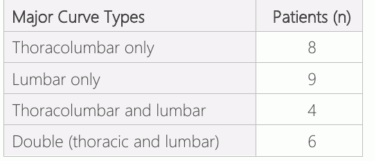
A total of 27 patients participated in the study, with 9 being male and 18 females. The mean age at the time of surgery was 68.2 years, with a range from 57 to 80 years. The average follow-up period lasted 28.3 months, ranging from 24 to 43 months, and the mean duration of hospitalization was 5.2 days, with a range of 2 to 7 days. Perioperative blood loss averaged 554 cc, ranging from 460 to 738 cc. The mean duration of the surgical procedure was 231 minutes, with a range of 187 to 345 minutes. The distribution of major curve types is detailed in Table 1.
According to patient recall, the average age at the time of AS diagnosis was 51.5 years, with a range from 43 to 72 years. The mean time elapsed between scoliosis diagnosis and surgery was 39.2 years, ranging from 9 to 67 years. Conversely, the average interval between the onset of symptoms (back pain and neurogenic claudication) and surgery was 5.4 years, with a range of 1 to 18 years. The patients were classified according to the Lenke-Silva classification, with 12 classified as Type 3, 8 as Type 4, 4 as Type 5, and 4 as Type 6. Schwab Type 3 osteotomy was performed in 4 patients with Lenke-Silva Type 6 deformity.
The upper level of fusion was T10 in 16 patients, T9 in 8 patients, and T2 in 4 patients. The fusions terminated at L5 in 8 patients, S1 in 13 patients, and the iliac bone in 7 patients. The mean Cobb angle measured 42.4° preoperatively, decreasing to 6.2° in the early postoperative period and further to 5.9° at the 2nd-year final follow-up (p<0.01). Similarly, the mean sagittal balance was 30.6 mm preoperatively, 15.4 mm in the early postoperative phase, and 9.7 mm at the 2nd-year final follow-up (p<0.01). The mean preoperative coronal balance was 22.3 mm preoperatively, 12.7 mm in the early postoperative period, and 10.5 mm at the 2nd-year final follow-up(p<0.01)(Table 2). ODI significantly improved from an initial score of 46.2, ranging from 38 to 49, to 25.8, with a range of 17 to 32, at the 2nd-year follow-up (p<0.01). Additionally, the preoperative Visual Analog Score (VAS) analogue score, averaging 7.8 with a range of 7 to 9, reduced to 2.8, ranging from 0 to 4, at the 2nd-year follow-up (p<0.01) (Table 3). Correlation analysis revealed no significant differences between changes in preoperative and 2nd-year follow-up Cobb angles and changes in preoperative and 2nd-year follow-up ODI and VAS values.
Notably, preoperative Cobb angle exhibited a strong correlation with both preoperative ODI (p=0.028) and postoperative ODI (p=0.014). Additionally, preoperative sagittal balance correlated with both preoperative and postoperative ODI. However, postoperative Cobb angle and sagittal balance showed no correlation with postoperative ODI (p<0.05). All patients who were employed before surgery were able to return to their previous jobs by the final follow-up assessment. None of the patients reported restrictions in their daily activities compared to their preoperative experiences. At the final follow-up assessment, all patients were physically independent in their daily activities. Only one patient among the cohort experienced a superficial infection, which was successfully treated with antibiotics and superficial debridement. At the end of the 2nd-year follow-up, no complications were observed in any of the patients.


Discussion
AS can manifest as a progression of adolescent scoliosis or develop de novo in adulthood. Unlike often-asymptomatic nature of idiopathic scoliosis, AS is typically characterized by pain as a predominant symptom. The main need for treatment mostly is for symptoms of excessive pain unlike balancing sagittal and coronal deformities in AS. The literature reports varying rates of pain incidence in AS, ranging from 40 to 90% (2, 19, 20). Smith et al. conducted a study of surgical and conservative treatments for AS and found that surgery significantly reduces pain (21). A systematic review by Yadla et al. demonstrated a decrease in the ODI score from 41.2 to 15.7, along with an average correction of 26.6 degrees in major curvature postoperatively (22). Another study by Ledonio et al. showed reductions in ODI and Visual Analog Score (VAS) from 34.2 and 4.5 to 20.1 and 1.98, respectively, after surgery, with an average Cobb angle reduction of 11.1 degrees (23). In our study, we observed a decrease in ODI scores from 46.2 to 25.8 and VAS scores from 7.8 to 2.8 postoperatively. Additionally, we achieved a substantial average correction of 36.5 degrees in the major curve.
Providing spinal balance is one of the primary goals in AS surgery. Coronal and sagittal balance were found to be associated with the patient's functional capacity, pain level and quality of life (24, 25). Ledonio et al. reported an average improvement of 7.7 degrees in coronal balance and 4.6 mm in sagittal balance in a review of 197 patients’ post-surgery (23). In our study, we observed a decrease in coronal balance from 22.3 mm to 10.5 mm and sagittal balance from 30.6 mm to 9.7 mm in the final follow-up.
The interest in surgical treatment for AS has grown over time due to the potential problems untreated adults may face, including spinal osteoarthritis, progressive deformity, spinal stenosis leading to radiculopathy, muscle pain due to coronal and sagittal imbalance, and cosmetic concerns. The objectives of surgical treatment include pain reduction, halting curve progression, restoring respiratory function, and enhancing cosmetic appearance. Surgery in patients presents unique challenges compared to adolescents, including issues such as osteo-penia or osteoporosis, disc degeneration, spinal rigidity, and sagittal-coronal imbalances, all of which necessitate thorough preoperative evaluation (16). An additional challenge is the spinal rigidity in adult patients, which can be mitigated through complete posterior decompression during surgery.
Spinal stenosis is a critical concern in the AS, leading to the nerve root ischemia due to compression (26). In the patients with scoliosis, spinal stenosis presents as a complex condition characterized by asymmetric collapse, rotation, lateral listhesis, spondylolisthesis, and facet joint hypertrophy. Notably, some studies have failed to establish a clear correlation between the severity of pain and the magnitude of the curvature (26, 27). The ideal treatment for spinal stenosis involves full decompression at the stenotic level, followed by rigid fixation. Clinical and radiological evaluation plays a primary role in determining the decompression level. In our study, effective posterior decompression facilitated correction and reduced spinal rigidity. Subsequently, rigid fixation was achieved by placing pedicle screws bilaterally at each level. Laminectomy performed in the stenotic region was beneficial in alleviating leg pain, and low back pain relief was achieved through reduction and stabilization using instrumentation. Our conclusion from these findings is that the extent of correction may not correlate with clinical outcomes in AS, but the success of the surgery can be attributed to the stabilization and decompression of stenotic areas.
The limitations of our study are its retrospective design and a relatively small number of cases. However, the strengths of this study lie in its single-surgeon, single-center approach, as well as meticulous data collection throughout the study period, with no lost follow-up cases. Future randomized prospective studies with larger sample sizes will further contribute to the literature on this subject.
In summary, despite the primary focus on restoring sagittal and coronal parameters in AS surgery, our study suggests that the extent of restoration does not necessarily correlate with improvements in clinical outcomes. This conclusion should be taken into account when planning surgery and patients should be well-informed in term of outcomes. This study demonstrates the strategic importance of several points in AS surgery.
Declarations
- Author contributions (CRediT)
- All authors contributed equally to all 14 CRediT roles (Conceptualization, Data curation, Formal analysis, Funding acquisition, Investigation, Methodology, Project administration, Resources, Software, Supervision, Validation, Visualization, Writing – original draft, Writing – review & editing).
- Funding
-
Funding information was not reported in the published version of this article.
- Conflicts of interest
- The Authors declare no conflict of interests.
- Ethics approval
- All patients had signed an informed consent.
- Data availability
- A data availability statement was not reported in the published version of this article.
References (27)
27 of 27 references are linked to their DOI.
- Bunnell WP. The natural history of idiopathic scoliosis before skeletal maturity. Spine. 1986;11(8):773-6. DOI Crossref PubMed Google Scholar
- Kostuik JP, Bentivoglio J. The incidence of low-back pain in adult scoliosis. Spine. 1981;6(3):268-73. DOI Crossref PubMed Google Scholar
- Pérennou D, Marcelli C, Hérisson C, Simon L. Adult lumbar scoliosis. Epidemiologic aspects in a low-back pain population. Spine. 1994;19(2):123-8. DOI Crossref PubMed Google Scholar
- Takahashi S, Delécrin J, Passuti N. Surgical treatment of idiopathic scoliosis in adults: an age-related analysis of outcome. Spine. 2002;27(16):1742-8. DOI Crossref PubMed Google Scholar
- Bradford DS, Tay BK, Hu SS. Adult scoliosis: surgical indications, operative management, complications, and outcomes. Spine. 1999;24(24):2617. DOI Crossref PubMed Google Scholar
- GRUBB SA, LIPSCOMB HJ, COONRAD RW. Degenerative adult onset scoliosis. Spine. 1988;13(3):241-5. DOI Crossref PubMed Google Scholar
- Grubb SA, Lipscomb HJ, Suh PB. Results of surgical treatment of painful adult scoliosis. Spine. 1994;19(14):1619-27. DOI Crossref PubMed Google Scholar
- Dick J, Boachie-Adjei O, Wilson M. One-stage versus two-stage anterior and posterior spinal reconstruction in adults. Comparison of outcomes including nutritional status, complications rates, hospital costs, and other factors. Spine. 1992;17(8 Suppl):S310-6. DOI Crossref PubMed Google Scholar
- Kim YJ, Lenke LG, Bridwell KH, Kim KL, Steger-May K. Pulmonary function in adolescent idiopathic scoliosis relative to the surgical procedure. JBJS. 2005;87(7):1534-41. DOI Crossref PubMed Google Scholar
- Lippman CR, Spence CA, Youssef AS, Cahill DW. Correction of adult scoliosis via a posterior-only approach. Neurosurgical focus. 2003;14(1):1-6. DOI Crossref PubMed Google Scholar
- Shimode M, Kojima T, Sowa K. Spinal wedge osteotomy by a single posterior approach for correction of severe and rigid kyphosis or kyphoscoliosis. Spine. 2002;27(20):2260-7. DOI Crossref PubMed Google Scholar
- Glassman SD, Schwab FJ, Bridwell KH, Ondra SL, Berven S, Lenke LG. The selection of operative versus nonoperative treatment in patients with adult scoliosis. Spine. 2007;32(1):93-7. DOI Crossref PubMed Google Scholar
- Glassman SD, Berven S, Kostuik J, Dimar JR, Horton WC, Bridwell K. Nonsurgical resource utilization in adult spinal deformity. Spine. 2006;31(8):941 -7. DOI Crossref PubMed Google Scholar
- Konieczny MR, Senyurt H, Krauspe R. Epidemiology of adolescent idiopathic scoliosis. Journal of children's orthopaedics. 2013;7(1):3-9. DOI Crossref PubMed Google Scholar
- Kelly A, Younus A, Lekgwara P. Adult degenerative scoliosis– A literature review. Interdisciplinary Neurosurgery. 2020; 20:100661. DOI Crossref PubMed Google Scholar
- Aebi M. The adult scoliosis. European spine journal. 2005; 14:925-48. DOI Crossref PubMed Google Scholar
- Silva FE, Lenke LG. Adult degenerative scoliosis: evaluation and management. Neurosurgical focus. 2010;28(3):E1. DOI Crossref PubMed Google Scholar
- Schwab F, Blondel B, Chay E, Demakakos J, Lenke L, Tropiano P, et al. The comprehensive anatomical spinal osteotomy classification. Neurosurgery. 2014;74(1):112-20. DOI Crossref PubMed Google Scholar
- Jackson RP, Simmons EH, Stripinis D. Incidence and severity of back pain in adult idiopathic scoliosis. Spine. 1983;8(7):749-56. DOI Crossref PubMed Google Scholar
- Nachemson A. Adult scoliosis and back pain. Spine. 1979;4(6):513-7. DOI Crossref PubMed Google Scholar
- Smith JS, Lafage V, Shaffrey CI, Schwab F, Lafage R, Hostin R, et al. Outcomes of operative and nonoperative treatment for adult spinal deformity: a prospective, multicenter, propensity-matched cohort assessment with minimum 2-year follow-up. Neurosurgery. 2016;78(6):851-61. DOI Crossref PubMed Google Scholar
- Yadla S, Maltenfort MG, Ratliff JK, Harrop JS. Adult scoliosis surgery outcomes: a systematic review. Neurosurgical focus. 2010;28(3):E3. 207 77 Correction and Outcome Correlation in Scoliosis Emre Bal and Onur Çetin DOI Crossref PubMed Google Scholar
- Ledonio CG, Polly Jr DW, Crawford III CH, Duval S, Smith JS, Buchowski JM, et al. Adult degenerative scoliosis surgical outcomes: A systematic review and meta-analysis. Spine Deformity. 2013;1(4):248-58. DOI Crossref PubMed Google Scholar
- Koller H, Pfanz C, Meier O, Hitzl W, Mayer M, Bullmann V, et al. Factors influencing radiographic and clinical outcomes in adult scoliosis surgery: a study of 448 European patients. European Spine Journal. 2016;25:532-48. DOI Crossref PubMed Google Scholar
- Cankaya D, Balci M, Deveci A, Yoldas B, Tuncel A, Tabak Y. Better life quality and sexual function in men and their female partners with short-segment posterior fixation in the treatment of thoracolumbar junction burst fractures. European Spine Journal. 2016;25:1128-34. DOI Crossref PubMed Google Scholar
- Lee BH, Moon S- H, Suk K-S, Kim H-S, Yang J-H, Lee H-M. Lumbar spinal stenosis: pathophysiology and treatment principle: a narrative review. Asian Spine Journal. 2020;14(5):682. DOI Crossref PubMed Google Scholar
- Amundsen T, Weber H, Lilleas F, Nordal HJ, Abdelnoor M, Magnaes B. Lumbar spinal stenosis: clinical and radiologic features. Spine. 1995;20(10):1178-86. DOI Crossref PubMed Google Scholar
Read the full text
Open the PDF here
Loading the PDF…
If the PDF does not show here, use the download button above.
How to cite this article
Bal E, Cetin O. The Degree of Correction Correlation with Clinical Outcomes in Adult Scoliosis Patients after Posterior Instrumentation Surgery. Ulutas Med J. 2023;9(3):202-207. doi:10.5455/umj.20230921092721
Bal, E., & Cetin, O. (2023). The Degree of Correction Correlation with Clinical Outcomes in Adult Scoliosis Patients after Posterior Instrumentation Surgery. The Ulutas Medical Journal, 9(3), 202–207. https://doi.org/10.5455/umj.20230921092721
Bal, E. and Cetin, O. (2023) 'The Degree of Correction Correlation with Clinical Outcomes in Adult Scoliosis Patients after Posterior Instrumentation Surgery', The Ulutas Medical Journal, 9(3), pp. 202–207. doi: 10.5455/umj.20230921092721.