The Ulutas Medical Journal · ISSN 2149-0430 (print), 2149-388X (online) · https://www.ulutasmedicaljournal.com/?mno=133517
Review Article · Open access
Profoundal Effects of Microcystin-LR Induced Cardiotoxicity in Mammals
Also available in: العربية · Türkçe · 中文
- Biochemistry Laboratory, Department of Zoology, Dr. Harisingh Gour Vishwavidyalaya, Sagar, Madhya Pradesh, India-470003 ROR
Abstract
Eutrophication and climate change have raised obnoxious cyanobacterial blooms, which have become a global public health problem. The most dangerous cyanotoxin found in water bodies is microcystin (MC), a byproduct of cyanobacterial metabolism. Several studies suggest that in addition to the liver, the heart may be another target organ for MCs poisoning. Recent research indicates a clear link between MC exposure and cardiotoxicity, posing a risk to human cardiovascular health. The cardiovascular system, which includes all tissues, cells, blood, and vascular tissues of the heart, can be unswervingly affected by MCs, resulting in abnormal cardiovascular system structure and/or function. This review concludes that MC cardiotoxicity may be linked to protein phosphatase suppression and the contractile unit, generation of contractile force, the role of intercellular junction proteins, and the role of the angiotensin signaling pathway in cardiomyocytes.
Keywords
Full text
Introduction
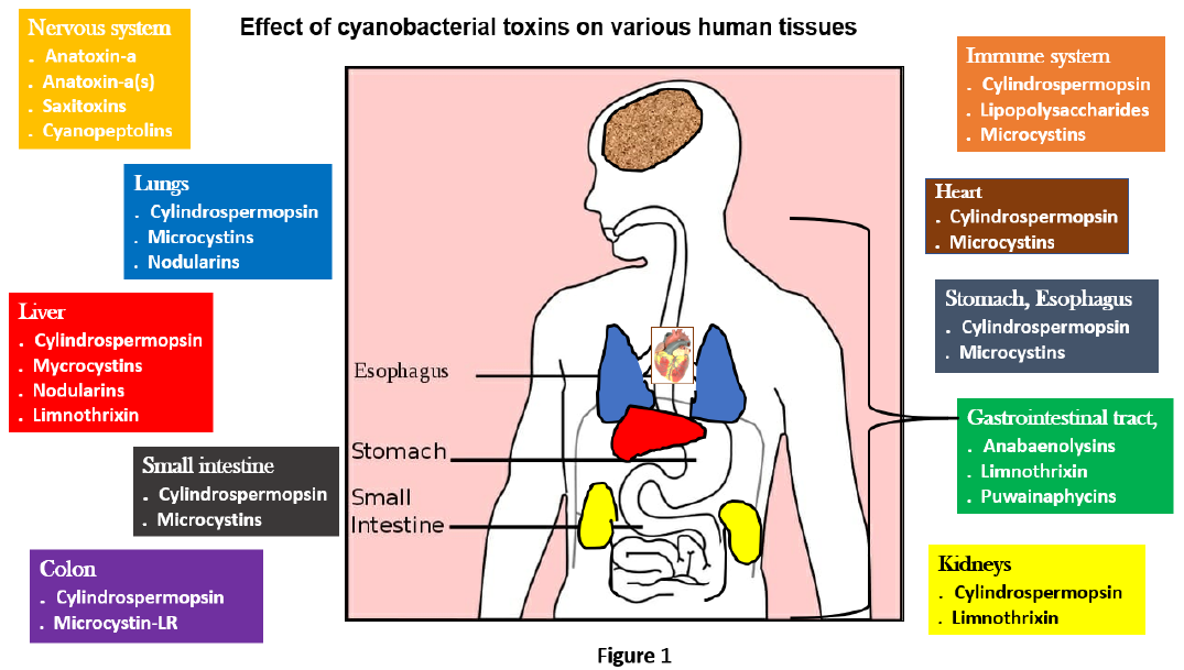
Cyanobacteria have a long evolutionary history that has allowed for the development of diverse adaptations and strategies that make them successful in many ecosystems worldwide (1). These adaptations allow cyanobacterial species to outcompete other algal species and dominate phytoplankton assemblages under certain conditions, resulting in harmful algal blooms (CyanoHABs). Cyanobacteria are found in fresh, brackish and marine water bodies worldwide. Toxins (cyanotoxins) are produced by some cyanobacteria species and have been found to be harmful to many tissues, including the heart (Figure 1). Microcystins (MCs) are thought to be the most frequent and harmful of these cyanotoxins (2). Cyanotoxins concentrate in aquatic organisms and are passed up the food chain through various trophic levels, which cause health problems to humans and animals (3). Monocyclic peptide microcystins (MCs) are the most prevalent, extensively dispersed, exceedingly poisonous and challenging to eliminate cyanotoxins by cyanobacterial blooms. Microcystis, Anabaena, Anabaenopsis, Oscillatoria and Nostoc produce MCs, a familyofcyclic heptapeptide cyanotoxins (4). The alteration in amino acids, peptide sequence, hydroxylation, epimerization and toxicity has resulted in the identification of more than 100 MCs isoforms (5). Microcystin-leucine arginine (MC-LR) is the most abundant and poisonous member of the MC family, between 46 and 99.8% of the total number of microcystins in natural waters are found in these microcystin forms. Water-soluble, stable in deionized or filtered water and stable even at temperatures up to 300 ℃ are all characteristics of MC-LR. The World Health Organization (WHO) has established a temporary limit of 1 μg/L MCs in drinking and bathing water for humans (6). By inhibiting protein phosphatase 1 and 2A, MCs act as hepatotoxins and tumour promoters (2). Protein phosphatase inhibition has been linked to MC cytotoxicity and tumor-promoting action, which is linked to gastric inflammation, hepatocellular carcinoma, lung cancer, kidney failure, cardiovascular disease and splenic illness (7–9). Eutrophication-induced cyanobacterial blooms and cyanotoxin pollution are becoming more common in aquatic habitats worldwide (10). Humans can come into contact with MCs in a variety of ways. These include consuming polluted water, coming into contact, breathing, eating tainted food, taking nutritional supplements containing algae and receiving hemodialysis (11).
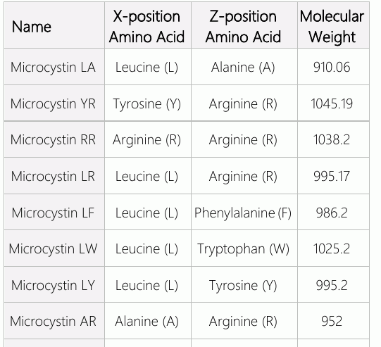
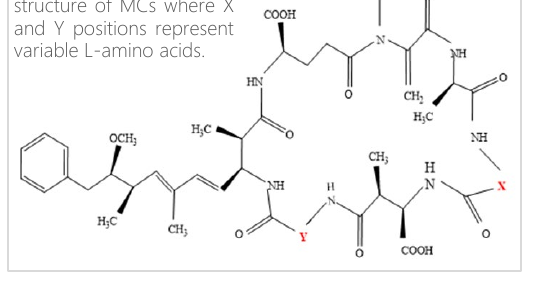
Microcystins are one of the most extensively studied cyanotoxins. Microcystin is a cyclic molecule with seven peptide-linked amino acids, with the terminal amino acids condensed (12). One of the riskiest microcystin variants is likely to be microcystin-LR (MC-LR). Protein phosphatases 1 and 2A (PP1 and PP2A) bind to the catalytic core of serine/threonine protein phosphatases (13). MCs are monocyclic peptide poisons with over 100 different forms (14). Seven amino acids are organized in a ring in the 1000 Da structure of MCs, each with a distinct side chain (ADDA group). The name of each variety is derived from two changeable positions that are invariably filled by L-amino acids (e.g. L-leucine and L-arginine in the case of MC-LR). The polarity of the variations varies; such as MC-LF (with phenylalanine) and MC- LW (with tryptophan), contain hydrophobic amino acid residues, whereas MC-LR is more hydrophilic (15). Many MC variants are due to amino acid changes in positions X and Y, as seen in figure 2. MC variants are the most prevalent, each containing distinct combinations of leucine (L), arginine (R) and tyrosine (Y) (Table 1) (16). The Adda moiety is essential for MC activity and is found in all variants (17). MCs cause toxicity due to the inhibition of protein phosphatases (PP1) and protein phosphatases (PP2A) (18). In animals and humans, MCs have been found to cause hepatotoxicity (19). However, there is evidence that MCs affect the colon, lung, heart, brain, testis and kidney (20–24).
Cardiomyocytes: the heart contractile unit
The capacity of the heart to constantly pump without interruption, alternating between relaxing (diastole) and contracting (systole), allows it to perform its job of maintaining a steady circulation of blood through the vascular system (systole) (25). Cardiomyocytes, which make up 30% of the myocardium cells, together with cardiac fibroblasts, endothelial cells and pericytes, are closely synchronized, which allows them to contract in this rhythm. Due to their huge size, cardiomyocytes are believed to make up around 85% of the volume of the myocardium while constituting just 30% of cell types (26). Specialized muscle cells known as cardiomyocytes are made of myofibrils that include repetitive patterns of myofilaments known as sarcomeres. Sarcomeres primary constituents are strong myosin filaments and thin actin filaments, which shrink sarcomere length by gliding past one another. This is the foundation for the coordinated multisarcomere shortening that causes the cardiomyocyte to contract, which is part of the sliding filament hypothesis of muscle contraction (27).
Generation of contractile force: excitation- contraction coupling
Cardiomyocytes possess a highly specialized T-tubular network that permits the action potential to pass inside the cell and assist in Ca2+ entry via L-type Ca2+ channels (LTCC) that open owing to membrane depolarization. Sarcoplasmic reticulum opening results in a considerably higher release of Ca2+ into the cell when the RyRs (ryanodine receptors) are activated through external Ca2+ binding (28). The procedure known as calcium-induced calcium release (CICR) causes a sharp rise in free cytosolic Ca2+, activating myofilaments and causes the heart to contract (29). The inhibitory troponin I is initially released due to Ca2+ binding to troponin C, which causes an allosteric shift, exposing the actin filaments and myosin binding sites. After ATP hydrolysis the thick filament myosin heads can create a cross bridge to the binding sites of the thin filaments and execute powerful contractions by sliding past one another, shrinking the sarcomere. The heart can reload with blood when the myocardium relaxes during the diastole phase (28). Ca2+ detaches from troponin C and is either eliminated from the cell by sodium-calcium exchangers (NCX) that exchange one Ca2+ for three sodium ions (Na+) or reabsorbed into sarcoplasmic reticulum via sarcoplasmic reticulum Ca2+ATPase (SERCA). Myocardial relaxation and cross-bridge formation are inhibited by a decrease in free Ca2+ (30). Any disruptions to the tightly regulated process of cardiac excitation-contraction coupling, which involves coordinated ion movement, calcium release and cross-bridge formation, can compromise the heart's ability to pump blood throughout the body by causing disturbances in cardiomyocyte contraction and cardiac rhythm. Most myocardial ECM proteins are produced by cardiac fibroblast cells, which are also essential for remodeling the heart and maintaining the extracellular matrix (ECM). Fibroblasts can affect the phenotypic of cardiomyocytes since they are close to cardiomyocytes and have the power to change the ECM's composition (31).
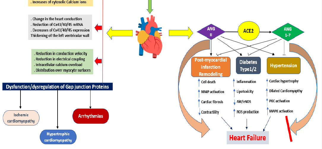
Following tissue damage, cardiac fibroblasts may also be involved in receiving and reacting to distress signals from nearby cardiomyocytes. The high metabolic demands of cardiomyocytes are mostly satisfied by the heart’s microvascular endothelial cells by supplying them with nutrients and oxygen (32). Other functions of cardiac endothelial cells include promoting intercellular communication, facilitating cell adhesion and preventing thrombus development (33). Any injury would negatively impact cardiac contractility as the supply of oxygen, nutrients and biological signalling components to cardiomyocytes would be impaired due to the critical function of cardiac microvascular endothelial cells. The researchers thoroughly investigated effects of continuous exposure to MC-LR on the rat cardiac tissues where chronic exposure to MC- LR is likely to pose risk to heart muscles. Histopathological experiments revealed cytoskeletal changes, including expansion of the cardiomyocytes, loss of cell cross-striations, fibrosis, and atypical nuclei (34). In addition, the administration of MC-LR causes enlarged cardiomyocytes, degenerative muscle fibers, accompanied by myocytolysis, and aberrant nuclei in the heart of rats (35). These findings have been confirmed in human cardio-myopathic heart tissue, where contractile failure & cytoskeletal abnormalities are related (36). Heart and the role of intercellular junction proteins Both homotypic (connections between the same cell type) and heterotypic connections take place at gap junctions, a significant kind of intercellular connection found on both cardiomyocytes and support cells. Gap junctions act as channels in all human cells that come into contact with one another, allowing ions and tiny molecules to be transferred directly between cells (37). Molecular weight of less than 1 kDa makes compounds permeable across gap junction channels. The connexin type and charge of the pore-forming molecule determine how permeable the material is. Gap junctions play a crucial role in the heart because the organ requires a quick transfer of oxygen and nutrients from the microvasculature to the cardiomyocytes (38). Gap junctions in the heart create the channels for intercellular current flow, which permits coordinated action potential propagation. Gap junction proteins of the connexin (Cx) family are essential for the rapid transmission of electrical impulses. Cx43, Cx40 and Cx45 are the main connexins expressed in cardiac myocytes, with Cx43 being the most prevalent (39). Due to high turnover rate of Cx43 in the heart, monitoring Cx43 degradation may be a strategy for controlling gap junctional communication in the organ both in healthy and pathological states (40). The most prevalent gap junction protein in cardiomyocytes is connexin-43 (Cx43) and the propagation of normal action potentials depends on its existence (41). In several cardiac disorders, connexin molecules that create gap junction channels, are depleted or relocated from intercalated discs to lateral cell boundaries. Action potential propagation may be disturbed and arrhythmias may develop from defects in gap junctional communication between nearby cardiomyocytes because the coordinated electrical activity is necessary for the synchronous contractile activity of the heart. Atrial fibrillation and abrupt new born death have been linked to Cx43 mutation (42). Gap junction remodeling is caused by microcystin, due to the changing number and distribution of gap junctions in diseased myocardium (Figure 3) (43). Cx43-containing spots moved to lateral cell borders, altering gap junction distribution in end-to-end intercalated discs (IDs). Normal, ischemic and hypertrophied left ventricles had equal-sized plaques, while pathological hearts exhibited 40% less amount of Cx43 (44). The lateralization of the gap junction appears to be a key feature of dysfunctional myocardium which results in 30% to 40% decline in gap junction area per ID unit of time. The MC-LR effect is thought to be responsible for the enlargement of intercellular gaps observed in mammals, which disrupts microtubules, microfilaments and cytokeratin intermediate filaments (45). The interaction of MC with protein phosphatases allows for the detection of cardiac muscles. The injections of MC are the fundamental processes, and based on the perplexing levels of cardiotoxicity that are displayed, they can provide information regarding whether a problem is involved (46).
Figu ca
Nitric oxide (NO) plays an important role in the physiology of heart which in turn is regulated by the enzyme nitric oxide synthase (NOS). Cardiac nitric oxide regulates vascular and cardiac functions by starting positive inotropy (increased myocardial contractility), altering intracardiomyocyte Ca2+ movements, vasodilation, accelerating ventricular relaxation and controlling cardiac metabolism (47). These NO synthases work together to adjust the effect as needed by increasing/decreasing its levels. Heart failure comes from cellular disruptions by losing this balance by producing excessive NO (48). High NO in tissues has been associated with membrane lipid peroxidation. The elevated LDH in MC-LR-treatedanimal may be attributed to energy blockage in the electron transport chain, since NO permanently inhibits complexes I-III and cyto-C-oxidase of the mitochondrial electron transport chain, reducing cellular respiration and ATP synthesis (49).
Role of angiotensin signalling pathway in cardiomyocytes
The primary set of cell surface receptors that play a significant role in cardiac signalling is the G-protein coupled receptors (GPCR). GPCRs include the angiotensin receptor, α- and β- adrenergic receptors and the ET1B receptors. Signalling mediated by these receptors can change the vascular tone, contractile force and heart rate (50). Serine/threonine kinases, significant protein kinases that add phosphate groups to either serine or threonine amino acid residues, are responsible for most signal transduction in eukaryotic cells and causes a conformational change in the protein (51). Voltage-dependent calcium channels that have been phosphorylated have lower intracellular Ca2+ levels, which makes it harder for vessel walls to contract (52). Protein phosphatases remove phosphate groups from their target proteins, reducing signal transduction through modulating kinase pathways (53). Angiotensin II promotes the activity of phospholipase C (PLC) by binding to the ATR1 in cardiovascular cells. This enzymatic activity hydrolyzes a membrane phospholipid to produce inositol (1, 4, 5)- trisphosphate (IP3) and diacylglycerol (DAG). Both intracellular Ca2+ levels and protein kinase C are raised by these second messengers (PKC), where intracellular proteins are phosphorylated, thereby triggering a biological reaction in response to the stimuli. ATR1 and ATR2 are 2 subtypes of angiotensin receptors (ATR), which are involved in this signalling pathway, and mediate the response of cardiac cells to angiotensin II. Angiotensin-II has a critical role in the cardiovascular system because it regulates minute variations in blood flow and vascular tone, maintaining sufficient perfusion of the organs. The adverse effects of angiotensin II, like hypertension and heart hypertrophy, can emerge from prolonged ATR1 activation (54). Interruption of this signalling is a recognized pharmacological target because of the consequent factors of angiotensin II on the heart and vascular system (Figure 3). Angiotensin receptor blockers (ARB) are helpful in the treatment of congestive heart failure (CHF) because they can stop the restructuring of the heart and lower blood pressure by preventing the adsorption of angiotensin II to the ATR1 (55). Evidence supports that RTKs and GPCRs expressed on cardiovascular system cells can "speak" to one another to coordinate intracellular signalling. Through a unique signalling mechanism known as transactivation, GPCRs can commander the signal transduction machinery of growth factor receptors for their functions. Angiotensin II can directly stimulate the expansion of cardiomyocytes by trans activating the EGFR, which sets off particular transduction pathways that control the expression of the hyperactive genes.
Distribution & metabolism of MC-LR in the heart According to reports, cardiomyocytes, which make up around 40% of cardiac cell volume is particularly high in mitochondria (56).
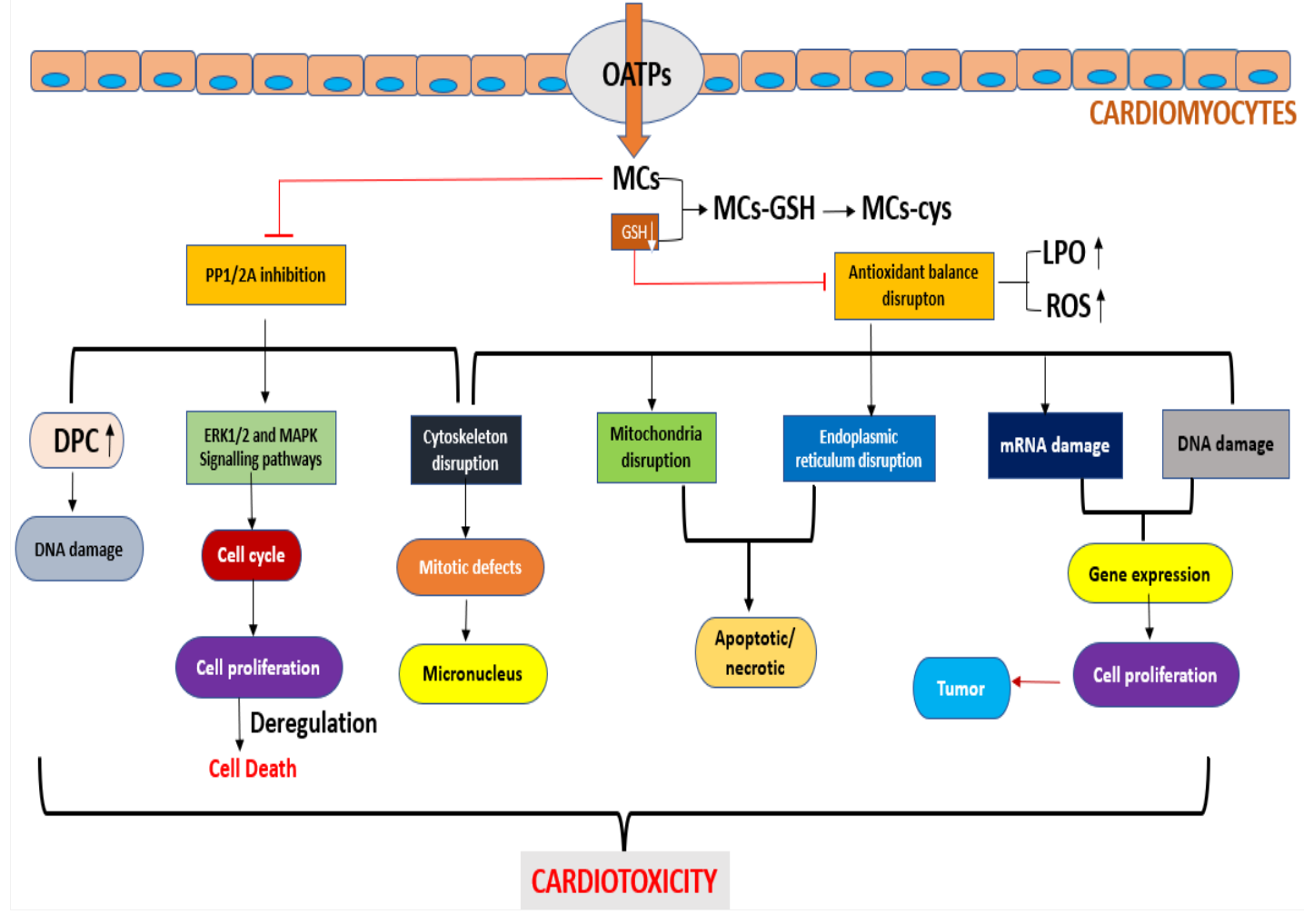
Numerous studies have demonstrated that MCs can lead to an imbalance in mitochondrial oxidative stress, which leads to neutrophil infiltration into tissues, increases protease production and produces significant oxidative intermediates, which promotes cell aging and ultimately death (57). During exposure, MCs can enter different cells via organic anion transporting polypeptides (OATP) (58). The OATP family of genes can be translated in almost all organs (20), albeit some OATP genes are expressed more frequently in particular tissues or even on a selective basis (59) and heart tissue is no exception (60). Heart tissue has been shown to express several OATP gene family, including OATP4A1, OATP2A1, OATP2B1 and OATP3A1 (61). This demonstrates that MCs can be delivered to and concentrate in heart tissue cells, possibly posing a threat to those cells. However, the methods by which MCs reach cardiomyocytes remain to be investigated (62). Furthermore, MCs enter the cell following the degree of blood perfusion, the types of OATP transporters and their expression levels (63).
The most prominent characteristic of the network is its metabolic flexibility to many stimuli, such as changes in maturation and starvation. In long-term pathological situations, the heart is also capable of remodeling metabolic pathways, which modifies myocardial enthalpies and contractile performance (64). Glutathione(GSH) is an endogenous detoxifying molecule that plays an important part in MC detoxification in both mammals and aquatic species. GSH pre-treatment was found to attenuate the harmful effects of MCs in mice in a previous investigation (65, 66).MC-LR-Cys and microcystins-glutathione conjugates (MC-LR- GSH) were discovered in the hearts of mice and rats (67).
Cardiotoxicity caused by MC-LR in mammals
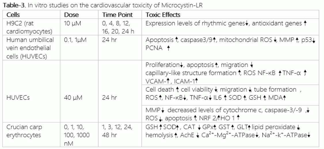
The cardiovascular system is a network of arteries and veins through which the heart pumps blood. Various cell types make up the heart where cardiomyocytes and myofilaments are the primary cells and organelles affected by MC-LR poisoning. Cardiotoxicity is caused by a high concentration of MC-LR, which results in hepatocyte necrosis, cytoskeletal alterations and cell death (68). MC-LR damages cardiac cells 24 hours after exposure, which further causes human oxidative stress-related diseases (69). Humans can be exposed to cyanotoxins in various ways, but oral exposure is the most harmful, mainly by drinking infected water or eating tainted food (70). The interplay of genetic, environmental and their effects on the regular operations of the cardiovascular system is thought to be a significant contributing factor to cardiovascular disease (CVD). Numerous studies have shown that MCs can harm the cardiovascular system in vivo and in vitro, as shown in Tables 2 and 3. It has been shown that acute exposure to MCs causes disruption of the cytoskeleton and dysfunction of the mitochondria. MCs can cause myocardial shrinkage and fibrosis in adults and directly cause cardiac deformity in larvae. MCs can influence different aspects of cardiomyocytes, including their shape, cell development and apoptosis, cytoskeleton and rhythm, ultrastructure, oxidative stress, membrane potential and mitochondrial respiratory chain enzyme activity (71). When MCs travel through the bloodstream and are delivered to the heart, they can cause heart damage.
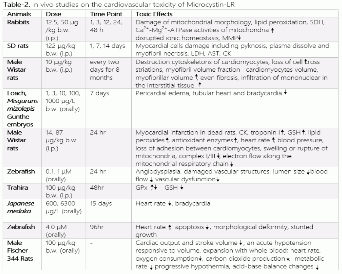
Pathophysiological alterations caused by MCs include decreased stroke volume and cardiac output, hypotension response to whole-blood volume expansion, decreased blood pressure and heart rate, decreased oxygen consumption and recruitment of the pancreatic and hepatic blood storage (72). This may lead to various diseases including cardiac arrest, hypovolemic shock and hypotension. Vascular endothelial cells are crucial mediators of vascular dynamic equilibrium because they control cell division, vascular regeneration, vascular osmolarity, inflammatory response and platelet activity (73). When massive amounts of MC are produced, a considerable amount of protein is generated in blood capillaries, causing peripheral pressure to increase. Furthermore, it was discovered that complexes I and III of the respiratory chain enzymes were suppressed, signifying that the mitochondrial electron transport chain was hindered. Increased levels of lipid peroxide, morphological alterations in heart mitochondria and succinate dehydrogenase activity were noted. The mitochondrial electron transport chain was further impacted by the inhibition of nicotinamide adenine dinucleotide dehydrogenase (NADH). Furthermore, MCs might change the functions of the Ca2+- Mg2+-ATPase in mitochondria, disrupting the ion homeostasis and perhaps leading to the loss of mitochondrial membrane potential (MMP) and ultimately succeeding in elevating the mitochondria of cardiac cells (74). The two effects of cardiotoxicity are reduced heart energy requirements and abnormalities in the mitochondria. Mitochondrial dysfunction in cardiac cells was reported in rats injected with MC-LR and decreased heart rate in zebrafish larvae exposed to MC-LR was associated with the accumulation of reactive oxygen species and apoptosis in the heart (75, 76).
Potential mechanism of action of MC-LR
Organic anion transporter peptides (OATP) such as OATP1B1, OATP1B2, OATP1B3 and OATP1A2 are especially involved in MC-LR absorption and the variation in their expression on organ tissues is what results in the unbalanced uptake across the body. According to previous studies, by employing omics technologies, the suppression of solubility and highly conserved protein serine/threonine phosphatase is largely accepted cellular/molecular processing mode of action of MC toxicity generated by the toxin in mammals and fishes (77). Protein phosphatase 1 (PP1) and 2A (PP2A) are primarily inhibited by MCs, which causes disruption of the dynamic equilibrium of regulatory protein expression and stimulation of their downstream proteins, as well as cytoskeletal reorganization (78). A potential mechanism of action for MCs is inhibition of protein phosphatases (PPs), which results in the hyper phosphorylation of cellular proteins. Exposure to MCs can also result in the excessive production of reactive oxygen species (ROS), which leads to oxidative damage. There is mounting evidence to support the hypothesis that Microcystins (MCs) are genotoxic because they cause damage to DNA which would indicate that mitochondria play a significant role as key mediators in the apoptosis resulting in the deregulation of cell cycle and mitosis (Figure 4) (79). In mammalian cells, PP regulate expression of genes, cell signalling, cell growth and cell division (80). According to studies, PP1 and PP2A dephosphorylate a variety of targeted proteins, including Cav1.2 and ATP-sensitive Na+/K+ channels, to control contractility (81). Upregulation of the PP2A-A subunit or a replacement for its myocardium expansion or HF were discovered to be caused by PP2B (82).
Conclusion
The human health risk is correlated with MC exposure pathways and dosages. Consumption of contaminated food and algal dietary supplements are also exposure modes, along with body contact, respiration, embolization and drinking contaminated water. However, it is emphasized that drinking polluted water and coming into contact with MCs cause the majority of exposure.
Microcystin has an adverse effect on the liver which is a primary target organ, as well as on other organs such as gastrointestinal, brain, renal, cardiac, respiratory and reproductive system via suppression of PP1, PP2A and oxidative stress. OATP channels transports MCs into cells, where they concentrate in primary target organs and tissues. MCs have the ability to suppress the activities of PP1 and PP2A, which results in the dephosphorylation of transcription factors, hence affecting cytoskeleton tissue, gene expression, cell death and CVD. A loss in cell viability and an increase in apoptosis, along with elevated level of oxidative stress and ROS in the mitochondria and ER of blood cells and vascular endothelial cells, may also be a cause of MC exposure. In addition of having a direct impact on the cardiovascular system, MCs can also cause CVD indirectly by damaging the liver, kidneys and gastrointestinal system. To combat the cardiotoxicity of MC-LR and to close the research gap, a standardized animal model strategy is required to explore both the chronic and acute toxic effects of MC-LR to provide convincing evidence of cardiotoxicity. Since there isn't much information about cardiotoxic effect of MC-LR, the role of cardiogenic complication is still unclear. This means that more research needs to be done that focuses solely on the cardiotoxic effect of MC-LR.
Declarations
- Author contributions (CRediT)
- All authors contributed equally to all 14 CRediT roles (Conceptualization, Data curation, Formal analysis, Funding acquisition, Investigation, Methodology, Project administration, Resources, Software, Supervision, Validation, Visualization, Writing – original draft, Writing – review & editing).
- Funding
-
This work was financially supported by a project from DST- SERB CRG/2018/003780 sanctioned to RKK.
- Conflicts of interest
- The authors do not have any relevant conflicts of interest.
- Ethics approval
- Ethics committee approval was not required for this article, as it is based on previously published literature and does not involve human participants, patient data or animals.
- Data availability
- Data sharing is not applicable to this article as no new data were created or analysed in this study.
References (82)
77 of 82 references are linked to their DOI.
- Paerl HW, Paul VJ. Climate change: links to global expansion of harmful cyanobacteria. Water Res. 2012;46(5):1349-1363 DOI Crossref PubMed Google Scholar
- Wang Q, Xie P, Chen J, Liang G. Distribution of microcystins in various organs (heart, liver, intestine, gonad, brain, kidney and lung) of Wistar rat via intravenous injection. Toxicon. 2008;52(6):721-727 DOI Crossref PubMed Google Scholar
- Florczyk M, Lakomiak A, Wozny M, Brzuzan P. Neurotoxicity of cyanobacterial toxins. Environ Biotechnol. 2014;10 DOI Crossref PubMed Google Scholar
- Puddick J, Prinsep MR, Wood SA, Kaufononga SA, Cary SC, Hamilton DP. High levels of structural diversity observed in microcystins from Microcystis CAWBG11 and characterization of six new microcystin congeners. Mar Drugs. 2014;12(11):5372- DOI Crossref PubMed Google Scholar
- Cao L, Massey IY, Feng H, Yang F. A review of cardiovascular toxicity of microcystins. Toxins. 2019;11(9):507 DOI Crossref PubMed Google Scholar
- Wei H, Yang F, Wang Y, Zhou Y, Yan Y, Liang G, Yin L, Pu Y. Electrospun polymer nanofibres as solid-phase extraction sorbents for extraction and quantification of microcystins. Environ Technol. 2015;36(21):2796-2802 DOI Crossref PubMed Google Scholar
- Miller TR, Xiong A, Deeds JR, Stutts WL, Samdal IA, Lovberg KE, Miles CO. Microcystin toxins at potentially hazardous levels in algal dietary supplements revealed by a combination of bioassay, Immunoassay and Mass Spectrometric Methods. J Agr Food Chem. 2020;68(30):8016-8025 DOI Crossref PubMed Google Scholar
- Zegura B. An overview of the mechanisms of microcystin-LR genotoxicity and potential carcinogenicity. Mini Rev Med Chem. 2016;16(13):1042-1062 DOI Crossref PubMed Google Scholar
- Tokodi N, Drobac D, Meriluoto J, Lujic J, Marinovic Z, Vazic T, Lazic G, Petrovic T. Cyanobacterial effects in Lake Ludos, Serbia-Is preservation of a degraded aquatic ecosystem justified? Sci Total Environ. 2018; 635: 1047-1062 DOI Crossref PubMed Google Scholar
- Merel S, Walker D, Chicana R, Snyder S, Baures E, Thomas O. State of knowledge and concerns on cyanobacterial blooms and cyanotoxins. Environ Int. 2013; 59: 303-327 DOI Crossref PubMed Google Scholar
- Melaram R. Microcystins and daily sunlight: Predictors of chronic liver disease and cirrhosis mortality. J Environ Sci. 2019;3(3):379-388 DOI Crossref PubMed Google Scholar
- Meneely JP, Elliott CT. Microcystins: measuring human exposure and the impact on human health. Biomarkers. 2013;18(8):639-649 DOI Crossref PubMed Google Scholar
- Welten RD, Meneely JP, Elliott CT. A comparative review of the effect of Microcystin-LR on the proteome. Expos Health. 2019; 12: 1-19 DOI Crossref PubMed Google Scholar
- Feurstein D, Stemmer K, Kleinteich J, Speicher T, Dietrich DR. Microcystin congener-and concentration-dependent induction of murine neuron apoptosis and neurite degeneration. Toxicol Sci. 2011;124(2):424-431 DOI Crossref PubMed Google Scholar
- Vesterkvist PS, Misiorek JO, Spoof LE, Toivola DM, Meriluoto JA. Comparative cellular toxicity of hydrophilic and hydrophobic microcystins on Caco-2 cells. Toxins. 2012;4(11):1008-1023 DOI Crossref PubMed Google Scholar
- Puerto M, Pichardo S, Jos A, Camean AM. Comparison of the toxicity induced by microcystin-RR and microcystin-YR in differentiated and undifferentiated Caco-2 cells. Toxicon. 2009;54(2):161-169 DOI Crossref PubMed Google Scholar
- Song W, Rein K, Cruz Ad1, O'Shea K. Ultrasonically-induced degradation of Microcystin LR and RR: Identification of by products and effect of environmental factors. Adv Exp Med Biol. 2008; 619: 305 PubMed Google Scholar
- Xu S, Yi X, Liu W, Zhang C, Massey IY, Yang F, Tian L. A review of nephrotoxicity of Microcystins. Toxins. 2020;12(11):693 103 77 Effect of MC-LR on the Heart of Mammals Roshni Rajpoot & Raj Kumar Koiri DOI Crossref PubMed Google Scholar
- Yang F, Wen C, Zheng S, Yang S, Chen J, Feng X. Involvement of MAPK/ERK1/2 pathway in microcystin-induced microfilament reorganization in HL7702 hepatocytes. J Toxicol Env Heal A. 2018;81(21):1135-1141 DOI Crossref PubMed Google Scholar
- Cao L, Huang F, Massey IY, Wen C, Zheng S, Xu S, Yang F. Effects of microcystin-LR on the microstructure and inflammation-related factors of jejunum in mice. Toxins. 2019;11(9):482 DOI Crossref PubMed Google Scholar
- Li X, Xu L, Zhou W, Zhao Q, Wang Y. Chronic exposure to microcystin-LR affected mitochondrial DNA maintenance and caused pathological changes of lung tissue in mice. Environ Pollut. 2016; 210: 48-56 DOI Crossref PubMed Google Scholar
- Hinojosa M, Gutierrez-Praena D, Prieto A, et al. Neuro toxicity induced by microcystins and cylindrospermopsin: A review. Sci Total Environ. 2019;668:547-565 DOI Crossref PubMed Google Scholar
- Liu Y, Yang M, Zheng L, Nguyen H, Ni L, Song S, Sui Y. Antioxidant responses of triangle sail mussel Hyriopsis cumingii exposed to toxic Microcystis aeruginosa and thermal stress. Sci Total Environ. 2020; 743: 140754 DOI Crossref PubMed Google Scholar
- Massey IY, Wu P, Wei J, Luo J, Ding P, Wei H, Yang F. A mini-review on detection methods of microcystins. Toxins. 2020;12(10):641 DOI Crossref PubMed Google Scholar
- Alexander W. Branko furst's radical alternative: Is the heart moved by the blood, rather than vice Versa? P & T. 2017; 42(1):33-39 PubMed Google Scholar
- Guo Y, Pu WT. Cardiomyocyte maturation: New phase in development. Circ Res. 2020;126(8):1086-1106 PubMed Google Scholar
- Squire J. The actin-myosin interaction in muscle: Background and overview. Int J Mol Sci. 2019; 20: 5715 DOI Crossref PubMed Google Scholar
- Ishii S, Oyama K, Shintani SA, Kobirumaki-Shimozawa F, Ishiwata SI, Fukuda N. Thermal activation of thin filaments in striated muscle. Front Physiol. 2020; 11: 278 DOI Crossref PubMed Google Scholar
- Rios E. Calcium-induced release of calcium in muscle: 50 years of work and the emerging consensus. J Gen Physiol. 2018;150(4):521-537 DOI Crossref PubMed Google Scholar
- Van Meer BJ, Krotenberg A, Sala L, Davis RP, Eschenhagen T, Denning C, Tertoolen LG, Mummery CL. Simultaneous measurement of excitation -contraction coupling parameters identifies mechanisms underlying contractile responses of hiPSC-derived cardiomyocytes. Nat commun. 2019;10(1):1-9 DOI Crossref PubMed Google Scholar
- Baudino TA, Carver W, Giles W, Borg TK. Cardiac fibroblasts: friend or foe? Am J Physiol-Heart C. 2006;291(3):H1015-H1026 DOI Crossref PubMed Google Scholar
- Talman V, Kivela R. Cardiomyocyte-endothelial cell interactions in cardiac remodeling and regeneration. Front Cardiovasc Med. 2018; 5: 101 DOI Crossref PubMed Google Scholar
- Jourde-Chiche N, Fakhouri F, Dou L, Bellien J, Burtey S, Frimat M, Jarrot PA, Kaplanski G, Le Quintrec M, Pernin V. Endothelium structure and function in kidney health and disease. Nat Rev Nephrol. 2019;15(2):87-108 DOI Crossref PubMed Google Scholar
- Milutinovic A, Zorc Pleskovic R, Petrovic D, Zorc M, Suput D. Microcystin-LR induces alterations in heart muscle. Folia Biol. 2006; 52: 116-118 DOI Crossref PubMed Google Scholar
- Zhang H, Zhang J, Chen Y, Zhu Y, 2007. Influence of intracellular Ca2+, mitochondria membrane potential, reactive oxygen species, and intracellular ATP on the mechanism of microcystin‐LR induced apoptosis in Carassius auratus lymphocytes in vitro. Environ. Toxicol. 2007; 22: 559-564 DOI Crossref PubMed Google Scholar
- Chen QM, Tu VC, Purdom S, Wood J, Dilley T, 2001. Molecular mechanisms of cardiac hypertrophy induced by toxicants. Cardiovasc. Toxicol. 2001; 1: 267-283 DOI Crossref PubMed Google Scholar
- Goodenough DA, Paul DL. Gap junctions. Csh Perspect Biol. 2009;1(1): a002576 PubMed Google Scholar
- Wang Y, Wang C, Ma J. Role of cardiac endothelial cell-derived MicroRNAs in cardiac remodeling. Discov Med. 2019; 28(152): 95-105 PubMed Google Scholar
- Severs NJ, Bruce AF, Dupont E, Rothery S. Remodelling of gap junctions and connexin expression in diseased myocardium. Cardiovasc Res. 2008;80(1):9-19 DOI Crossref PubMed Google Scholar
- Bacova BS, Vinczenzova C, Zurmanova J, Kasparova D, Knezl V, Benova TE, Pavelka S, Soukup T, Tribulova N. Altered thyroid status affects myocardial expression of connexin -43 and susceptibility of rat heart to malignant arrhythmias that can be partially normalized by red palm oil intake. Histochem Cell Biol. 2017;147(1):63-73 DOI Crossref PubMed Google Scholar
- Delmar M, Makita N. Cardiac connexins, mutations and arrhythmias. Curr Opin Cardiol. 2012;27(3):236-241 DOI Crossref PubMed Google Scholar
- Van Norstrand DW, Asimaki A, Rubinos C, Dolmatova E, Srinivas M, Tester DJ, Saffitz JE, Duffy HS, Ackerman MJ. Connexin43 mutation causes heterogeneous gap junction loss and sudden infant death. Circulation. 2012;125(3):474-481 DOI Crossref PubMed Google Scholar
- Jeong MH, Kim HJ, Pyun JH, Choi KS, Lee DI, Solhjoo S, O'Rourke B, Tomaselli GF, Jeong DS, Cho H. Cdon deficiency causes cardiac remodeling through hyperactivation of WNT/β-catenin signaling. P Natl A Sci. 2017;114(8): E1345-E1354 DOI Crossref PubMed Google Scholar
- Kontaridis MI, Geladari EV, Geladari CV. Structural alterations in the hypertensive heart disease result in intercalated disc remodeling and arrhythmias. In: Hypertension and Cardiovascular Disease. Springer. 2016 DOI Crossref PubMed Google Scholar
- Zhou M, Tu WW, Xu J. Mechanisms of microcystin-LR-induced cytoskeletal disruption in animal cells. Toxicon. 2015; 101: 92-100 DOI Crossref PubMed Google Scholar
- Alosman M, Cao L, Massey IY, Yang F., 2021. The lethal effects and determinants of microcystin-LR on heart: A mini review. Toxin Rev. 2021; 40: 517-526 DOI Crossref PubMed Google Scholar
- Sztormowska-Achranowicz K, Jankowski Z, Kocic I. Protective effect of nicotinamide and L-arginine against monocrotaline-induced pulmonary hypertension in rats: gender dependence. Pharmacol Rep. 2020;72(5):1334-1346 DOI Crossref PubMed Google Scholar
- Ziolo MT, Kohr MJ, Wang H. Nitric oxide signalling and the regulation of myocardial function. J Mol Cell Cardiol. 2008;45(5):625-632 DOI Crossref PubMed Google Scholar
- Lone Y, Bhide M, Koiri RK. Amelioratory effect of coenzyme Q10 on potential human carcinogen Microcystin-LR induced toxicity in mice. Food Chem. Toxicol. 2017; 176-185 DOI Crossref PubMed Google Scholar
- Bencivenga L, Liccardo D, Napolitano C, Visaggi L, Rengo G, Leosco D. β-adrenergic receptor signaling and heart failure: From Bench to Bedside. Heart Fail Clin. 2019;15(3):409-419 104 77 Effect of MC-LR on the Heart of Mammals Roshni Rajpoot & Raj Kumar Koiri DOI Crossref PubMed Google Scholar
- Bononi A, Agnoletto C, De Marchi E, Marchi S, Patergnani S, Bonora M, Giorgi C, Missiroli S, Poletti F, Rimessi A, Pinton P. Protein kinases and phosphatases in the control of cell fate. Enzyme Res. 2011;329098 DOI Crossref PubMed Google Scholar
- Chasseigneaux S, Moraca Y, Cochois-Guegan V, Boulay AC, Gilbert A, Le Crom S, Blugeon C, Firmo C, Cisternino S, Laplanche JL. Isolation and differential transcriptome of vascular smooth muscle cells and mid-capillary pericytes from the rat brain. Sci Rep. 2018;8(1):1-12 DOI Crossref PubMed Google Scholar
- Wheeler-Jones CP. Cell signalling in the cardiovascular system: an overview. Heart. 2005;91(10):1366-1374 DOI Crossref PubMed Google Scholar
- Mehta PK, Griendling KK. Angiotensin II cell signaling: physiological and pathological effects in the cardiovascular system. Am J Physiol Cell Physiol. 2007;292(1):C82-C97 DOI Crossref PubMed Google Scholar
- Barreras A, Gurk-Turner C. Angiotensin II receptor blockers. Proceedings (Baylor University Medical Cn). 2003;16(1):123-126 DOI Crossref PubMed Google Scholar
- Varga I, Kyselovic J, Galfiova P, Danisovic L. The non-cardiomyocyte cells of the heart. Their possible roles in exercise- induced cardiac regeneration and remodeling. In: Exercise for Cardiovascular Disease Prevention and Treatment. Springer. 2017 DOI Crossref PubMed Google Scholar
- Zhang J, Xie Y, Fan Q, Wang C. Effects of karanjin on dimethylhydrazine induced colon carcinoma and aberrant crypt foci are facilitated by alteration of the p53/Bcl2/BAX pathway for apoptosis. Biotech Histochem. 2020:1-11 DOI Crossref PubMed Google Scholar
- Steiner K, Zimmermann L, Hagenbuch B, Dietrich D. Zebrafish Oatp- mediated transport of microcystin congeners. Arch Toxicol. 2016;90(5):1129-1139 DOI Crossref PubMed Google Scholar
- Li TT, An JX, Xu JY, Tuo BG. Overview of organic anion transporters and organic anion transporter polypeptides and their roles in the liver. World J Clin Cases. 2019;7(23):3915 DOI Crossref PubMed Google Scholar
- Moraes JG, Behura SK, Geary TW, Spencer TE. Analysis of the uterine lumen in fertility-classified heifers: I. Glucose, prostaglandins, and lipids. Biol Reprod. 2020;102(2):456-474 DOI Crossref PubMed Google Scholar
- Bakos E, Tusnady GE, Nemet O, Patik I, Magyar C, Nemeth K, Kele P, Ozvegy-Laczka C. Synergistic transport of a fluorescent coumarin probe marks coumarins as pharmacological modulators of Organic anion-transporting polypeptide, OATP3A1. Biochem Pharmacol. 2020; 182: 114250. DOI Crossref PubMed Google Scholar
- Foss AJ, Aubel MT, Gallagher B, Mettee N, Miller A, Fogelson SB. Diagnosing microcystin intoxication of canines: Clinicopathological indications, pathological characteristics, and analytical detection in postmortem and antemortem samples. Toxins. 2019;11(8):456 DOI Crossref PubMed Google Scholar
- Valerio E, Barreiros S, Rodrigues S, Turkina MV, Vasconcelos VM, Campos A: New insights in Saccharomyces cerevisiae response to the cyanotoxin Microcystin-LR, revealed by proteomics and gene expression. Toxins. 2020;12(10):667 DOI Crossref PubMed Google Scholar
- Kolwicz Jr SC, Purohit S, Tian R. Cardiac metabolism and its interactions with contraction, growth, and survival of cardiomyocytes. Circ. Res. 2013; 113: 603-616 DOI Crossref PubMed Google Scholar
- Guzman- Guillen R, Puerto M, Gutierrez-Praena D, Prieto AI, Pichardo S, Jos A, Campos A, Vasconcelos V, Camean AM. Potential use of chemoprotectants against the toxic effects of cyanotoxins: A review. Toxins. 2017;9(6):17 DOI Crossref PubMed Google Scholar
- Hermansky S, Stohs S, Eldeen Z, Roche VF, Mereish K. Evaluation of potential chemoprotectants against microcystin‐ LR hepatotoxicity in mice. J Appl Toxicol. 1991;11(1):65-73 DOI Crossref PubMed Google Scholar
- Buckingham M, Meilhac S, Zaffran S. Building the mammalian heart from two sources of myocardial cells. Nat Rev Genet. 2005;6(11):826-835 DOI Crossref PubMed Google Scholar
- McLellan NL, Manderville RA: Toxic mechanisms of microcystins in mammals. Toxicol Res. 2017;6(4):391-405 DOI Crossref PubMed Google Scholar
- Martins ND, Colvara WA, et al. Microcystin-LR: how it affects the cardio-respiratory responses to hypoxia in Nile tilapia, Oreochromis niloticus. Chemosphere. 2011;84(1): 154-159 DOI Crossref PubMed Google Scholar
- Funari E, Testai E. Human health risk assessment related to cyanotoxins exposure. Crit Rev Toxicol. 2008; 38: 97-125 DOI Crossref PubMed Google Scholar
- Vecoli C, Pulignani S, Foffa I, Grazia Andreassi M. Congenital heart disease: the crossroads of genetics, epigenetics and environment. Curr genomics. 2014; 15: 390-399 DOI Crossref PubMed Google Scholar
- Lone Y, Bhide M, Koiri RK. Amelioratory effect of coenzyme Q10 on potential human carcinogen microcystin-LR induced toxicity in mice. Food Chem Toxicol. 2017; 102: 176-185 DOI Crossref PubMed Google Scholar
- Curry FRE, Adamson RH. Vascular permeability modulation at the cell, microvessel, or whole organ level: towards closing gaps in our knowledge. Cardiovasc Res. 2010;87(2):218-229 DOI Crossref PubMed Google Scholar
- Liu Y, Song L, Li X, Liu T. The toxic effects of microcystin-LR on embryo-larval and juvenile development of loach, Misguruns mizolepis Gunthe. Toxicon. 2002;40(4):395-399 DOI Crossref PubMed Google Scholar
- Diez-Quijada Jimenez L, Medrano Padial C, Llana Ruiz-Cabello M, Catunescu GM, Moyano Salvago R, Risalde Md1A, Camean Fernandez AM, Jos Gallego AM. Cylindrospermopsin - microcystin-LR combinations may induce genotoxic and histopathological damage in rats. Toxins. 2020; 12: 348 DOI Crossref PubMed Google Scholar
- Zeng C, Sun H, Xie P, Wang J, Zhang G, Chen N, Yan W, Li G. The role of apoptosis in MCLR-induced developmental toxicity in zebrafish embryos. Aquat Toxicol. 2014;149:25-32 DOI Crossref PubMed Google Scholar
- Buratti FM, Manganelli M, Vichi S, Stefanelli M, Scardala S, et al. Cyanotoxins: producing organisms, occurrence, toxicity, mechanism of action and human health toxicological risk evaluation. Arch Toxicol. 2017;91(3):1049-1130 DOI Crossref PubMed Google Scholar
- Massey IY, Yang F, Ding Z, Yang S, Guo J, l-Osman M, Kamegni RB, Zeng W. Exposure routes and health effects of microcystins on animals and humans: A mini-review. Toxicon. 2018; 151: 156-162 DOI Crossref PubMed Google Scholar
- Chen L, Xie P. Mechanisms of microcystin -induced cytotoxicity and apoptosis. Mini Rev Med Chem. 2016;16(13): 1018-1031 DOI Crossref PubMed Google Scholar
- Samarasimhareddy M, Mayer D, Metanis N, Veprintsev D, Hurevich M, Friedler A. A targeted approach for the synthesis of multi- phosphorylated peptides: a tool for studying the role of phosphorylation patterns in proteins. Org Biomol Chem. 2019;17(42):9284-9290 DOI Crossref PubMed Google Scholar
- Heijman J, Ghezelbash S, Wehrens XH, Dobrev D. Serine/threonine phosphatases in atrial fibrillation. J Mol Cell Cardiol. 2017; 103:110-120 105 77 Effect of MC-LR on the Heart of Mammals Roshni Rajpoot & Raj Kumar Koiri DOI Crossref PubMed Google Scholar
- Gergs U, Kirchhefer U, Bergmann F, Kunstler B, Miblinger N, Au B, Mahnkopf M, Wache H, Neumann J. Characterization of stressed transgenic mice overexpressing H2-histamine receptors in the heart. J Pharmacol Exp Ther. 2020;374(3):479- DOI Crossref PubMed Google Scholar
Respond to this article
Comments on a published article are welcome as a Letter to the Editor.
Get an e-mail when new articles in your fields are published
Questions to the authors
Questions are moderated: an editor approves each question, the authors answer, and an editor approves the answer before both appear here.
No answered questions yet.
Ask the authors a question
Discussion
Comments are moderated: they appear after the writer confirms the e-mail address and an editor approves them. Each comment carries a conflict-of-interest statement.
No comments yet.
Write a comment
Read the full text
Open the PDF here
Loading the PDF…
If the PDF does not show here, use the download button above.
How to cite this article
Rajpoot R, Koiri RK. Profoundal Effects of Microcystin-LR Induced Cardiotoxicity in Mammals. Ulutas Med J. 2023;9(2):92-105. doi:10.5455/umj.20221222071638
Rajpoot, R., & Koiri, R. K. (2023). Profoundal Effects of Microcystin-LR Induced Cardiotoxicity in Mammals. The Ulutas Medical Journal, 9(2), 92–105. https://doi.org/10.5455/umj.20221222071638
Rajpoot, R. and Koiri, R.K. (2023) 'Profoundal Effects of Microcystin-LR Induced Cardiotoxicity in Mammals', The Ulutas Medical Journal, 9(2), pp. 92–105. doi: 10.5455/umj.20221222071638.
@article{umj133517,
  title = {Profoundal Effects of Microcystin-LR Induced Cardiotoxicity in Mammals},
  author = {Rajpoot, Roshni and Koiri, Raj Kumar},
  journal = {The Ulutas Medical Journal},
  year = {2023},
  volume = {9},
  number = {2},
  pages = {92--105},
  doi = {10.5455/umj.20221222071638},
  url = {https://www.ulutasmedicaljournal.com/?mno=133517},
  issn = {2149-388X}
}