The Ulutas Medical Journal · ISSN 2149-0430 (print), 2149-388X (online) · https://www.ulutasmedicaljournal.com/?mno=268878
Review Article · Open access
Role of Vascular Endothelial Growth Factor and Transforming Growth Factor-β in Hepatocellular Carcinoma
Also available in: العربية · Türkçe · 中文
- Biochemistry Lab, Department of Zoology, Dr. Harisingh Gour Central University, Sagar - 470003, Madhya Pradesh, India ROR
Abstract
Hepatocellular carcinoma is one of the most common malignant tumor and leading cause of death worldwide. Tumor genes, signals from tumor stroma, recruited inflammatory cells and hypoxia has been reported to switch on angiogenesis. Vascular endothelial growth factor is a pleiotropic factor that regulates cell proliferation, vascular permeability and plays an important role in tumor angiogenesis through its different angiogenic receptors. Multifunctional regulatory cytokine transforming growth factor-β also controls cell proliferation, differentiation, extracellular matrix production and survival/or apoptosis of many types cells. By raising level of epithelial-mesenchymal transition, transforming growth factor-β creates a favourable microenvironment for tumor cell growth. Depending upon the stage of cancer progression it can either initiate tumor development, progression and metastasis or can act as a tumor suppressor. In the present review, an attempt has been made to comprehensively define the role of Vascular Endothelial Growth Factor and transforming growth factor-β in the development and progression of hepatocellular carcinoma.
Keywords
Full text
Introduction
Hepatocellular carcinoma (HCC) has been reported as the seventh most common cancer worldwide and the third leading cause of cancer-related deaths (1). High mortality rate has been observed in patients with HCC due to lack of effective treatment options, especially at the advanced stages. Although surgery and percutaneous ablation can achieve long-term control in patients with early HCC, recurrence rates are high (approximately 50% at 3 years)(2). Moreover, because of asymptomatic nature of early HCC, lack of awareness and poorly defined screening strategies, most patients (approximately 80%) present with advanced or unresectable disease (3).
Etiological factors associated with HCC include chronic liver diseases, hepatitis virus (HBV & HCV) and alcoholism. Irrespective of the cause liver cirrhosis has emerged as one of the major risk factor for tumor development and it has been observed that almost 80% of patients with HCC pass through this stage before developing tumor (4). Further, aflatoxin has been reported to have a synergistic effect on Hepatitis C virus (HCV) induced liver cancer (5). From epidemiological studies, it is evident that contamination of food with AFB1 is the major risk factor for human liver cancer (6, 7).
HCC is considered an extreme type of vascular tumor growth and is dependent upon angiogenesis, which is growth of new blood vessels. During the pathogenesis and progression of cancer from the preinvasive and dormant form to the invasive and metastatic phase, angiogenesis has been observed to play a critical role by yielding angiogenic factors necessary for tumor growth, invasion and metastasis. Multiple studies have demonstrated that the degree of tumor vascularity correlates positively with disease stage, the likelihood of metastases, and cancer recurrence (8).
Vascular endothelial growth factor (VEGF) is a well known, potent angiogenic factor that has been reported to enhance vascular permeability, promotes neovascularization and endothelial cell proliferation in HCC due to its endothelial cell mitogen property (9). In addition to VEGF, further during the development of liver diseases, transforming growth factor-b (TGF-b) has been reported to play an important role in fibrosis of the lesions (10). Although in normal liver TGF-β inhibits hepotocyte cell growth but has been observed to get activated in those with injured livers and promote the development of hepatocellular carcinoma (11). During cancer progression, activation of TGF-β signalling leads to the adherence of TGF-β ligand to its receptor on the cell membrane (12).
VEGF as Angiogenesis Biomarkers of Hepatocellular Carcinoma
Described for the first time in 1989 (13), VEGF protein family currently comprises several members: VEGF (or VEGF-A), VEGF-B, VEGF-C and VEGF-D, VEGF-F,placental growth factor (PlGF), and their receptors VEGFR-1, VEGFR-2 and VEGFR-3 (14). VEGF is a ligand of the VEGF tyrosine kinase receptor superfamily and includes VEGF-A, -B, -C and -D, with splice variants of VEGF-A resulting in several different isoforms (15, 16).
The VEGF family ligands bind to tyrosine kinase receptors VEGFR1, VEGFR2 and VEGFR3 (Figure-1); each receptor contains an extracellular domain of approximately 750 amino acid residues, arranged within seven immunoglobulin like folds (17). Additionally, heparin sulphate proteo-glycans (HSPGs) as well as neuropilins (NRP-1 and NRP-2) can act as coreceptors for VEGF and promote VEGFR activation (18, 19). VEGF/VEGFR pathway is influenced and regulated by many factors like local environmental hypoxia, various hormones, growth factors and cytokines. Hor-mones such as androgens upregulate stromal cell and malignant cell VEGF production and angiogenesis, enhancing prostate cancer growth (20, 21).
VEGF has been described as the main angiogenic growth factor and the level of its expression has been used as a biomarker in the identification of angiogenic diseases. Expression level of VEGF has been reported to increase with the progression and pathogenesis of HCC and has been reported to be very high at advanced stages of cancer. In solid tumors due to neovascularization, VEGF facilitates tumor growth and metastasis by supplying continuous nutrient flow and its expression correlates with tumour progression (22). It has been identified as one of the important angiogenic factor in HCC. Its expression has been reported to increase gradually from low-grade dysplastic nodules to high-grade dysplastic nodules to early HCC (23). During the early stages of HCC, higher ment of HCC has been observed to correlate with micro vascular density, unpaired arteries (i.e., arteries not accompanied by bile ducts, indicative of angiogenesis) and with CD34 staining as a marker of sinusoidal capillarization in HCC (23). VEGF-C gene polymorphism resulting into its over expression has been status of neo-angiogenesis and cell observed to occur frequently in HCC and proliferation activity has been observed as compared to advanced form of HCC (23). The degree of VEGF expression during develophas been suggested to be a predictive factor for advanced stage of HCC (24).
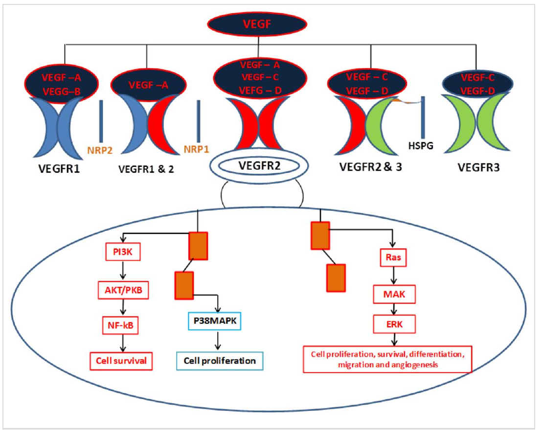
VEGF has been accounted as the chief angiogenic factor responsible for the neoangiogenesis, development and/or progression/spread of HCC tumour (25) and HCC patients with over expression of VEGF in serum and tissue has been associated with poor survival rate (26). VEGF induced formation of new microvessels, is critical for the growth and progression of various human solid tumors as it enables delivery of oxygen and nutrients to the rapidly growing tumors (27). Some of these rapidly growing tumors concomitantly also secrete angiopoietins (28).
Tumor Suppressive and Oncogenic Functions of TGF-β in HCC
TGF-β is a cytokine with multiple functions like regulation of cell proliferation, differentiation and extracellular matrix production (29). Deregulation of TGF-β expression or signalling has been implicated in the pathogenesis of a variety of diseases, including cancer and fibrosis. Expression of TGF-β has been repor- ted to be very low that increases dramatically during the later stages of cancer development and is thus actively secreted by tumor cells and thus it does not act merely as a bystander but rather contributes to cell growth, invasion, metastasis and decreases host-tumor immune response during advanced stages (30). TGF-β is the prototypic member of a large family of secreted proteins, which can be further divided into two subfamilies, depending on the signal pathways they activate - the TGF-β/Activin/Nodal subfamily and the Bone Morphogenetic Protein (BMP) subfamily (31).
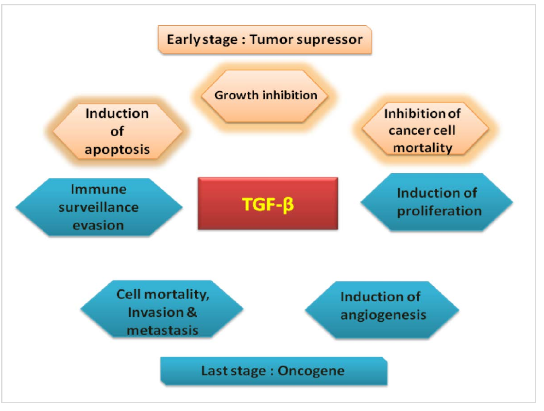
TGF-βs are able to regulate tumor initiation, progression, and metastatic development. It is widely accepted that TGF-βs act as both tumor suppressors and tumor promoters, depending on the cancer type and tumor development timing (32, 33). During the early stage of tumor development, TGF-β1 has been reported to acts as a tumor suppressor mainly due to its ability to inhibit the proliferation of epithelial cells. Further it can also cause G1 cell cycle arrest by declining expression of oncogenes such as c-Myc and ID, and/or by inducing the expression of CDK inhibitors such as p21Cip1 and p15lnk4b (34, 35). TGF-β has been reported to play a dual role of tumor suppressor as well as tumor promoter (Figure 2), however till date the mechanism is not very clear. In HCC expression of TGF-β has been reported to increase concomitantly with collagen deposition and the reduction in proteolytic degradation (36).
Tumor suppressor function of TGF-β is evident by the frequent mutational defects of Smad4, TβRI or TβRII (TGF-β type II receptor) observed in many malignancies such as cancers of the stomach, colon, prostate, breast, lung, liver and pancreas (37, 38). Dual role of TGF-β has been observed in epithelial cell of liver during the development and progression of HCC. In the initial phase it exerts a tumour-suppressive, which is quite evident by the enhanced susceptibility to HCC because of reduced availability of TGF-β type II receptor (TβRII) due to the ectopic expression of soluble TβRII in hepatocytes (39) or reduced TβRII expression in heterozygous mice has confirmed the tumor suppressive function of TGF-β signalling pathway (40).
In most of cancers that arise from epithelial cells, TGF-β suppressed malignant cell growth by inducing cell cycle arrest through the upregulation of cyclin-dependent protein kinase (CDK) inhibitors such as p15 and p21, activating apoptosis through induction of proapoptotic proteins like Death associated protein kinase (DAP) (41). Contrary to this expression in case of prostate and colorectal cancers, expression of TGF-β1 in cancer cells was found to strongly correlate with progression and metastasis (42–44). Most commonly mutated TGF-β pathway genes in cancer being TGFBR2, TGFBR1, SMAD4 and SMAD2.
Conclusion
HCC exhibits numerous molecular abnormalities, which may be involved in the process of cancer development and progression. Thus, it is important to identify key players which may be involved in the pathogenesis and progression of cancer, so that accurate prognosis can be made and a reasonable selection criterion can be applied to patients with HCC, particularly during the early stages. It has been observed that in various tumors including HCC, whenever there is local invasion of cancer cells or metastasis to distal parts take place, there is elevated levels of VEGF in blood through angiogenesis. Further tumor suppressive function of TGF-β plays an important role. Thus, both VEGF and TGF-β regulates angiogenesis, tumor progression and production of the extracellular matrix and immune suppression in HCC via different mechanism. In such scenario, better understanding of VEGF and TGF-β pathway will lead to the development of modalities that can target cancer cells in HCC. Till date the exact mechanisms involved in the malignant transformation of TGF-β is not clear and subject to future research with therapeutic point of view.
Declarations
- Author contributions (CRediT)
- All authors contributed equally to all 14 CRediT roles (Conceptualization, Data curation, Formal analysis, Funding acquisition, Investigation, Methodology, Project administration, Resources, Software, Supervision, Validation, Visualization, Writing – original draft, Writing – review & editing).
- Funding
-
Funding information was not reported in the published version of this article.
- Conflicts of interest
- The authors declare that there is no conflict of interests.
- Ethics approval
- Ethics committee approval was not required for this article, as it is based on previously published literature and does not involve human participants, patient data or animals.
- Data availability
- Data sharing is not applicable to this article as no new data were created or analysed in this study.
References (44)
38 of 44 references are linked to their DOI.
- Simbaqueba Ferlay J, Shin HR, Bray Forman FD, Mathers C, Parkin DM. Estimates of worldwide burden of cancer in 2008: GLOBOCAN 2008, Int J Cancer 2010;127: 2893– 2917. DOI Crossref PubMed Google Scholar
- Mulcahy MF. Management of hepatocellular cancer. Curr Treat Options Oncol 2005;6: 423–435. DOI Crossref PubMed Google Scholar
- Thomas MB, Abbruzzese JL. Opportunities for targeted therapies in hepatocellular carcinoma. J Clin Oncol 2005;23: 8093– 8108. DOI Crossref PubMed Google Scholar
- Minguez B, Tovar V, Chiang D, Villanueva A, Llovet JM. Pathogenesis of hepatocellular carcinoma and molecular therapies. Curr Opin Gastroenterol 2009;25: 186-194. DOI Crossref PubMed Google Scholar
- 5. Kirk GD, Lesi OA, M Mendy et al. 249 (ser) P53 mutation in plasmid DNA, hepatitis B viral infection, and risk of hepatocellular carcinoma. Oncogene 2005;24:5858 5867. DOI Crossref PubMed Google Scholar
- Li Y, SU JJ, Qin LL, Yang C, Ban KC, Yan RQ. Synergistic effect of hepatitis B virus and aflatoxin B1 in hepato carcinogenesis in tree shrews. Ann Acad Med Singapore 1999;28: 67-71. DOI Crossref PubMed Google Scholar
- Madden CR, Finegold MJ, Slagle BL. Altered DNA mutation spectrum in aflatoxin b1-treated transgenic mice that express the hepatitis B virus x protein. J Virol 2002;76: 11770-11774. DOI Crossref PubMed Google Scholar
- Weidner N. Angiogenesis as a predictor of clinical outcome in cancer patients. Hum Pathol 2000;31: 403– 405. DOI Crossref PubMed Google Scholar
- Dvorak HF, Brown IF, Detmar MA, Dvorak M. Vascular permeability factor/vascular endothelial growth factor, microvascular hypermeability, and angiogenesis. Am J Pathol 1995;146:1029–1039. PubMed Google Scholar
- Gressner AM, Weiskirchen R, Breitkopf K, Dooley S. Roles of TGF-beta in hepatic fibrosis. Front Biosci 2002;7:d793– d807. DOI Crossref PubMed Google Scholar
- Carr BI, Hayashi I, Branum EL, et al. Inhibition of DNA synthesis in rat hepatocytes by platelet-derived type beta transforming growth factor. Cancer Res 1986;46: 2330– 2334. PubMed Google Scholar
- Nakamura T, Tomita Y, Hirai R et al. Inhibitory effect of transforming growth factor-beta on DNA synthesis of adult rat hepatocytes in primary culture. Biochem Biophys Res Commun 1985;133:1042– 1050. DOI Crossref PubMed Google Scholar
- Tischer E, Gospodarowicz D, Mitchell Silva RM, Schilling Lau JK, Crisp T, Fiddes JC, Abraha JA. Vascular endothelial growth factor: a new member of the platelet-derived growth factor gene family. Biochem Biophys Res Commun 1989;165:1198-1206. DOI Crossref PubMed Google Scholar
- Tammela TM, Enholm B, Alitalo K, Paavonen K. The biology of vascular endothelial growth factors. Cardiovas Res 2005;65: 550-563. DOI Crossref PubMed Google Scholar
- Clifford RL, Deacon K, Knox AJ. Novel regulation of vascular endothelial growth factor-A (VEGF-A) by transforming growth factor β1: requirement for Smads, β - catenin, and GSK3 β. J Biol Chem 2008;283:35337– 35353. DOI Crossref PubMed Google Scholar
- Fan F, Wey JS, McCarty MF et al. Expression and function of vascular endothelial growth factor receptor-1 on human colorectal cancer cells. Oncogene 2005;24:2647– 2653. DOI Crossref PubMed Google Scholar
- Olsson A, Dimber KA, Kreuger J, et al. VEGF receptor signalling—in control of vascular function. Nat Rev Mol Cell Biol 2006;7:359– 371. DOI Crossref PubMed Google Scholar
- Ferrara N, Gerber HP, LeCoute JR. The biology of VEGF and its receptors. Nature Medicine 2003;9: 669–676. DOI Crossref PubMed Google Scholar
- Takahashi H, Shibuya M. The vascular endothelial growth factor (VEGF)/VEGF receptor system and its role under physiological and pathological conditions. Clin Sci 2005;109:227–241. DOI Crossref PubMed Google Scholar
- Levine A, Liu C, Greenberg XH PD, et al. Androgen induce the expression of vascular endothelial growth factor in human fetal prostatic fibroblasts. Endocrinology 1998; 139:4672– 4678. PubMed Google Scholar
- Jackson MW. A potential autocrine role for vascular endothelial growth factor in prostate cancer. Cancer Res 2002;62:854– 859. PubMed Google Scholar
- Fukumura D, Xavier R, Suguira T, et al. Tumor induction of VEGF promoter activity in stromal cells. Cell 1998;94:715-725. DOI Crossref PubMed Google Scholar
- Park YN, Kim YB, Yang KM, Park C. Increased expression of vascular endothelial growth factor and angiogenesis in the early stage of multistep hepatocarcinogenesis. Arch Pathol Lab Med 2000;124:1061– 1065. DOI Crossref PubMed Google Scholar
- Hsieh MC, Hsu HT, Hsiao PC, et al. Role of VEGF-C gene polymorphisms in susceptibility to hepatocellular carcinoma and its pathological development. J Clin Lab Anal 2014;28:237-244. DOI Crossref PubMed Google Scholar
- Spinzi G, Paggi S. Sorafenib in advanced hepatocellular carcinoma. N Engl J Med 2008;359:2497-2498. VEGF and TGF-β in Hepatocellular Carcinoma Saurabh Kumar Chhonker, et al. DOI Crossref PubMed Google Scholar
- Zhou L, Liu J, Luo F. Serum tumor markers for detection of hepatocellular carcinoma. World J Gastroenterol 2006;12: 1175-1181. DOI Crossref PubMed Google Scholar
- Leung DW, Carchianes G, Kuang WJ, Goeddel DV, Ferrara N. Vascular endothelial growth factor is a secreted angiogenic mitogen. Science 1989;246:1306– 1309. DOI Crossref PubMed Google Scholar
- Fox SB, Gatter KC, Harris AL. Tumor angiogenesis. J Pathol 1996;179:232–237. PubMed Google Scholar
- Jennings MT, Pietenpol JA. The role of transforming growth factor in glioma progression. J Neurooncol 1998;36:123-140. DOI Crossref PubMed Google Scholar
- Lindholm D, Castren E, Kiefer R, Zafra F, Thoenen H. Transforming growth factor-beta1 in rat brain: increase after injury and inhibition of astrocyte proliferation. J Cell Biol 1992;117: 395-400. DOI Crossref PubMed Google Scholar
- Heldin CH, Miyazono K, Dijke PT. TGF-beta signaling from cell membrane to nucleus through SMAD proteins. Nature 1997;390:465-471. DOI Crossref PubMed Google Scholar
- Roberts AB, Wakefield LM. The two faces of transforming growth factor beta in carcinogenesis. Proc Natl Acad Sci USA 2003;100:8621–8623. DOI Crossref PubMed Google Scholar
- Wang J, Shao N, Ding X, Tan B, Song Wang QN, Jia Y, Ling H, Cheng Y. Crosstalk between transforming growth factor-β signaling pathway and long non-coding RNAs in cancer. Cancer Lett 2016;370:296– 301. DOI Crossref PubMed Google Scholar
- Pardali K, Kurisaki A, Morén A, Ten Dijke P, Kardassis D, Moustakas A. Role of Smad proteins and transcription factor Sp1 in p21(Waf1/Cip1) regulation by transforming growth factor-beta. J Biol Chem 2000;275:29244–29256. DOI Crossref PubMed Google Scholar
- Seoane J, Le HV, Shen L, Anderson SA, Massagué J. Integration of Smad and forkhead pathways in the control of neuroepithelial and glioblastoma cell proliferation. Cell 2004;117:211–223. DOI Crossref PubMed Google Scholar
- Murawaki Y, Ikuta Y, Nishimura Y Koda, M, Kawasaki H. Serum markers for fibrosis and plasma transforming growth factor-beta 1 in patients with hepatocellular carcinoma in comparison with patients with liver cirrhosis, J Gastroenterol Hepatol 1996;11:443–450. DOI Crossref PubMed Google Scholar
- Elliott RL, Blobe GC. Role of transforming growth factor Beta in human. J Oncol Clin 2005;23:2078-2093. DOI Crossref PubMed Google Scholar
- Levy L, Hill CS. Alterations in components of the TGF-beta superfamily signaling pathways in human cancer. Cytokine Growth Factor Rev 2006;17: 41-58. DOI Crossref PubMed Google Scholar
- Kanzler S, Meyer E, Lohse AW, Schirmacher P, Henninger J, Galle PR, Blessing M. Hepatocellular expression of a dominant negative mutant TGF-β type II receptor accelerates chemically induced hepatocarcinogenesis. Oncogene 2001;20:5015–5024. DOI Crossref PubMed Google Scholar
- Im YH, Kim HT, Kim IY, Factor VM, Hahm KB, Anzano M, Jang JJ, Flanders K, Haines DC, Thorgeirsson SS, Sizeland A, Kim SJ. Heterozygous mice for the transforming growth factor-β type II receptor gene have increased susceptibility to hepatocellular carcinogenesis. Cancer Res 2001;61: 6665–6668. PubMed Google Scholar
- Ohgushi M, Kuroki S, Fukamachi H, O'Reilly LA, Kuida K, Strasser A, et al. Transforming growth factor beta-dependent sequential activation of Smad, Bim, and caspase-9 mediates physiological apoptosis in gastric epithelial cells. Mol Cell BioL 2005;25:10017–10028. DOI Crossref PubMed Google Scholar
- Tsushima H, Kawata S, Tamura S, Ito N, Shirai Y, Kiso S, Imai Y, Shimomukai H, Nomura Y, Matsuda Y, et al. High levels of transforming growth factor beta 1 in patients with colorectal cancer: association with disease progression. Gastroenterology 1996;110:375-382. DOI Crossref PubMed Google Scholar
- Wikstrom P, Stattin P, Franck-Lissbrant I, Damber JE, Bergh A. Transforming growth factor beta1 is associated with angiogenesis, metastasis, and poor clinical outcome in prostate cancer. Prostate 1998;37:19-29. DOI Crossref PubMed Google Scholar
- Friedman E, Gold LI, Klimstra D, Zeng ZS, Winawer S, Cohen A. High levels of transforming growth factor beta 1 correlate with disease progression in human colon cancer. Cancer Epidemiol Biomarkers Prev 1995;4:549-554. DOI Crossref PubMed Google Scholar
Respond to this article
Comments on a published article are welcome as a Letter to the Editor.
Get an e-mail when new articles in your fields are published
Questions to the authors
Questions are moderated: an editor approves each question, the authors answer, and an editor approves the answer before both appear here.
No answered questions yet.
Ask the authors a question
Discussion
Comments are moderated: they appear after the writer confirms the e-mail address and an editor approves them. Each comment carries a conflict-of-interest statement.
No comments yet.
Write a comment
Tell me when this article is cited
Leave your e-mail address and confirm it. These notices are not being sent yet; your confirmed address is kept for them and used for nothing else.
Read the full text
Open the PDF here
Loading the PDF…
If the PDF does not show here, use the download button above.
How to cite this article
Chhonker SK, Rawat D, Koiri RK. Role of Vascular Endothelial Growth Factor and Transforming Growth Factor-β in Hepatocellular Carcinoma. Ulutas Med J. 2017;3(2):25-31. doi:10.5455/umj.20170608022309
Chhonker, S. K., Rawat, D., & Koiri, R. K. (2017). Role of Vascular Endothelial Growth Factor and Transforming Growth Factor-β in Hepatocellular Carcinoma. The Ulutas Medical Journal, 3(2), 25–31. https://doi.org/10.5455/umj.20170608022309
Chhonker, S.K., Rawat, D. and Koiri, R.K. (2017) 'Role of Vascular Endothelial Growth Factor and Transforming Growth Factor-β in Hepatocellular Carcinoma', The Ulutas Medical Journal, 3(2), pp. 25–31. doi: 10.5455/umj.20170608022309.
@article{umj268878,
  title = {Role of Vascular Endothelial Growth Factor and Transforming Growth Factor-β in Hepatocellular Carcinoma},
  author = {Chhonker, Saurabh Kumar and Rawat, Divya and Koiri, Raj Kumar},
  journal = {The Ulutas Medical Journal},
  year = {2017},
  volume = {3},
  number = {2},
  pages = {25--31},
  doi = {10.5455/umj.20170608022309},
  url = {https://www.ulutasmedicaljournal.com/?mno=268878},
  issn = {2149-388X}
}