The Ulutas Medical Journal · ISSN 2149-0430 (print), 2149-388X (online) · https://www.ulutasmedicaljournal.com/?mno=153797
Case Report · Open access
An Unusual Mid-Line Nasal Mass in a Child: A Case Report
Also available in: العربية · Türkçe · 中文
- Department of Clinical Pharmacology, Osh State University, International Medical Faculty, Osh city, Kyrgyzstan ROR
- Department of Otorhinolaryngology, Osh State University, International Medical Faculty, Osh city, Kyrgyzstan ROR
- Department of Otorhinolaryngology, Mersin Private Su Hospital, Mersin, Turkey
- Department of General Surgery, Osh State University, International Medical Faculty, Osh city, Kyrgyzstan ROR
Abstract
Congenital midline nasal masses are rare anomalies with a prevalence of 1 in 20,000 to 40,000 births. These masses include nasal glioma, dermoid cyst, lipoma, hemangioma, and encephalocele. Timely diagnosis and appropriate surgical management are essential for optimal outcomes. We report the case of a 3-year-old girl presenting with a firm, non-fixed, slightly erythematous midline nasal mass, which was present at birth but increased in size over time, and a right nasal encephalocele. Radiological evaluation, including computed tomography (CT) and magnetic resonance imaging (MRI), confirmed the presence of both lesions with no connection between them. Initially, the right nasal encephalocele was endoscopically repaired. Subsequently, an incision was made in the midline nasal dorsum, and the nasal dorsal mass was successfully excised. Postoperative recovery was uneventful, and the patient was discharged without complications. The pathological examination of the excised nasal dorsum sample revealed a dark-colored, thick, malodorous material and hair follicles on macroscopic inspection. Microscopic inspection revealed the presence of skin appendages and hair follicles, findings consistent with a dermoid cyst. This case report highlights the co-occurrence of an encephalocele and a dermoid cyst, emphasizing the importance of thorough radiographic evaluation for accurate diagnosis. The surgical management, a novel technique, included skin closure using an advancement flap without grafting, resulting in a faster patient recovery time and a reduced hospital stay.
Keywords
Full text
Introduction
Pediatric nasal masses represent a diagnostic challenge due to their relatively rare occurrence and diverse differential diagnosis (1). An unusual mid-line nasal mass in a child may be indicative of various underlying conditions, which could range from benign congenital anomalies, such as a dermoid cyst or encephalocele, to more serious malignancy (2). The clinical presentation can be ambiguous, often including symptoms like nasal obstruction, epistaxis, or a visible external deformity. However, for instance, these masses may remain asymptomatic, leading to delayed diagnosis and treatment (3–5).
Case Presentation
A 3-year-old girl presented on the midline nasal dorsum with an atypical firm, non-fixed, slightly erythematous mass measuring approximately 2x2 cm, an uncommon size for such mass. Physical examination revealed a mass explicitly located in the anterior region of the right side of the nasal cavity, causing an obstruction. The patient experienced symptoms such as open-mouth sleeping, snoring, attention deficit, constant nasal congestion, nasal discharge, and difficulty learning due to the growth of the mass on the nasal dorsum. The mass had been present since birth and had progressively increased in size, noticed as a 2x3 mm lesion on the nasal dorsum at birth.
Visual examination was regular and confirmed that the mass did not obstruct the visual field, thus eliminating the potential risk for amblyopia. Imaging studies, including a computed tomography (CT) scan, revealed a mass on the nasal dorsum with smooth surfaces, solid components, and microcysts but caused no bone defects on the nasal bone, a relatively unusual finding. Furthermore, magnetic resonance (MR) imaging with contrast identified a right nasal encephalocele. Interestingly, the mass on the nasal dorsum was independent of encephalocele. A neurosurgery consultation was obtained as necessary.
The patient underwent excision of the nasal dorsal mass and endoscopic encephalocele repair. No graft was used following the nasal dorsal mass excision—the encephalocele repair involved harvesting a graft from the fascia lata. The patient experienced no postoperative complications, and no cerebrospinal fluid (CSF) leakage was observed during the 3-day follow-up. Pre- and postoperative photographs of the patient are shown in Figure 1.
Pathological examination of the excised nasal dorsum sample revealed hypertrophied fibrous tissue containing a foreign body reaction and eccrine gland structures in the dermis. The skin tissue was covered with stratified squamous epithelium. These findings are consistent with a dermoid cyst.
Materials and Methods
The surgical procedure was initiated with an endoscopic repair of the nasal encephalocele. The region of the encephalocele was cauterized using a nasal bipolar coagulator, facilitating a reduction in the mass size towards the cranium. Subsequently, a graft was harvested from the fascia lata and applied to the anterior ethmoid area's defect using fibrin tissue adhesive. Attention was then shifted to the mass on the nasal dorsum. A regional hydrodissection was performed to isolate the mass.
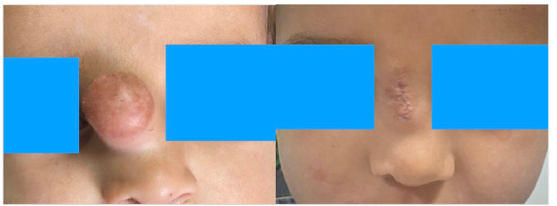
The mass excision was carried out from the upper to lower midline of the nasal dorsum. The defect was closed using an advancement flap with 5/0 polydioxanone sutures. Throughout the procedures, meticulous attention was paid to minimizing trauma to surrounding tissues. The patient was then closely monitored during the postoperative period for any potential complications, including cerebrospinal fluid leakage. The excised tissue sample from the nasal dorsum was sent for histopathological examination to confirm the diagnosis and further understand the nature of the mass.
Discussion
Congenital midline nasal masses are rare, with a prevalence of 1 in 20,000 to 40,000 births (1). Differential diagnosis of midline nasal masses includes nasal glioma, dermoid cyst, lipoma, hemangioma, and encephalocele (2). The nasal dermoid was commonly present as a midline mass and may be located on the nasal dorsum or the nasoglabellar region (3).
An exciting aspect observed during the treatment was a tissue resembling granulation tissue seen in foreign body reactions. A foreign body reaction is a complex process where the body responds to a foreign object, such as surgical sutures, splinters, or implants, forming granulation tissue. Further study on occurrence and implications of this in congenital nasal mass treatment could be a topic of interest.
Computed tomography (CT) and magnetic resonance imaging (MRI) scans are the gold standards for radiographic evaluation of nasal dermoids. MRI may be sufficient to determine intracranial extension (4, 5). Advancement flaps are important reconstructive options after skin mass removal. The flaps are mainly used for defects located on the superior two-thirds region of the nose (6, 7). A notable aspect of this case is the closure of the skin using an advancement flap without a graft following tumor excision. This approach facilitated a faster recovery for the child compared to grafted reconstruction.The length of stay was reduced due to the accelerated recovery (8). The treatment of congenital nasal dorsal masses is typically surgical. It's crucial to approach surgeries, especially in children, with great caution, given the potential impact on their psychological development. The findings from this case highlight the importance of meticulous surgical techniques.
An encephalocele and a dermoid cyst co-occurred incidentally in this patient, as there is no physical connection between two lesions. This presents unique learning point and extends our understanding of the complexity and variability of congenital nasal dorsal masses. In the one-year follow-up period, no recurrence or growth was observed at the excision site. In conclusion, this case report demonstrates the co-occurrence of an encephalocele and a dermoid cyst of the nasal dorsum, with no connection between the two. The successful surgical management, which included skin closure with an advancement flap without grafting, highlights the potential benefits of this technique in reducing patient recovery time and length of hospital stay. The careful handling of such cases, including long-term follow-up, adds to our understanding of congenital nasal dorsal masses and can potentially improve the overall outcomes for affected children.
Declarations
- Author contributions (CRediT)
- All authors contributed equally to all 14 CRediT roles (Conceptualization, Data curation, Formal analysis, Funding acquisition, Investigation, Methodology, Project administration, Resources, Software, Supervision, Validation, Visualization, Writing – original draft, Writing – review & editing).
- Funding
-
Funding information was not reported in the published version of this article.
- Conflicts of interest
- The authors have no conflicts of interest.
- Ethics approval
- The research conducted and reported in this case study, "An unusual midline nasal mass in a child: A Case Report", was carried out in full compliance with accepted international and national ethical standards for clinical research. The research procedures adhered to the principles of the Declaration of Helsinki and its subsequent amendments, which provide guidelines on the ethical aspects of medical research involving human subjects, including
- Data availability
- A data availability statement was not reported in the published version of this article.
References (8)
7 of 8 references are linked to their DOI.
- Verney Y, Zanolla G. Midline nasal mass in infancy: a nasal glioma case report. Eur J Pediatr Surg 2001;11:324-7 DOI Crossref PubMed Google Scholar
- Salati SA, Rather AA. Congenital intranasal glioma. Case Rep Surg. 2011;175-209 DOI Crossref PubMed Google Scholar
- Rahbar R, Shah P, Mulliken JB, et al. The presentation and management of nasal dermoid: a 30-year experience. Arch Otolaryngol Head Neck Surg 2003;129:464–471 DOI Crossref PubMed Google Scholar
- Bloom DC, Carvalho DS, Dory C, et al. Imaging and surgical approach of nasal dermoids. Int J Pediatr Otorhinolaryngol 2002; 62:111– 122 DOI Crossref PubMed Google Scholar
- Penslar JM, Bauer BS, Naidich TP. Craniofacial dermoids. Plast Renconstr Surg 1988;82:953– 959 PubMed Google Scholar
- Salmon PJ. Repair of the nasal sidewall. Br J Dermatol. 2014; 171:17–22 DOI Crossref PubMed Google Scholar
- Vinciullo C. Reconstructing the nasal dorsum. Br J Dermatol. 2014; 171:7–16 DOI Crossref PubMed Google Scholar
- Ogawa Y, Ogawa Y. Lateral nasal advancement flap for reconstruction of the nasal sidewall and dorsum. Arch Plast Surg. 2020 Jan;47(1):102-105 DOI Crossref PubMed Google Scholar
Respond to this article
Comments on a published article are welcome as a Letter to the Editor.
Get an e-mail when new articles in your fields are published
Questions to the authors
Questions are moderated: an editor approves each question, the authors answer, and an editor approves the answer before both appear here.
No answered questions yet.
Ask the authors a question
Discussion
Comments are moderated: they appear after the writer confirms the e-mail address and an editor approves them. Each comment carries a conflict-of-interest statement.
No comments yet.
Write a comment
Read the full text
Open the PDF here
Loading the PDF…
If the PDF does not show here, use the download button above.
How to cite this article
Zhanbaeva AK, Guvenmez O, Keskin H, Zhanbaev A. An Unusual Mid-Line Nasal Mass in a Child: A Case Report. Ulutas Med J. 2023;9(2):106-109. doi:10.5455/umj.20230518033004
Zhanbaeva, A. K., Guvenmez, O., Keskin, H., & Zhanbaev, A. (2023). An Unusual Mid-Line Nasal Mass in a Child: A Case Report. The Ulutas Medical Journal, 9(2), 106–109. https://doi.org/10.5455/umj.20230518033004
Zhanbaeva, A.K. et al. (2023) 'An Unusual Mid-Line Nasal Mass in a Child: A Case Report', The Ulutas Medical Journal, 9(2), pp. 106–109. doi: 10.5455/umj.20230518033004.
@article{umj153797,
  title = {An Unusual Mid-Line Nasal Mass in a Child: A Case Report},
  author = {Zhanbaeva, Anara Keneshovna and Guvenmez, Oguz and Keskin, Huseyin and Zhanbaev, Adylbek},
  journal = {The Ulutas Medical Journal},
  year = {2023},
  volume = {9},
  number = {2},
  pages = {106--109},
  doi = {10.5455/umj.20230518033004},
  url = {https://www.ulutasmedicaljournal.com/?mno=153797},
  issn = {2149-388X}
}