The Ulutas Medical Journal · ISSN 2149-0430 (print), 2149-388X (online) · https://www.ulutasmedicaljournal.com/?mno=96651
Original Article · Open access
Adult Medulloblastoma: Clinical Profile and Treatment Result of 25 Patients
Also available in: العربية · Türkçe · 中文
Abstract
- Introduction
- The study aimed to discuss the clinical presentation of radiological findings, surgical treatments, and postoperative complications as well as the radiotherapy and chemotherapy options for adult patients with medulloblastoma.
- Materials and Methods
- A total of 25 patients (14 males and 11 females) between the ages of 17 and 63 who underwent an operation at our clinic between 2006-2016 are discussed in this article. The degree of resection in the patients and tumor sizes after radiotherapy (RT) and chemotherapy (Cht) was compared using the patients' preoperative and postoperative magnetic resonance (MR) images.
- Results
- The average age of the patients was 30.9 years. Gross total resection was achieved in 20 patients, whereas subtotal resection was achieved in the remaining 5 patients. Three patients developed hydrocephalus during the 6th month after resection, which was treated with a V-P shunt. Another four patients developed a cerebrospinal fluid (CSF) fistula. Among the five patients who only achieved subtotal resection, two died during the 24th month of the post-operative period, and the other three died during the 18th month. Eleven patients presented pathology of the desmoplastic type, and 14 patients presented classical medulloblastoma. Spinal metastasis was detected in three patients during the pre-operative follow-up and in three patients during the post-operative follow-up. The average survival rate of the cohort was 60% during the 3 to 5-year follow-up period.
- Conclusion
- Medulloblastoma development is a risk not only for children and adolescents but also for adults. Maximum tumor removal during treatment affects patient outcomes, and RT and Cht administration during the post-operative period can extend the life span of these patients.
Keywords
Full text
Introduction
Medulloblastomas are malignant tumors that often metastasize locally to the posterior fossa and occupy significant regions within the central nervous system of children (1). However, approximately 20% of individuals diagnosed with medulloblastoma are over 16 years of age. Although adult medulloblastomas are similar to pediatric medulloblastomas, they differ from the pediatric subtype via a higher incidence of the desmoplastic variant and more frequent lateral localization (2). Patients with desmoplastic histology always have better survival rates than those with either classical or large-cell histology. Additionally, the lateral (i.e., non-vermian) location of the tumor allows for a greater likelihood of total resection. However, there is no prominent consensus related to the treatment of medulloblastomas in adolescents and adults, and there is no complete consensus on the application of chemotherapy (Cht) for adult medulloblastoma because of the high risk of toxicity associated with Cht and reduced number of cases requiring an emergency decrease in the craniospinal radiotherapy (RT) dosage (3–5). Notwithstanding these factors, administering Cht for adult medulloblastoma can delay relapse and inhibit systematic metastasis (6–8).
The purpose of this article is to discuss the treatment and follow-up of adult medullo blastoma patients based on the findings of 25 cases monitored at our clinic.
Materials and Methods
Data from 25 adult medulloblastoma patients followed in our clinic between 2006 and 2016 were evaluated in this study. The average age of the patients was 30.9 years old (range 17-63); 11 of them were female, and 14 of them were male. The most frequent reason for seeking clinical consultation was headache followed by complaints of nausea, vomiting, and ataxia. Craniospinal imaging was performed on all the patients during the pre-operative period. Metastasis, tumor localization, intensity and the presence of hydrocephalus was evaluated using imaging of the patients. All the patients were placed in a sitting position during surgery. Subtotal resection was achieved in 5 patients, whereas gross total resection was achieved in 20 patients. Two of the patients who achieved subtotal resection underwent the second operation 1 year later. Craniospinal imaging of patients was performed during the post operative follow-up period. The amount of resected tissue, instances of relapse, and metastasis were evaluated using follow-up imaging. All the patients received RT and Cht.
Results
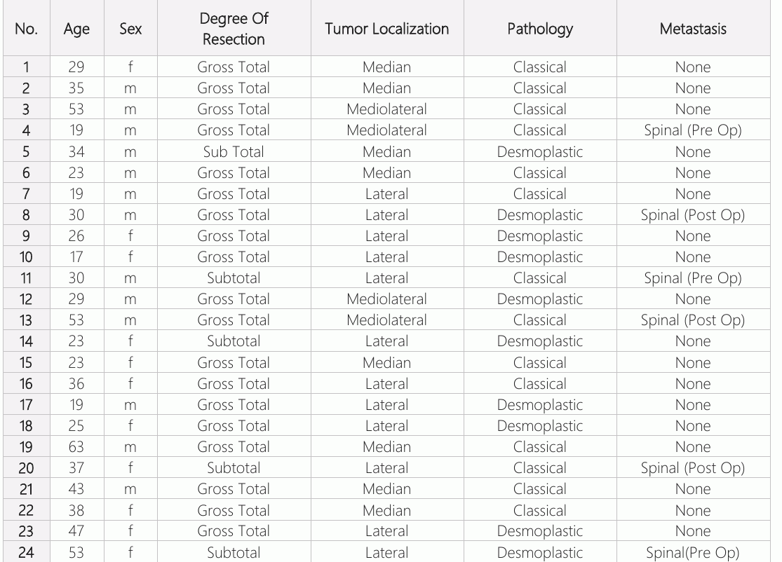
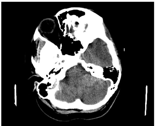
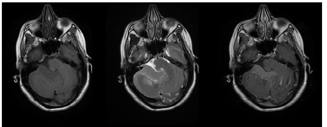
Computerized brain tomography (CBT) of the lesions revealed a generally iso-hypodense appearance (Fig. 1). The lesions were generally iso-hypodense in the T1-weighted images and hyperintense in the T2-weighted images; In addition, pre-operative magnetic resonance image (MRI) showed prominent homogeneous contrast (Fig. 2). Among 25 lesions, 9 were at the midline, 4 were lateral from the midline, and 12 were laterally localized. Hydrocephalus was observed in 10 patients, and spinal metastasis was detected in 3 patients (Table 1).
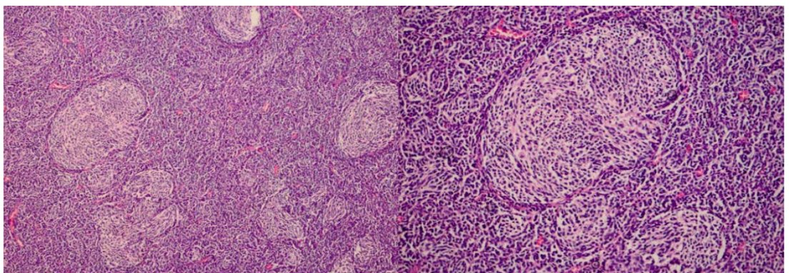
While in the post-operative recovery unit, one of the patients developed a hematoma within 6 hours after surgery completion, which qualified as an early-period complication. The patient underwent surgery again to drain hematoma. On a postoperative day 3, another 3 patient developed hydrocephalus, which was treated with a V-P shunt. Cerebrospinal fluid (CSF) leakage from the incision site was observed in four patients within the first week during the post-operative period; these patients underwent repair of the incision site and dura. Two of the patients who achieved subtotal resection underwent a second tumor resection 1 year after the initial surgery. The pathology results of 14 patients showed classical type medullo blastoma, whereas the pathology of the remaining 11 indicated desmoplastic medullo blastoma (Fig. 3). The average survival rate of the cohort was 60% during the 3 to 5-year follow-up. Three patients developed spinal metastasis during the post-operative period. 2 patients who achieved subtotal resection and underwent a second operation died in the 24th month of the post-operative period after the first surgery, and the other 3 patients who only achieved subtotal resection died in the 18th month of the post-operative period.
Discussion
Medulloblastoma is a tumor that is mostly observed during childhood and originates from transient undifferentiated neuroepithelial cells in either the cerebellum or the roof of the 4th ventricle (1). These cells can differentiate into astroglial or neuroblastic subtypes. The embryonic origin of these cells is the primary reason that medulloblastoma is frequently observed in childhood; additionally, it is the second most frequently observed tumor of the central nervous system in childhood.
Many features of adult medulloblastomas are similar to those in pediatric medulloblastomas; however, adult and pediatric medulloblastomas have different demographic, morphological, and molecular characteristics. Medulloblastoma is categorized based on gene expression profiles and can be divided into 4 subgroups: Wingless (WNT), Sonic hedgehog (SHH), Group 3, and Group 4. Among these groups, the WNT subtype has the best patient prognosis. The SHH and Group 4 subtypes have mediocre prognoses, and Group 3 has a poor prognosis (9). A meta-analysis showed that the majority of patients in the SHH subgroup (57%) were in the adult group. The percentage of adults within the other subgroups was 13% for WNT, 4% for Group 4, and 2% for Group 3 (10–12). Using cytogenetic analyses, Northcott et al. (9) showed that the frequency of deletions on chromosome 10q was lower among adult SHH patients than among pediatric patients. These four subgroups also differ in the prognosis rates among pediatric and adult patients. The prognosis of adult Group 4 and WNT patients is worse than that in corresponding pediatric patients. In the SHH subgroup, both pediatric and adult groups have similar prognoses. Among pediatric patients, metastasis is more frequent in Group 3 and Group 4 patients, whereas adult patients in each subgroup exhibit similar metastasis rates; however, metastasis in adults, groups do not affect prognosis (10). Adult medulloblastoma is less prone to metastasis; additionally, pulmonary metastasis is more common in adult medulloblastoma, whereas liver metastasis is more commonly observed in pediatric medulloblastomas (12, 13). Metastasis is less frequent in the desmoplastic and lipomatous medulloblastoma, melanotic medulloblastoma, medullomyoblastoma, and large cell medulloblastoma subtypes but is more commonly observed in classic medullo blastoma type (14). The desmoplastic subtype has reduced metastasis compared to classical medulloblastoma with a better clinical course. The observation frequency of medulloblastoma among primary brain tumors in adults is approximately 1%. It is most often diagnosed between 45-50 years of age and is rarely observed after the 5th decade (4, 5, 15). It is male dominant in adults as well as in children (3, 6). The patients in our series were between 17 and 63 years old with an average age of 30.9. There was a pattern of male dominance within our cohort, which is following the literature. Performing a craniospinal MRI on patients is significant for determining the optimal surgical strategy for the patients, examining the patient, and detecting spinal metastasis. Although the MRI findings of adult medulloblastomas do not reveal distinctive characteristics, studies reporting these findings indicate that medullo blastomas in adults tend to develop more laterally compared to the localization observed in children. No prominent distinguishable characteristics regarding the intensity and contrast relating to the tumor were detected. Generally, isodense regions are observed in T1- weighted images, and hyperdensity is observed in T2-weighted images. Contrast of the tumor may be homogeneous or inhomogeneous. The fact that the tumor may contain cystic or necrotic areas may affect the specified findings. In a study of 13 patients conducted by Bezircioğlu et al., (16) T1-weighted imaging was hypointense in 4 patients and isointense in 9 patients; however, all the patients showed hyperintense lesions in T2-weighted images. Contrast involvement was homogeneous in 5 of the patients and inhomogeneous in 8 of them. In 5 of the patients, the lesion was located in the midline. In 5 of the patients the lesion was located in lateral to the midline and in 3 of the patients was located in lateral (16). In a cohort of 12 patients published by Koci et al. (17), 8 patients showed lateral tumor localization, 2 were medial to lateral and 2 were medial. In our series, 9 of the lesions in the patients were at the midline, 4 of them were lateral across the midline and 12 of them were laterally localized. Homogeneous involvement was observed after performing iso-hypointense contrast in T2- weighted images and hyperintense contrast in T2-weighted images. Spinal metastasis was observed in 3 patient, and hydrocephalus was observed in 8 patients.
The percentage of resected tumor is an important parameter in the study of cancer patients. According to the risk criteria set by the University of California San Francisco, resections below 75% constitute a significant risk factor for poor prognosis. However, it should be noted that sequelae, which may occur while performing resection, can affect the study outcomes. In the long-term study of 27 patients by Riffaud et al. (18), the 5-year survival rate was 85% for patients who achieved total resection and 67% for patients who either experienced subtotal resection or only underwent a biopsy. In a study conducted by Jiang et al.,(19) the 5- year survival rate within a cohort of 33 patients was approximately 50-60% among patients who underwent gross total resection and 25% among patients who only achieved subtotal resection (15). In a report by Lai et al. (20) that evaluated the prognosis of a cohort of 454 adult patients with medulloblastoma at 17 centers, gross total resection was revealed to be among the best prognostic factors. In our study, we achieved a gross total resection in 20 of the 25 patients and subtotal resection in the remaining 5 patients. Among the latter patients, two underwent a second operation 1 year later; unfortunately, these patients died 24 months after the first operation. The other three patients who only achieved subtotal resection died 18 months after the operation.The average 3 to 5-year survival rate was calculated as 60%. As common as the classical medulloblastoma subtype is observed among pediatric patients, the desmoplastic variant is observed more frequently among adults. The desmoplastic type tends to develop in a more lateral location than the classical subtype. Although lateral localization of classical medulloblastoma often plateaus at a rate of 12%, approximately 71% of desmoplastic cases exhibit lateral localization (2). The term desmoplasia is used based on the observation of excessive collagenases within the connective tissue surrounding the tumor. The tumor tissue is macroscopically observed as a benign limited lobule leptomeningeal mass. A 13-patient study by Bezircioğlu et al. described 2 patients classified with classical medulloblastoma and 11 patients classified with desmoplastic medulloblastoma (2). In a A 27-patient study by Riffaud et al. (19), 21 patients were identified with classical disease, and 6 patients were classified as desmoplastic medulloblastoma. In a larger cohort, Lai et al. stratified their cohort of 454 patients with medulloblastoma as follows: 397 patients with classical medulloblastoma; 52 patients with desmoplastic medulloblastoma; 2 patients with medullomyoblastoma; and 3 patients with large cell medulloblastoma (20). The cohort described by Jiang et al. included 17 patients with classical medulloblastoma, 13 patients with desmoplastic type medulloblastoma, and 3 patients with anaplastic medulloblastoma. The frequency of the desmoplastic type compared to that of the classical type differs in the various studies of adult medulloblastoma. In our study, 14 of the 25 patients were classified as classical type and the remaining 11 had the desmoplastic type.
RT application is highly recommended for patients during the post-operative period. In a study conducted by Abacıoğlu et al. of 30 patients with adult medulloblastoma, low dosages of RT were observed to control the tumor occurrence in the posterior fossa (3). RT administered during the post-operative period minimizes the development of residual lesions as well as prevents spinal metastasis. The main subject under discussion is the requirement of administering Cht to these patients. Because the toxicity of established Cht agents is higher in adults than in children, there is increased risk in administering Cht. The recent discovery of agents such as temozolomide, which has low toxicity, minimizes these associated risks. In the studies performed, adjuvant Cht delays recurrence among patients who are at high risk for recurrence. The prospective study conducted by Brandes et al. (7) reported that among patients who underwent only RT during the postoperative period, the incidence of recurrence increased prominently over a 7.6-year follow-up period. In a meta-analysis conducted by Kocakaya et al. (21) on 907 patients, adult medulloblastoma patients who were administered Cht had an increased long-term prognosis. We administered RT and Cht to all the patients in our study during the post-operative period. The average survival rate of our cohort was 60% during the 3 to 5-year follow-up. Additionally, three patients suffered from spinal metastasis during the postoperative follow-up period.
Conclusion
Although medulloblastoma is more commonly observed during childhood, they also affect 1% of adults with central nervous system tumors. They primarily tend to settle in the lateral region, which is different from the tumors that develop in children. The desmoplastic variant of medulloblastomas was observed more frequently among adults than among children. Achieving total resection is critical in terms of treatment, and administering RT to patients during the postoperative period is important in terms of eliminating residue lesions and preventing spinal metastasis. Additionally, CT significantly reduces relapse rates. Finally, scanning the spinal region of patients during the postoperative follow-up period is essential to identify spinal metastasis.
Declarations
- Author contributions (CRediT)
- All authors contributed equally to all 14 CRediT roles (Conceptualization, Data curation, Formal analysis, Funding acquisition, Investigation, Methodology, Project administration, Resources, Software, Supervision, Validation, Visualization, Writing – original draft, Writing – review & editing).
- Funding
-
Funding information was not reported in the published version of this article.
- Conflicts of interest
- The authors declared no conflict of interest.
- Ethics approval
- The Ethical Committee and Institutional Review Board of Adana City Training a nd Research Hospital, where the study was conducted, approved the study design.
- Data availability
- A data availability statement was not reported in the published version of this article.
References (21)
16 of 21 references are linked to their DOI.
- Russell DS, Rubinstein LJ. Pathology of tumors of the nervous system. 5th ed. London: Edward Arnold, 1989 PubMed Google Scholar
- Katsetos CD, Herman MM, Frankfurter A, Gass P, Collins VP, Walker CC, et al. Cerebellar desmoplastic medulloblastomas. A further immunohistochemical characterization of the reticulin-free pale islands. Arch Pathol Lab Med. 1989;113(9): 1019-1029 PubMed Google Scholar
- Abacioglu U, Uzel O, Sengoz M, Turkan S, Ober A. Medulloblastoma in adults: treatment results and prognostic factors. Int J Radiat Oncol Biol Phys, 2002;54(3):855–860 DOI Crossref PubMed Google Scholar
- Brandes AA, Paris MK. Review of the prognostic factors in medulloblastoma of children and adults. Crit Rev Oncol Hematol. 2004;50(2):121-128 DOI Crossref PubMed Google Scholar
- Chan AW, Tarbell NJ, Black PM, Louis DN, Frosch MP, Ancukiewicz M, et al. Adult medulloblastoma: prognostic factors and patterns of relapse. Neurosurgery. 2000;47(3), 623-31 DOI Crossref PubMed Google Scholar
- Brandes AA, Ermani M, Amista P, Basso U, Vastola F, Gardiman M, et al. The treatment of adults with medulloblastoma: a prospective study. Int J Radiat Oncol Biol Phys. 2003;57(3):755–761 DOI Crossref PubMed Google Scholar
- Brandes AA, Franceschi E, Tosoni A, Blatt V, Ermani M. Long-term results of a prospective study on the treatment of medulloblastoma in adults. Cancer. 2007;110(9):2035– 2041 DOI Crossref PubMed Google Scholar
- Friedrich C, von Bueren AO, von Hoff K, Kwiecien R, Pietsch T, Warmuth- Metz M, et al. Treatment of adult nonmetastatic medulloblastoma patients according to the paediatric HIT 2000 protocol: A prospective observational multicentre study. Eur J Cancer. 2013;49(4):893–903 DOI Crossref PubMed Google Scholar
- Northcott PA, Hielscher T, Dubuc A, Mack S, Shih D, Remke M, et al. Pediatric and adult sonic hedgehog medullo blastomas are clinically and molecularly distinct. Acta Neuropathol. 2011;122(2):231–240 DOI Crossref PubMed Google Scholar
- Kool M, Korshunov A, Remke M, Jones DT, Schlanstein M, Northcott PA, et al. Molecular subgroups of medullo blastoma: an international meta- analysis of transcriptome, genetic aberrations, and clinical data of WNT, SHH, Group 3, and Group 4 medulloblastomas. Acta Neuropathol. 2012; 123(4):473-484 DOI Crossref PubMed Google Scholar
- Remke M, Hielscher T, Northcott PA, Witt H, Ryzhova M, Wittmann A, et al. Adult medulloblastoma comprises three major molecular variants. J Clin Oncol. 2011;29(19):2717–2723 DOI Crossref PubMed Google Scholar
- Birch JM, Marsden HB, Jones PH, Pearson D, Blair V. Improvements in survival from childhood cancer: results of a population based survey over 30 years. Brit Med J. 1988;296 (6633):1372–1376 DOI Crossref PubMed Google Scholar
- Brandes AA, Franceschi E, Tosoni A, Reni M, Gatta G, Vecht C, et al. Adult neuroectodermal tumors of posterior fossa Adult Medulloblastoma Arslan et al. (medulloblastoma) and of supratentorial sites (stPNET). Crit Rev Oncol Hematol. 2009;71(2):165‑179 DOI Crossref PubMed Google Scholar
- Resenbium MK,Bilbao JM,Ang LC. Neuromuscular system. In: Ackerman`s surgical pathology. Volume 2, 8th ed. St Louis: Mosby. 1996;p.231-2315 PubMed Google Scholar
- Taylor MD, Northcott PA, Korshunov A, Remke M, Cho YJ, Clifford SC, et al. Molecular subgroups of medulloblastoma: the current consensus. Acta Neuropathol. 2012;123(4):4 DOI Crossref PubMed Google Scholar
- Bezircioglu H, Sucu HK, Minoglu M, Tunakan M,Olmezoglu A. Adult medulloblastoma: clinical profile and treatment result of 13 patients. J Neurol Sci. 2009;26:(4),463-471. PubMed Google Scholar
- Koci TM, Chiang F, Mehringer CM, Yuh WT, Mayr NA, Itabashi H, et al. Adult cerebellar medulloblastoma: imaging features with emphasis on MR findings. AJNR. 1993;14(4), 929-939. PubMed Google Scholar
- Riffaud L, Saikali S, Leray E, Hamlat A, Haegelen C, Vauleon E, et al. Survival and prognostic factors in a series of adults with medulloblastomas. J NeuroSurg. 2009;111(3):478-487 DOI Crossref PubMed Google Scholar
- Jiang T, Zhu J, Dong J, Wang JM, Du J, Li CD, et al. Clinical outcomes of adult medulloblastoma: A retrospective analysis at a single institute. Transl Neurosci Clin. 2015;1(1):17–24 DOI Crossref PubMed Google Scholar
- Lai R. Survival of patients with adult medulloblastoma: a population-based study. Cancer. 2008;112(7):1568-1574 DOI Crossref PubMed Google Scholar
- Kocakaya S, Beier CP, Beier D. Chemotherapy increases long-term survival in patients with adult medulloblastoma-a literature-based meta- analysis. Neuro Oncol. 2016;18(3): 408-416 DOI Crossref PubMed Google Scholar
Respond to this article
Comments on a published article are welcome as a Letter to the Editor.
Get an e-mail when new articles in your fields are published
Questions to the authors
Questions are moderated: an editor approves each question, the authors answer, and an editor approves the answer before both appear here.
No answered questions yet.
Ask the authors a question
Discussion
Comments are moderated: they appear after the writer confirms the e-mail address and an editor approves them. Each comment carries a conflict-of-interest statement.
No comments yet.
Write a comment
Tell me when this article is cited
Leave your e-mail address and confirm it. These notices are not being sent yet; your confirmed address is kept for them and used for nothing else.
Read the full text
Open the PDF here
Loading the PDF…
If the PDF does not show here, use the download button above.
How to cite this article
Arslan A, Acik V. Adult Medulloblastoma: Clinical Profile and Treatment Result of 25 Patients. Ulutas Med J. 2020;6(3):131-138. doi:10.5455/umj.20200712045718
Arslan, A., & Acik, V. (2020). Adult Medulloblastoma: Clinical Profile and Treatment Result of 25 Patients. The Ulutas Medical Journal, 6(3), 131–138. https://doi.org/10.5455/umj.20200712045718
Arslan, A. and Acik, V. (2020) 'Adult Medulloblastoma: Clinical Profile and Treatment Result of 25 Patients', The Ulutas Medical Journal, 6(3), pp. 131–138. doi: 10.5455/umj.20200712045718.
@article{umj96651,
  title = {Adult Medulloblastoma: Clinical Profile and Treatment Result of 25 Patients},
  author = {Arslan, Ali and Acik, Vedat},
  journal = {The Ulutas Medical Journal},
  year = {2020},
  volume = {6},
  number = {3},
  pages = {131--138},
  doi = {10.5455/umj.20200712045718},
  url = {https://www.ulutasmedicaljournal.com/?mno=96651},
  issn = {2149-388X}
}