The Ulutas Medical Journal · ISSN 2149-0430 (print), 2149-388X (online) · https://www.ulutasmedicaljournal.com/?mno=85435
Original Article · Open access
Comparison of Pre-treatment and Post-treatment Auditory Brainstem Response Results in Children with Serous Otitis
- Department of Otorhinolaryngology, Sutcu Imam University, Faculty of Medicine, Kahramanmaraş, Turkey ROR
- Department of Pediatrics, Giresun University, Gynecology and Child Training and Research Hospital, Giresun, Turkey ROR
- Department of Pediatrics, Sutcu Imam University, Faculty of Medicine, Kahramanmaraş, Turkey ROR
- Department of Pediatrics, Giresun University, Faculty of Medicine, Giresun, Turkey ROR
Abstract
- Introduction
- The present study aimed to investigate the effect of middle ear effusion on the ABR test through a comparison of clinical auditory brainstem response (ABR) results in pre-treatment and post-treatment in patients diagnosed with otitis media with effusion (OME).
- Materials and Methods
- This retrospective study included 42 children aged 3 months-12 months, who failed the otoacoustic emissions (OAE) test in the hearing screening program and were diagnosed with serous otitis.
- Results
- Following treatment of the patients, the right ears of 76.9% of patients and the left ears of 69.04% of patients passed the distortion product otoacoustic emissions test. In the OAE tests post-treatment, in 23.8% of patients, the right ear, and 30.95% patients, the left ear was negative, and a decrease was seen in the hearing thresholds in clinical ABR. The latencies of the ABR 1st and 3rd waves were observed to be significantly prolonged pre-treatment compared to post-treatment in all patients.
- Conclusion
- The appropriate intervention in the early stage eliminates or reduces the negative effects of hearing loss. To avoid a very long follow-up period of infants diagnosed with serous otitis media in the first years of life, prompt treatment should be applied to patients who have not responded to medical treatment, and if the thresholds after treatment are above normal values, a hearing aid should be applied as soon as possible and family therapy should be provided.
Keywords
Full text
Introduction
Otitis media is a generic definition for any inflammatory process in the middle ear cavity behind the healthy ear membrane and there are two main types of acute otitis media and otitis media with effusion (OME). OME can be defined as an inflammatory table characterized by fluid accumulation behind healthy tympanic membrane without concomitant findings of local or systemic acute infection (1–3).
As it is generally seen in childhood, the main challenges in diagnosis are technical difficulties in the physical examination and very young children cannot verbalize their complaints, so symptoms may not be noticeable and reduced hearing may remain hidden. Otomicroscopy, otoscopy, or pneumatic otoscopy are the most important diagnostic methods. Audiological methods are diapazon tests, tympanometry, acoustic reflex, pure sound audiometry, brain stem audiometry, and audio acoustic emissions (4, 5). Tympanometry is a useful method in the diagnosis of OME in children aged 2-24 months, but together with younger age, there is reduced sensitivity of the method. Despite the difficulty of use, high-frequency tympanometry should be used in the middle ear evaluation of new borns.Treatment is considered in 3 main groups of conservative, medical and surgical. The treatment modality is determined according to the otoscopic appearance, tympanometry type, response to conservative treatment, and hearing level (2, 6).
The auditory brainstem response (ABR) test records the electrical activity occurring as a result of the response of the VIII nerve and brainstem neurons to an auditory stimulus (7). In the determination of hearing thresholds and the diagnosis of hearing loss and retrocochlear pathologies in the elderly, unconscious patients, and young children, ABR is used, because patient compliance is not required. In these two situations, peak latencies and inter-peak latencies (IPL) of waves forming the response are important. Classically, ABR is formed from 7 peaks seen within the first 10 min but in clinic, the first 5 peaks are of importance (8–10).
This study aimed to compare the clinical ABR results taken pre and post-treatment to determine the effect on the ABR test of effusion in the middle ear in patients with no response to OAE in the neonatal hearing screening who was then diagnosed with OME from clinical examination and tympanometric examination.
Materials and Methods
In this retrospective study, the records were examined of patients applied with a hearing test in the neonatal hearing screening program in our clinic between January 2015 and June 2017. Of the patients followed up because of the neonatal hearing screening, the files were reviewed of patients diagnosed with OME and applied with clinical ABR before and after treatment. A total of 100 ears of 48 patients (26 male, 22 female) were included for evaluation.
Ethical Statement
Approval for the study was granted by the Ethic Committee of Sutcu Imam University, Medical Faculty (session no: 2017/04, decision no: 03, dated 15.03.2017). A total of 16 patients were excluded from the study because of a syndrome diagnosis, familial hearing problem, auditory pathway anomaly, or incomplete information. The final evaluation was made of 84 ears of 42 patients (28 males, 14 females). The diagnosis of OME was made from the anamnesis and history of hearing loss with in ear examination with an otoscope (Heine Beta 100 otoscope, Germany) of a matte appearance of the tympanic membrane, vascularisation and air-fluid level behind the membrane, and type B or typeC tympanogram (Interacoustics, AT235, Denmark). When the hearing screening was required, distortion product otoacoustic emissions (DPOAE) and transient-evoked oto acoustic emission(TEOAE) otoacoustic emission tests and screening ABR tests (Madsen Capella GN Otometrics, Denmark) were negative. The OME diagnosis was made and to determine hearing thresholds in cases thought to have advanced hearing loss, clinical ABR was applied (Neurosoft, Neuro-Audio, Ivanovo, Russia). After recovery of OME with medical and/or surgical treatment, ABR has applied again. The ABR tests were applied using adhesive electrodes with patient supine on a comfortable bed in a quiet, dark room that would not have any visual stimuli, in the Audiology Laboratory. The negative electrodes were placed on the two mastoid regions, the positive electrode on the vertex and the earth electrode on the forehead. Care was taken the electrode skin impedances were below 5 kΩ. The tests were applied with a click stimulus at a frequency of 27/1s, 100µs alternating. For each ear, at least 2 records were made at the intensities applied and care was taken that the latencies of the waves were consistent with each other. Sedation agents were not used in any child. The tests were applied during natural sleep. The pre and post-treatment ABR latency data of the patients were recorded on a standard data form, then transferred to statistics program and analyzed.
Statistical Analysis
The data obtained in the study were analyzed using IBM SPSS for Windows v22 software (IBM statistics Windows, IBM Corporation, Armonk, New York, USA). Conformity of the data to normal distribution was examined (histogram) with analytical methods. Descriptive analyses for normal distribution were stated as mean± standard deviation (SD) and significance was set at the level of p<0.05. For the relationship between two groups, the Pearson Correlation coefficient was calculated. In the comparison of 2 groups, Wilcoxon t-test and the Independent Samples t-test were used. Results were given in a 95% confidence interval and a value of p<0.05 was accepted as statistically significant.
Results
In all the patients who did not pass the OAE tests in the hearing screening program follow-up and who were diagnosed with serious otitis, the latencies of the clinical ABR waves were prolonged before treatment. When evaluated according to gender, the pre to post-treatment difference in latency was greater in males. In the right ear examination of 3 patients and the left ear examination of 4 patients, the otoscopic examination and tympanogram were normal, so OAE was not applied and the hearing thresholds on clinical ABR recovered during follow-up. In 82% of the patients, the middle ear recovered with medical treatment and follow-up, and 18% with surgical treatment. In all the patients surgically applied with grommet tubes, glue-type fluid was aspirated from the middle ear after parasynthesis. One patient had postoperative complaints of otorrhea but recovered in a short time after removal of the tube.
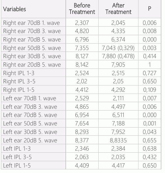
Following treatment of the patients, the right ears of 32 (76.9%) patients and the left ears of 29 (69.04%) patients passed the DPOE test and the hearing thresholds were within the normal limits on clinical ABR. In the OAE tests post-treatment, in 10 (23.8%) patients, the right ear, and in 13(30.95%) patients, the left ear was negative, and a decrease was seen in the hearing thresholds in clinical ABR. Following these results, a hearing aid was recommended. In the results of the clinical ABR applied before OME treatment, a mean 45dB was observed, with the highest 60dB and lowest 40dB in the right ear, and mean 40dB with highest 70dB and lowest 30dB in the left ear. After treatment, these values were highest 50dB, lowest 15dB in the right ear, and highest 60dB, and lowest 40 dB in the left ear. Just as in the decrease in thresholds before and after treatment, a statistically significant reduction was observed in the latencies of the first, third, and fifth 70dB waves and the fifth 50dB and 30dB waves (p<0.05)(Table-1). No significant difference was observed between the pre and post-treatment interpeak latencies (IPL 1-3, IPL 3-5, IPL 1-5). Pre-treatment DPOAE was taken in the right ear of 6 patients (24 could not be taken) and in the left ear of 5 patients (25 could not be taken), and post-treatment could not be taken in the right ear of 4 patients and the left ear of 3. The change in DPOAE from before medical treatment to after was found to be significant in both ears. The clinical ABR threshold values were determined to have significantly reduced post-treatment compared to pre-treatment.
Discussion
OME is one of the diseases most often seen in childhood that leads to reduced hearing. This is a situation that requires correction and follow-up because of importance of communication, and psychosocial and mental development at this age. Middle ear effusions are seen in 50% of all children in the first year of life and this is something that can very often be overlooked (11). It is not greatly affected as measurement cannot be made from the external ear canal when there is amniotic fluid, debris, or middle ear fluid in the external ear canal. However, as the stimulus is given from the external auditory canal, if the external and middle ear problem is of a level serious enough to cause hearing loss to develop, then the ABR results are affected with prolonged latency in the first wave (12). Middle ear dysfunctions prevent the recording of otoacoustic emissions by negatively affecting the pathways traversed until the recording of cochlear emissions on microphones (13). In the current study, DPOAE and TEOAE could not be taken from OME patients. When the middle ear is full of effusion, compliance is low, and the tympanogram obtained was flat,in other words, type B configuration according to the Jegger definitions. 14 In the determination of OME, a type B curve obtained on tympanometry has been reported to have >80% sensitivity. Thus a type B curve cannot indicate the presence of OME in all conditions. On type A, the presence of OME is discounted as there is only a 5% probability. However, in infants below the age of 7 months, as the external auditory canal is extremely elastic and the tympanic ring has not yet fully developed, a normal tympanogram may be obtained even if OME is present (15, 16). In the current study, 3 patients with type A tympanogram and negative OAE were applied with a detailed examination, and OME was diagnosed.Ahigh-frequency tympanogram was used in all cases.
OME is a disease requiring close follow-up and has a high rate of spontaneous resolution. In the diagnosis and follow-up, evaluation is made with the otoscopic, pneumatic otoscopic examination, tympanogram, and OAE together. In cases with a high degree of hearing loss that does not respond to medical treatment, the application of a ventilation tube should be considered(11). In the study, a ventilation tube was applied to 9 patients with high hearing thresholds who did not respond to medical treatment.
The ABR test is the most frequently used objective test that plays a major role in the evaluation of hearing in childhood. Latency is one of the most important data in the determination of the threshold (17). Previous studies have reported delays in the first, third, and fifth waves and prolonged 3-5 IPL and 1-5 IPL in patients with OME compared to control groups (12, 13). In the current study, prolonged wave latencies and IPL values were observed pre-treatment. Evidence has shown that great changes in brain organization occur because of middle ear problems in the early stages of life (18, 19). Matschke et al. Reported 39 human brains were studied from the 29th week of pregnancy until the age of 70 years, and it was revealed that the necessary functional maturation of myelinization depended on sufficient stimulus in the first year of life (18). Physiologically, intervals between prolonged waves are reflected in the slowing down of the rate of central transmission in the inferior brain stem (19).
In our clinic, to determine the thresholds of all patients diagnosed with OME who have not passed OAE, ABR is applied as soon as possible and thresholds are determined with wave latencies and IPL. Following treatment, if the thresholds are above normal, even at a very mild degree, a hearing aid is recommended for the patient. Those with thresholds within normal limits and latencies above our clinic standards are kept under close follow-up and family therapy is provided. In previous studies, while prolonged first, third, and fifth wave latencies and IPL have been observed in OME patients, there has been reported to be no change in thresholds (12, 13, 20).
In this current study, a significant reduction was observed in the hearing thresholds of all the OME patients after treatment. A limitation of the study can be said to be that because of the low number of patients, there was little variability in the hearing loss of the children with OME. In patients with OME, the lengthening of wave latencies and reduced hearing can cause a negative effect on the hearing system. To avoid a very long follow-up period of infants diagnosed with SOM in the first years of life, prompt treatment should be applied to patients who have not responded to medical treatment, and if the thresholds after treatment are above normal values, a hearing aid should be applied as soon as possible and family therapy should be provided.
Conclusion
Hearing loss is an invisible health problem, and whatever the amount of hearing loss, it leads to delays in speech, cognitive and social development in infants and children. With appropriate early intervention, negative effects of hearing loss can be reduced or eliminated. Therefore, neonatal hearing screening should be applied in the first month, diagnostic examinations of infants that fail the screening should be completed within 3 months for the diagnosis of permanent hearing loss and until the age of 6 months, the infant should be applied with a hearing aid and hearing rehabilitation should be started. If permanent hearing loss is identified and appropriate intervention is made within the first 6 months, there is an increased chance that the infant will reach the same level of speech and language development as those with normal hearing. If OME is also present in infants with hearing loss, it is important that OME treatment is applied and real thresholds are determined before the use of a hearing aid. In addition, it is important for the child’s development that hearing screening is continued according to the risk status of the child during childhood healthcare monitoring (pre-school and school-age).
Declarations
- Author contributions (CRediT)
- All authors contributed equally to all 14 CRediT roles (Conceptualization, Data curation, Formal analysis, Funding acquisition, Investigation, Methodology, Project administration, Resources, Software, Supervision, Validation, Visualization, Writing – original draft, Writing – review & editing).
- Funding
-
Funding information was not reported in the published version of this article.
- Conflicts of interest
- A conflict of interest statement was not reported in the published version of this article.
- Ethics approval
- Approval for the study was granted by the Ethic Committee of Sutcu Imam University, Medical Faculty (session no: 2017/04, decision no: 03, dated 15.03.2017).
- Data availability
- A data availability statement was not reported in the published version of this article.
References (20)
13 of 20 references are linked to their DOI.
- Williamson I. Otitis media with effusion in children. BMJ Clinical Evidence. 2011:05-02 PubMed Google Scholar
- Rosenfeld RM, Shin JJ, Schwartz SR, Coggins R, Gagnon L, Hackell JM, et al. Clinical practice guideline: otitis media with effusion. Otolaryngol Head Neck Surg 2016;154:1-41 DOI Crossref PubMed Google Scholar
- Zhang Q, Wei J, Xu M, Zhang Q, Zhang X, Zhang Z, et al. Prevalence of otitis media with effusion among children in Xi'an, China: A randomized survey in China's mainland. Ann Otol Rhinol Laryngol. 2011:120:617–621 DOI Crossref PubMed Google Scholar
- Linsk R, Blackwood A, Cooke J, Harrison V, Lesperance M, Hildebrandt HM. University of Michigan Health System Otitis Media Guideline for Clinical Care. UMHS Otitis Media Guideline, 2013 PubMed Google Scholar
- Muderrıs T, Yazıcı A, Bercın S, Yalcıner G, Sevıl E, Kırıs M. Consumer acoustic reflectometry: accuracy in diagnosis of otitis media with effusion in children. Int J Pediatric Otorhino laryngology. 2013;77(10):1771–1774 PubMed Google Scholar
- Handler SD, Magardino TM. Otitis Media with effusion. in: Canalis RF, Lambert PR, eds. The Ear. Philadelphia: Lippincott Williams & Wilkins, 2000:383-396 PubMed Google Scholar
- Silva DD, Lopez P, Mantovani JC. Auditory brainstem response in term and preterm infants with neonatal complications: the importance of the sequential evaluation. International Arch Otorhinol. 2015;19 (2):161-165 DOI Crossref PubMed Google Scholar
- Ferraro JA, Durrant JD. Auditory evoked potentials: overview and basic principals. In: Katz J. Handbook of clinical Audiology. 4th edition 1994. p.317-338 PubMed Google Scholar
- Liang S, Soli SD, Zheng Y, Li G, Meng Z. Initial classification of pediatric hearing impairment using behavioral measures of early prelingual auditory development. Int J Audiol. 2016;55 (4):224–231 DOI Crossref PubMed Google Scholar
- Farias VB, Sleifer P, Pauletti LF, Krimberg CFD. Correlation of the findings of auditory steady-state evoked potential and of beha- vioral hearing assessment in infants with sensorineural hearing loss. CoDAS. 2014;26(03):226–230 DOI Crossref PubMed Google Scholar
- Robb PJ, Williamson I. Otitis media with effusion in children: current management. Paediatr Child Health. 2016;26.1: 9-14 DOI Crossref PubMed Google Scholar
- Conijn EAJG, Van Der Drift JFC, Brocaar MP, Van Zanten GA. Conductive hearing loss assessment in children with otitis media with effusion. A comparison of pure tone and BERA results. Clin Otolaryngol Allied Sci. 1989;14.2: 115-120 DOI Crossref PubMed Google Scholar
- Kumral TL, Veyseller B, Ulusan M, Açıkalın RM, Cengiz K, Kablan N. [Value of otoacoustic emission tests in the diagnosis and follow-up of the otitis media with effusion]. Kulak Burun Bogaz Ihtis Derg. 2010;20:123-128 PubMed Google Scholar
- Jerger J. Clinical experience with impedance audiometry. Arc Otolarygol. 1970;92:311-324 DOI Crossref PubMed Google Scholar
- Gaihede M, Bramstoft M, Thomsen LT, Fogh A. Accuracy of tympanometric middle ear pressure determination in secretory otitis media: dose-dependent overestimation related to the viscosity and amount of middle ear fluid. Otol Neurotol. 2005;26:5-11 DOI Crossref PubMed Google Scholar
- Gates GA, Avery CA, Prihoda TJ, Cooper JC Jr. Effectiveness of adenoidectomy and tympanostomy tubes in the treatment of chronic otitis media with effusion. N Engl J Med. 1987;317: 1444-1451 DOI Crossref PubMed Google Scholar
- Baldwin M, Watkin P. Predicting the degree of hearing loss using click auditory brainstem response in babies referred from newborn hearing screening, Ear Hear. 2013;34:361-369 DOI Crossref PubMed Google Scholar
- Matschke RG, Stenzel C, Plath P, Zilles K. Maturational aspects of the human auditory pathway: anatomical and electro physiological findings. ORL J Otorhinolaryngol Relat Spec. 1994;56:68-72 PubMed Google Scholar
- Maruthy S, Mannarukrishnaiah J. Effect of early onset otitis media on the brainstem and cortical auditory processing. Behav Brain Funct. 2008,2;4:17 DOI Crossref PubMed Google Scholar
- Suzuki HG, Dewez JE, Nijman RG, Yeung S. Clinical practice guidelines for acute otitis media in children: a systematic review and appraisal of European national guidelines. BMJ Open. 2020; 10(5):035343 DOI Crossref PubMed Google Scholar
Respond to this article
Comments on a published article are welcome as a Letter to the Editor.
Get an e-mail when new articles in your fields are published
Questions to the authors
Questions are moderated: an editor approves each question, the authors answer, and an editor approves the answer before both appear here.
No answered questions yet.
Ask the authors a question
Discussion
Comments are moderated: they appear after the writer confirms the e-mail address and an editor approves them. Each comment carries a conflict-of-interest statement.
No comments yet.
Write a comment
Read the full text
Open the PDF here
Loading the PDF…
If the PDF does not show here, use the download button above.
How to cite this article
Sarica S, Argama HI, Kirik S, Bulut M. Comparison of Pre-treatment and Post-treatment Auditory Brainstem Response Results in Children with Serous Otitis. Ulutas Med J. 2021;7(3):203-208. doi:10.5455/umj.20210603085155
Sarica, S., Argama, H. I., Kirik, S., & Bulut, M. (2021). Comparison of Pre-treatment and Post-treatment Auditory Brainstem Response Results in Children with Serous Otitis. The Ulutas Medical Journal, 7(3), 203–208. https://doi.org/10.5455/umj.20210603085155
Sarica, S. et al. (2021) 'Comparison of Pre-treatment and Post-treatment Auditory Brainstem Response Results in Children with Serous Otitis', The Ulutas Medical Journal, 7(3), pp. 203–208. doi: 10.5455/umj.20210603085155.