The Ulutas Medical Journal · ISSN 2149-0430 (print), 2149-388X (online) · https://www.ulutasmedicaljournal.com/?mno=330036
Original Article · Open access
Association of High Positive End-Expiratory Pressure with Intra-Abdominal Pressure and Urine Output in Critically III Patients: A Prospective Observational Study
Also available in: العربية · Türkçe · 中文
- Department of Anesthesiology and Reanimation, Faculty of Medicine, Van Yuzuncu Yil University, Türkiye ROR
Abstract
- Background
- This study evaluated the association between high positive end-expiratory pressure (PEEP) and hourly urine output and intra-abdominal pressure (IAP) in critically ill patients receiving mechanical ventilation.
- Materials and Methods
- This single-center, prospective observational study included 40 adult patients who were expected to require invasive mechanical ventilation for at least 24 hours. We classified patients into a low-PEEP group (4–6 cmH₂O; n=20) and a high-PEEP group (14–16 cmH₂O; n=20) based on the PEEP level prescribed as part of routine clinical care. We recorded body weight-normalized hourly urine output and measured IAP using the intravesical technique at baseline and at 12 and 24 hours. We analyzed data using two-way repeated-measures analysis of variance.
- Results
- No significant group effect [F(1,38)=0.61; p=0.441], time effect [F(2,76)=0.22; p=0.804], or group-by-time interaction [F(2,76)=0.25; p=0.781] was observed for hourly urine output. IAP showed a significant group effect [F(1,38)=5.99; p=0.019], with an estimated marginal mean difference of 2.17 mmHg between the high- and low-PEEP groups. IAP also increased over time [F(2,76)=6.65; p=0.002]. Recurrent intra-abdominal hypertension was more frequent in the high-PEEP group than in the low-PEEP group (45% vs 10%; p=0.031).
- Conclusion
- High PEEP was not associated with a significant reduction in hourly urine output during the 24-hour observation period; however, it was associated with higher IAP.
Keywords
Full text
Introduction
Positive end-expiratory pressure (PEEP) is applied during mechanical ventilation to prevent alveolar collapse, increase functional residual capacity, and improve oxygenation (1, 2). However, the increase in intrathoracic pressure induced by PEEP may reduce venous return and cardiac output while increasing right ventricular afterload and the systemic venous pressure. These hemodynamic alterations may adversely affect renal perfusion, glomerular filtration, and urine output (3–5).
The renal effects of PEEP are multifactorial. In addition to reduced cardiac output, increases in central venous and renal venous pressures may decrease the renal perfusion gradient (6, 7). Mechanical ventilation may also affect renal function by activating the sympathetic nervous system and the renin–angiotensin–aldosterone system and by increasing antidiuretic hormone release. The magnitude of these responses may vary with intravascular volume status, lung recruitability, right ventricular function, and applied PEEP level (7, 8).
Classical physiological studies have shown reductions in renal blood flow, glomerular filtration rate, and urine output during PEEP application (7). More recently, increasing PEEP from 5 to 15 cmH₂O was associated with an increase in the renal resistive index (8). Nevertheless, short-term changes in urine output may not fully reflect alterations in renal hemodynamics or kidney function. PEEP may also increase intra-abdominal pressure (IAP) by transmitting intrathoracic pressure to the abdominal compartment and caudally displacing the diaphragm. Higher PEEP levels have been associated with increased IAP in patients with acute lung injury or acute respiratory distress syndrome (9). In contrast, changes in PEEP between 5 and 15 cmH₂O have not consistently affected IAP in stable patients without pulmonary disease (10, 11). Discrepant findings suggest that the relationship between PEEP and IAP may depend on characteristics and thoracoabdominal compliance.
Sustained or repeated IAP≥12 mmHg is defined as intra-abdominal hypertension, whereas abdominal compartment syndrome is defined as IAP>20 mmHg accompanied by new-onset organ dysfunction (12). Intra-abdominal hypertension may increase renal venous pressure, impair renal perfusion, and contribute to oliguria and acute kidney injury (13–15). Evidence regarding the association between clinically prescribed high PEEP and short-term hourly urine output remains limited. This study primarily aimed to evaluate the association between high PEEP and hourly urine output in critically ill patients receiving mechanical ventilation. The secondary aims were to examine associations between high PEEP and IAP, intra-abdominal hypertension, and basic hemodynamic variables.
Materials and Methods
Study Design and Ethical Approval
This single-center, prospective observational study was conducted in the intensive care unit of a tertiary university hospital between June 1, 2018, and December 31, 2019.
The Clinical Research Ethics Committee of Van Yüzüncü Yıl University Faculty of Medicine approved the study protocol on May 9, 2018 (Decision No. 05). The study was conducted in accordance with the principles of the Declaration of Helsinki. We informed the legal representatives of all patients about the study and obtained written informed consent before enrollment.
PEEP levels were neither randomized nor modified for research purposes. The attending intensivist determined the PEEP level for each patient as part of routine clinical care, taking into account oxygenation status, ventilatory requirements, clinical tolerance, and hemodynamic condition.
Patient Selection
Patients aged 18–80 years who were receiving invasive mechanical ventilation, were expected to require mechanical ventilation for at least 24 hours, remained within the same PEEP range throughout the observation period, had an indwelling urinary catheter, and were suitable for IAP measurement in the supine position were eligible. The exclusion criteria were a history of open abdominal, urological, gynecological, or obstetric surgery; marked intra-abdominal hypertension at baseline; renal replacement therapy; neurogenic bladder; contraindication to urinary catheterization; increased intracranial pressure; morbid obesity; clinically significant hemodynamic instability; and any change in PEEP level during the 24- hour observation period.
Study Groups and Mechanical Ventilation
Patients receiving PEEP levels of 4–6 cmH₂O were classified as the low-PEEP group (Group L; n=20), whereas those receiving PEEP levels of 14–16 cmH₂O were classified as the high-PEEP group (Group H; n=20). The groups were formed according to the PEEP level prescribed as part of routine clinical care rather than through randomization. All patients were mechanically ventilated using the synchronized intermittent mandatory ventilation mode. PEEP was titrated according to individual oxygenation targets, carbon dioxide levels, clinical tolerance, and hemodynamic status. Patients remained within the same PEEP range throughout the observation period.
Measurement Time Points
Measurements were performed at baseline (hour 0; T1), 12 hours (T2), and 24 hours (T3).
Measurement of Urine Output
Hourly urine output was measured through an indwelling Foley urinary catheter, normalized to body weight, and recorded as mL/kg/h. The isotonic solution instilled into the bladder for IAP measurement was completely drained after each measurement and was excluded from the recorded urine volume.
Measurement of Intra-Abdominal Pressure
Intra-abdominal pressure was measured using a closed intravesical pressure-monitoring system (AbViser® AutoValve® ABV320, Wolfe Tory Medical, Utah, USA). Under the sterile conditions, the system was connected to the indwelling Foley urinary catheter and flushed with 0.9% sodium chloride solution to remove air from the tubing. With the patient in the supine position, the pressure transducer was zeroed at the level of the pubic symphysis, as prespecified in the study protocol. Twenty milliliters of sterile 0.9% sodium chloride solution was instilled into the bladder through the closed system. After an equilibration period of at least 1 minute, the stable pressure value displayed on the monitor was recorded in mmHg at end-expiration (12). After each measurement, the bladder was drained, and the instilled saline volume was excluded from the urine output calculation.
Outcome Measures
The primary outcome was the comparison of body weight-normalized hourly urine output between the high- and low-PEEP groups during the 24-hour observation period. Secondary outcomes were changes in IAP between groups and across measurement time points; the frequency of measurements with IAP ≥12 mmHg; the occurrence of IAP≥12 mmHg at two or more measurement time points; changes in mean arterial pressure and heart rate; and the development of IAP>20 mmHg. For the exploratory analysis, we classified IAP≥12 mmHg at two or more separate measurement time points as recurrent intra-abdominal hypertension.
Statistical Analysis
Continuous variables are presented as mean±standard deviation, and categorical variables as number and percentage. We compared baseline continuous variables using the independent-samples t-test. We compared categorical variables using Pearson’s chi-square test or Fisher’s exact test, as appropriate. We analyzed repeated continuous measurements using a two-way analysis of variance with one repeated-measures factor. The PEEP group was the between-subject factor, and measurement time was the within-subject factor. The main effects of group and time and the group-by-time interaction were reported.
Effect sizes were expressed as partial eta squared (ηp²). Differences between estimated marginal means were reported with 95% confidence intervals (CIs). The proportions of patients with recurrent intra-abdominal hypertension were compared using Fisher’s exact test. A two-sided p<0.05 was considered significant. Statistical analyses were performed using IBM SPSS Statistics for Windows, v20.
Results
Patient Characteristics
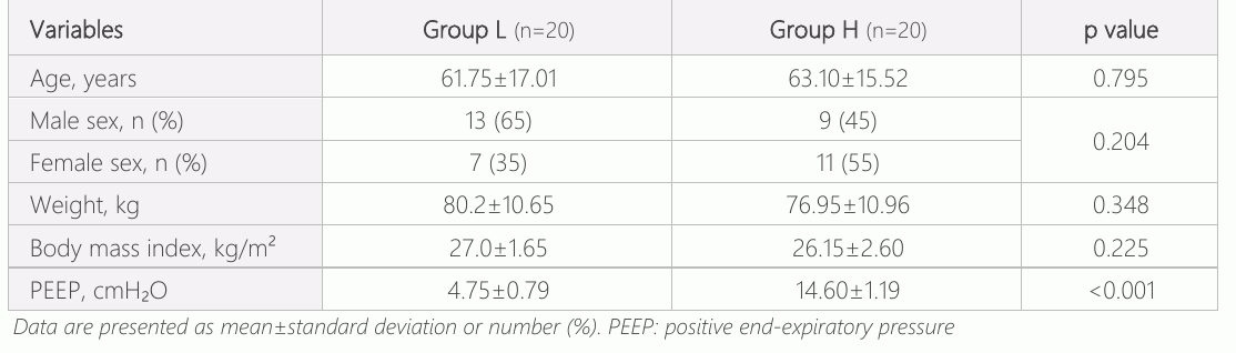
A total of 40 patients were included, with 20 patients in each PEEP group. No data were missing for the primary or secondary outcome variables. Group L included 13 men (65%) and seven women (35%), whereas Group H included nine men (45%) and 11 women (55%). The sex distribution did not differ between the groups (p=0.204). The groups were also comparable with respect to age, body weight, and body mass index. As expected, the mean PEEP level was higher in Group H (Table 1).
Hourly Urine Output
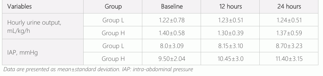
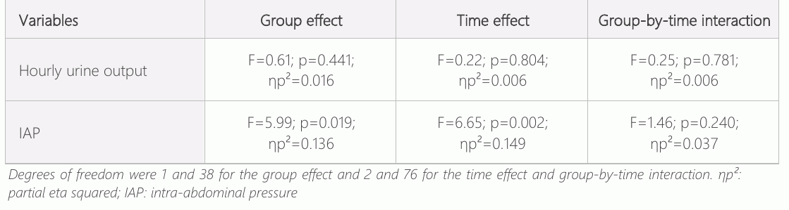
Table 2 presents hourly urine output values at the 3 measurement time points. No significant group effect [F(1,38)=0.61; p=0.441; ηp²=0.016], time effect [F(2,76)=0.22; p=0.804; ηp²=0.006], or group-by-time interaction [F(2,76)=0.25; p=0.781; ηp²=0.006] was observed (Table 3). The estimated marginal mean across the three measurement time points was 1.23 mL/kg/h in Group L and 1.36 mL/kg/h in Group H. The mean difference between Group H and Group L was 0.13 mL/kg/h (95% CI, −0.20 to 0.45).
Intra-Abdominal Pressure
Table 2 presents intra-abdominal pressure values at the three measurement time points. A significant group effect was observed [F(1,38)= 5.99; p=0.019; ηp²=0.136]. The estimated mean difference across the measurement time points was 2.17 mmHg, with higher values in Group H (95% CI, 0.37–3.96). We also observed a significant time effect [F(2,76)=6.65; p=0.002; ηp²=0.149]. The group-by-time interaction was not significant [F(2,76)=1.46; p=0.240; ηp²=0.037], indicating that IAP increased overall during the observation period, but the pattern of change did not differ significantly between groups.
Intra-Abdominal Hypertension
At baseline, IAP≥12 mmHg was observed in two patients (10%) in Group L and four patients (20%) in Group H. At 12 hours, the frequencies were two patients (10%) and seven patients (35%), respectively; at 24 hours, they were six (30%) and 11 patients (55%), respectively. In the exploratory analysis, recurrent intra-abdominal hypertension, defined as IAP ≥12 mmHg at two or more measurement time points, was identified in two patients (10%) in Group L and nine patients (45%) in Group H. The between-group difference was significant (Fisher’s exact test, p=0.031). The unadjusted risk ratio was 4.50. The highest recorded IAP was 17 mmHg, and no patient developed IAP>20 mmHg.
Hemodynamic Variables
No significant group effect, time effect, or group-by-time interaction was observed for mean arterial pressure. Although heart rate was generally lower in Group H, the pattern of change over time did not differ significantly between the groups. Because the groups were not randomized, patient diagnoses were heterogeneous, and illness-severity scores were unavailable, we did not interpret the between-group difference in heart rate as a direct effect of PEEP.
Discussion
The principal finding was that PEEP levels of 14–16 cmH₂O were not associated with a significant reduction in hourly urine output during the 24-hour observation period. In contrast, IAP was significantly higher in patients receiving high PEEP, and recurrent intra-abdominal hypertension occurred more frequently in this group.
The absence of a significant difference in urine output should not be interpreted as evidence that high PEEP had no effect on kidney function. We evaluated urine output at only three time points, and we did not assess serum creatinine, blood urea nitrogen, glomerular filtration, renal Doppler parameters, electrolyte excretion, or acute kidney injury criteria. Furthermore, the 95% CI for the between-group difference ranged from −0.20 to 0.45 mL/kg/h and did not exclude small effects in either direction.
PEEP may affect renal function through hemodynamic/neurohormonal mechanisms. Increased intrathoracic pressure may reduce venous return and cardiac output, whereas elevated central venous and renal venous pressures may decrease the renal perfusion gradient. Activation of the sympathetic nervous system, renin–angiotensin–aldosterone system, and antidiuretic hormone secretion may also contribute to sodium and water retention (6, 7). Annat et al. (7) reported reductions of approximately 34% in urine output, 32% in renal blood flow, and 19% in glomerular filtration rate during PEEP application. However, that study was a short-term physiological intervention in which different ventilatory conditions were evaluated within the same patients. In the present study, we determined PEEP according to clinical requirements, and groups were not randomized. Consequently, confounding by indication and the possibility that patients in Group H had more severe respiratory failure cannot be excluded.
Fogagnolo et al. (8) found that increasing PEEP from 5 to 15 cmH₂O increased the renal resistive index from 0.62 to 0.66 and increased the proportion of patients with an abnormal renal resistive index from 18% to 41%. These findings suggest that PEEP may affect renal vascular resistance or venous congestion without immediately reducing urine output. Chraibi et al. (16) reported increased urine output in 77.5% of patients within 48 hours after PEEP reduction, with a mean increase of approximately 0.46 mL/kg/h. Their findings are not directly comparable with ours because their study included patients with acute respiratory distress syndrome and impaired baseline urine output, used a within-patient design, and assessed cumulative 24-hour urine output. The present study compared two independents and diagnostically heterogeneous groups and evaluated separate hourly urine output.
An important secondary finding was the significantly higher IAP in Group H. The estimated marginal mean difference of 2.17 mmHg was consistent with Verzilli et al. (9), who reported an approximately 2–3 mmHg increase in IAP at higher PEEP. Conversely, Harnisch et al. (10) found that increasing PEEP from 5 to 15 cmH₂O did not significantly affect IAP or splanchnic perfusion markers in stable neurosurgical patients without pulmonary disease. Differences in pulmonary disease, fluid status, thoracoabdominal compliance, and illness severity may account for these discrepant findings.
In an exploratory analysis, recurrent intra-abdominal hypertension occurred more frequently in Group H than in Group L (45% vs 10%). This finding should be interpreted cautiously because the analysis was based on a small number of patients and only three IAP measurement time points. Nevertheless, the observation is consistent with the higher overall IAP values observed in patients receiving high PEEP. Increased renal venous and interstitial pressures may impair renal perfusion even when systemic arterial pressure is maintained. In IAH-WK study, intra-abdominal hypertension was independently associated with a composite outcome of kidney injury, kidney replacement therapy, or death (13). Multicenter and serial-measurement studies showed associations between intraabdominal hypertension, adverse renal outcomes, and mortality (14, 17–19).
Several factors may explain why hourly urine output remained preserved despite the higher IAP in Group H. Most IAP values were in the mild-to-moderate range, mean arterial pressure was comparable between groups, and fluid and inotropic treatments were adjusted to individual clinical needs. Mild alterations in renal perfusion may also be insufficient to produce detectable short-term changes in hourly urine output.
The strengths of the present study include normalizing urine output to body weight, repeated IAP measurements using a closed intravesical system, complete outcome data, and maintaining patients within the same PEEP range throughout the observation period. This study has several limitations. First, the groups were not randomized, resulting in a substantial risk of confounding by indication. Second, we did not record renal biomarkers, renal Doppler findings, acute kidney injury stage, fluid balance, diuretic exposure, or the need for renal replacement therapy. Third, illness-severity scores, PaO₂/FiO₂ ratio, tidal volume, plateau pressure, driving pressure, total PEEP, and intrinsic PEEP were unavailable. Fourth, the diagnostic heterogeneity of the study population may have independently affected urine output and IAP. Finally, the single-center design and small sample size limit the generalizability and precision of the findings. In addition, patients with clinically hemodynamic instability and those requiring a change in PEEP during the 24-hour observation period were excluded. These criteria may have selected a relatively more stable population and may have reduced the ability to detect renal or hemodynamic consequences associated with higher PEEP. They also limit the generalizability of the findings to patients with rapidly changing ventilatory or circulatory requirements.
Conclusion
Compared with PEEP levels of 4–6 cmH₂O, PEEP levels of 14–16 cmH₂O were not associated with a significant reduction in hourly urine output during the 24-hour observation period. However, high PEEP was associated with higher IAP and a greater frequency of recurrent intra-abdominal hypertension. Preserved hourly urine output during high- PEEP ventilation should not be interpreted as evidence that renal hemodynamics or kidney function remain unaffected. These findings suggest that attention to intra-abdominal pressure and renal function may be warranted in patients requiring higher PEEP, particularly when additional risk factors for intra-abdominal hypertension are present. Larger prospective studies incorporating serial renal biomarkers, cumulative fluid balance, diuretic exposure, and renal Doppler measurements are required. Ethical Approval: The study protocol was approved by the Clinical Research Ethics Committee of University Faculty of Medicine on May 9, 2018 (Decision No. 05).
Declarations
- Author contributions (CRediT)
- All authors contributed equally to all 14 CRediT roles (Conceptualization, Data curation, Formal analysis, Funding acquisition, Investigation, Methodology, Project administration, Resources, Software, Supervision, Validation, Visualization, Writing – original draft, Writing – review & editing).
- Funding
-
No institution/organization provided financial support for this study.
- Conflicts of interest
- The authors declare no conflicts of interest.
- Ethics approval
- The study protocol was approved by the Clinical Research Ethics Committee of University Faculty of Medicine on May 9, 2018 (Decision No. 05). Informed Consent: Written informed consent was obtained from the legal representatives of all patients.
- Data availability
- De-identified data supporting the findings of this study may be made available by the corresponding author upon reasonable request, subject to ethical and institutional regulations.
References (19)
18 of 19 references are linked to their DOI.
- Edginton S, Kruger N, Stelfox HT et al. Methods for determining optimal positive end-expiratory pressure in patients undergoing invasive mechanical ventilation: a scoping review. Can J Anaesth 2024;71(11):1535-55. DOI Crossref PubMed Google Scholar
- Battaglini D, Roca O, Ferrer R. Positive end-expiratory pressure optimization in ARDS: physiological evidence, bedside methods and clinical applications. Intensive Care Med 2024;50(5):762-5. DOI Crossref PubMed Google Scholar
- Lambermont BC, Detry O, D’Orio V et al. Effects of PEEP on systemic venous capacitance. Arch Physiol Biochem 1997;105(4):373-8. DOI Crossref PubMed Google Scholar
- Algera AG, Pisani L, Chaves RCF et al. Effects of PEEP on lung injury, pulmonary function, systemic circulation and mortality in animals with uninjured lungs—a systematic review. Ann Transl Med 2018;6(2):25. DOI Crossref PubMed Google Scholar
- Hemmer M, Suter PM. Treatment of cardiac and renal effects of PEEP with dopamine in patients with acute respiratory failure. Anesthesiology 1979;50(5):399-403. DOI Crossref PubMed Google Scholar
- Benites MH, Suarez-Sipmann F, Kattan E, Cruces P, Retamal J. Ventilation-induced acute kidney injury in acute respiratory failure: do PEEP levels matter? Crit Care 2025;29(1):130. DOI Crossref PubMed Google Scholar
- Annat G, Viale JP, Bui Xuan B et al. Effect of PEEP ventilation on renal function, plasma renin, aldosterone, neurophysins and urinary ADH, and prostaglandins. Anesthesiology 1983;58(2):136-41. DOI Crossref PubMed Google Scholar
- Fogagnolo A, Grasso S, Morelli E et al. Impact of positive end-expiratory pressure on renal resistive index in mechanical ventilated patients. J Clin Monit Comput 2024;38(5):1145-53. DOI Crossref PubMed Google Scholar
- Verzilli D, Constantin JM, Sebbane M et al. Positive end-expiratory pressure affects the value of intra-abdominal pressure in acute lung injury/acute respiratory distress syndrome patients: a pilot study. Crit Care 2010;14(4):R137. DOI Crossref PubMed Google Scholar
- Harnisch LO, Tampe D, Wieditz J, Moerer O. Effect of positive end-expiratory pressure levels on splanchnic perfusion and intra-abdominal pressure: a prospective clinical-experimental study in lung-healthy patients. J Anesth Analg Crit Care 2026;6(1):75. DOI Crossref PubMed Google Scholar
- Dumanlıdağ US, Yüzkat N, Soyalp C, Gülhas N. Effects of increasing positive end-expiratory pressure values on intraabdominal pressure and hemodynamics: a prospective clinical study. East J Med 2019;24(2):235-41. DOI Crossref PubMed Google Scholar
- Kirkpatrick AW, Roberts DJ, De Waele J et al. Intra-abdominal hypertension and the abdominal compartment syndrome: updated consensus definitions and clinical practice guidelines from the World Society of the Abdominal Compartment Syndrome. Intensive Care Med 2013;39(7):1190-206. 48 4- DOI Crossref PubMed Google Scholar
- Suphatheerawatr N, Jaturapisanukul S, Prommool S, Kurathong S, Pongsittisak W. Intra-abdominal hypertension among medical septic patients associated with worsening kidney outcomes: the IAH-WK study. Medicine (Baltimore) 2023;102(4):e32807. DOI Crossref PubMed Google Scholar
- Sarı Öztürk A, Konur K, Kara E. Association of serial intra-abdominal pressure measurements with renal outcomes and mortality in critically ill adults. J Clin Med 2026;15(14):5742. DOI Crossref PubMed Google Scholar
- Rocha NN, Samary CS, Antunes MA et al. The impact of fluid status and decremental PEEP strategy on cardiac function and lung and kidney damage in mild-moderate experimental acute respiratory distress syndrome. Respir Res 2021;22(1):214. DOI Crossref PubMed Google Scholar
- Chraibi H, De Longeaux K, L’her E. Diuresis increase after positive end-expiratory pressure weaning during acute respiratory distress syndrome. Sci Rep 2025;15:35272. DOI Crossref PubMed Google Scholar
- Reintam Blaser A, Regli A, De Keulenaer B et al. Incidence, risk factors, and outcomes of intra-abdominal hypertension in critically ill patients: a prospective multicenter study. Crit Care Med 2019;47(4):535-42. DOI Crossref PubMed Google Scholar
- Sugrue M, Jones F, Deane SA, Bishop G, Bauman A, Hillman K. Intra-abdominal hypertension is an independent cause of postoperative renal impairment. Arch Surg 1999;134(10):1082-85. DOI Crossref PubMed Google Scholar
- Malbrain MLNG, Chiumello D, Cesana BM et al. A systematic review and individual patient data meta-analysis on intra-abdominal hypertension in critically ill patients: the WAKE-Up project. Minerva Anestesiol 2014;80(3):293-306. PubMed Google Scholar
Respond to this article
Comments on a published article are welcome as a Letter to the Editor.
Get an e-mail when new articles in your fields are published
Questions to the authors
Questions are moderated: an editor approves each question, the authors answer, and an editor approves the answer before both appear here.
No answered questions yet.
Ask the authors a question
Discussion
Comments are moderated: they appear after the writer confirms the e-mail address and an editor approves them. Each comment carries a conflict-of-interest statement.
No comments yet.
Write a comment
Tell me when this article is cited
Leave your e-mail address and confirm it. These notices are not being sent yet; your confirmed address is kept for them and used for nothing else.
Read the full text
Open the PDF here
Loading the PDF…
If the PDF does not show here, use the download button above.
How to cite this article
Yuzkat N. Association of High Positive End-Expiratory Pressure with Intra-Abdominal Pressure and Urine Output in Critically III Patients: A Prospective Observational Study. Ulutas Med J. 2026;12(3):41-48. doi:10.5455/umj.20260803090348
Yuzkat, N. (2026). Association of High Positive End-Expiratory Pressure with Intra-Abdominal Pressure and Urine Output in Critically III Patients: A Prospective Observational Study. The Ulutas Medical Journal, 12(3), 41–48. https://doi.org/10.5455/umj.20260803090348
Yuzkat, N. (2026) 'Association of High Positive End-Expiratory Pressure with Intra-Abdominal Pressure and Urine Output in Critically III Patients: A Prospective Observational Study', The Ulutas Medical Journal, 12(3), pp. 41–48. doi: 10.5455/umj.20260803090348.
@article{umj330036,
  title = {Association of High Positive End-Expiratory Pressure with Intra-Abdominal Pressure and Urine Output in Critically III Patients: A Prospective Observational Study},
  author = {Yuzkat, Nureddin},
  journal = {The Ulutas Medical Journal},
  year = {2026},
  volume = {12},
  number = {3},
  pages = {41--48},
  doi = {10.5455/umj.20260803090348},
  url = {https://www.ulutasmedicaljournal.com/?mno=330036},
  issn = {2149-388X}
}