The Ulutas Medical Journal · ISSN 2149-0430 (print), 2149-388X (online) · https://www.ulutasmedicaljournal.com/?mno=167883
Original Article · Open access
The Role of the FDG-PET in the Staging of Patients with Differentiated Thyroid Cancer
- Department of Nuclear Medicine, University of Gaziantep, School of Medicine, Gaziantep, Turkey ROR
Abstract
- Introduction
- This study aims to determine the role of the FDG-PET in the staging of patients with Differentiated Thyroid Cancer (DTC) who have high Tg levels after thyroidectomy. Material and methods: Thirty-three DTC patients who performed FDG-PET scans after thyroidectomy were selected for this retrospective study. All patients had Tg levels over 10 ng/ml. FDG-PET was performed on patients six weeks after surgery. 2 weeks later FDG-PET imaging, patients received 131-I radioiodine treatment (RIT) except those with lymph node involvement. After RIT, post-ablation therapy whole body scan (TWBS) was performed on the patients.
- Results
- Twelve patients (36.4%) without involvement outside the thyroid bed, both on the FDG-PET and TWBS, were remissed completely. In 21 patients (63.4%), there was involvement outside the thyroid bed on the FDG-PET and/or TWBS. Eleven patients (33.3%) had distant organ involvement in the FDG-PET and/or TWBS. Patients with involvement outside the thyroid bed had higher Tg levels than others. The disease was completely or partially regressed in the patients with distant organ involvement who had negative FDG-PET scans and positive TWBS results. The disease tended to progress in patients with distant organ involvement who had higher SUVmax levels on FDG-PET.
- Conclusion
- Our data suggest that the FDG-PET scan, performed on the DTC patients with high Tg levels at the time of staging, would be the right approach. This approach will be useful, especially in demonstrating lymph node involvement and predicting prognosis. The results of FDG-PET may also change the treatment management.
Keywords
Full text
Introduction
Differentiated thyroid cancer (DTC) is the most common endocrine cancer with good prognosis (1, 2). The main treatment method for DTC is total/nearly total thyroidectomy followed by 131-I radioiodine treatment (RIT)(3). After the thyroidectomy and before the RIT; patients are evaluated with some laboratory and imaging technics for detecting residual thyroid tissue and remnant tumor tissue. The main laboratory tool and most sensitive biochemical method for evaluating of DTCs are the measurement of serum thyroglobulin (Tg) with anti-Tg antibody (Tg-Ab) and thyroid-stimulating hormone (TSH) (4). Tg is important tumor marker for follow-up of DTC patients. Despite the residual thyroid tissue, Tg level which measured before the RIT can be useful and high Tg levels are correlated with local and distant metastases (5–8). Detailed neck ultrasound and thyroid scintigraphy are imaging modalities which routinely used at the staging of DTC. F-18-fluorodeoxyglucose position emission tomography (FDG-PET) is not routine imaging tool for staging of DTC. However, FDG PET is strongly recommended in the follow-up of patients with high/elevated serum Tg and negative whole body radioiodine scanning (WBS). On the other hand, there are some studies that provide the benefit of FDG- PET in the staging of selected DTC patients who have intermediate-high risk (9–13).
The aim of this study is to determine the role of the FDG-PET in the staging of DTC patients who have high Tg levels after thyroidectomy.
Material and Methods
Ethical Statement
Approval was obtained from the institutional review board of Gaziantep University. All patients signed an informed consent form that allows the participating institution to use their clinical data (Ethics approval No: 2023/172 and date: 24/05/2023).
Study Design
Thirhty three DTC patients (19 women, 14 men with mean age 39.9±12 years) who performed FDG-PET after total/nearly total thyroidectomy between January 2019 and January 2023 were selected for this retrospective study. Histopathological examination revealed papillary thyroid carcinoma (PTC) in 32 patients and follicular thyroid carcinoma (FTC) in 1 patient. All patients had Tg over 10 ng/ml with negative Tg-Ab (Tg-Ab<0.2 IU/mL) when TSH>30 IU/ml. Tg levels were measured using immune-radiometric analyser that uses paramagnetic microparticles and chemiluminescent detection technology with a lower detection limit of 0.2 ng/ml. FDG-PET was performed to patients 6 weeks after surgery. And 2 weeks after the FDG- PET imaging, patients received RIT except who had head-neck lymph node involvoment. The patients with head-neck lymph node involvement went to neck dissection or fine needle aspiration biopsy and histopathological data were evaluated. After the RIT, post-ablation therapy whole body scan (TWBS) was performed to patients. FDG-PET and TWBS findings were correlated. Mean follow up time of the patients was 16.2±4.2 months (min 6-max 36). On the follow up, TWBS and DWBS were performed some patients, and stimulated/non-stimulated Tg levels were measured.
FDG-PET Imaging
After 12-h fasting, patients whose glucose levels under 150 mg/ dL were injected with intravenous (iv) 5 Mbq/kg 18FDG. The PET/CT imaging was acquired with Siemens Biograph 2 LSO combined PET/CT device (Biograph LSO/Somatom Emotion; Siemens Medical Solutions) 60 minutes after the injection. Sequential transmission and emission images of patient’s body between the base of the skull and the proximal femur with the patient in a supine position were obtained. The PET/CT images were evaluated by two experienced nuclear medicine specialists. Uptake areas higher than ground activity which calculated as maximum standard uptake (SUVmax) were detected. The patients were classified according to whether they had pathological FDG uptake outside the thyroid bed or not.
Whole Body Radioiodine Scan
Patients except who had head-neck lymph node involvoment, were given RIT after total or near-total thyroidectomy. Before RIT patients did not take levothyroxine, and they had a low-iodine diet for 2 weeks (12 days before and 2 days after I-131 administration). Patients were treated according to fixed dosage protocol: 50-100 mCi 131-I in cases without known lymph node or distant metastasis, 150 mCi 131-I in cases with lymph node involvement detected pre-or perioperatively and 200 mCi 131-I in cases of known distant metastasis. TWBS was performed a week after RIT. The patients were classified according to whether they had pathological radioiodine uptake outside the thyroid bed or not. For the DWBS, patients discontinued taking thyroxine and followed a low-iodine diet during 3 weeks. Just before DWBS, stimulated Tg and Tg-Ab levels were measured when TSH>30 IU/ml. After a TSH stimulation, 5 mCi (185 MBq) 131-I was given orally to the patients and 2-3 days later, DWBS was performed.
Statistical Analysis
Data of the patients were obtained from the computer system of the hospital (electronic record system of health data) and PET/CT images in our clinic. Data obtained were analysed with SPSS 18.0 statistical software programme. The descriptive values such as mean, standard deviation and percentage were used for age, gender and sensitivity. Quantitative variables were expressed as mean±standard deviation in the case of normal distribution, and median [IQR, interquartile range] in the case of non-normal distribution. The means between 2 groups were compared using Student’s t-test for normal distributions and Mann–Whitney U-test for abnormal distributions. Spearman’s correlation analysis was used to reveal the relationship between biochemical tumour markers and imaging findings. P<0.05 was considered significant.
Results
Mean Tg level of all of patients was 166.6±324 ng/ml (min 10.5-max 515). 12 patients (36.4%) had no pathological FDG uptake on the FDG- PET. Mean Tg of these patients was 31.5±18.8 ng/ml (min 10.5-max 88). RIT was given to the patients. On TWBS performed after RIT, patients had radiotracer involvement only in the thyroid bed. There was not extrathyroidal involvement. For all these 12 patients, DWBS which performed 6 months after RIT were normal. Also stimulated Tg level measured before DWBS was under 2 ng/ml for all patients. Tg-Abs were negative. For this reason, these patients were considered in remission.
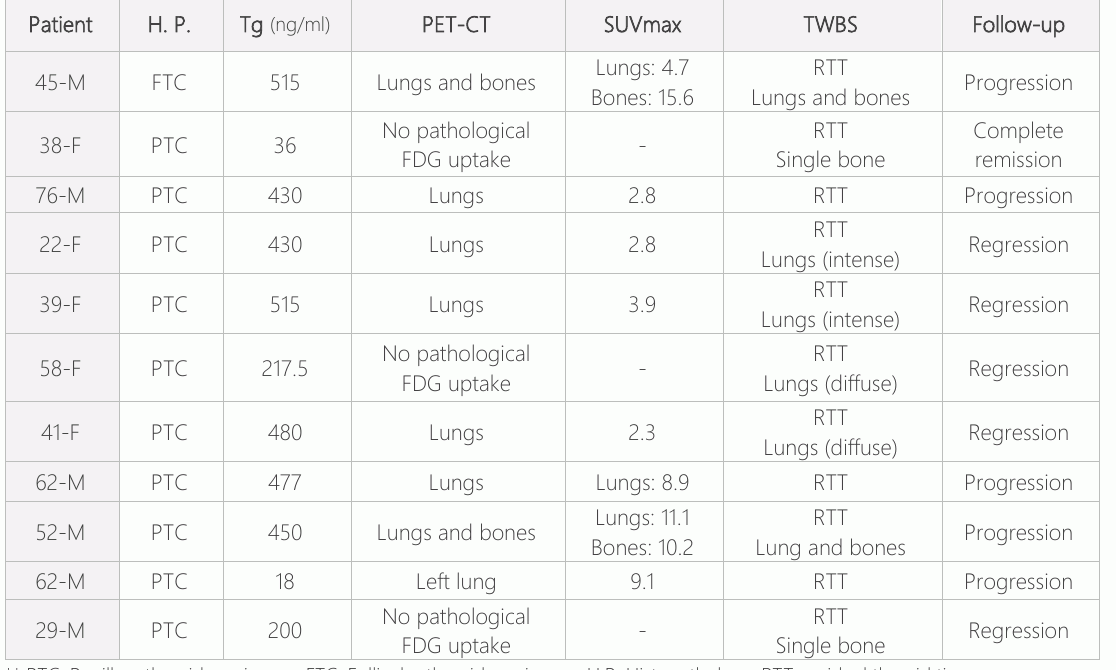
In twenty-one patients (63.4%), there were pathological involvements outside the thyroid bed on FDG-PET and/or TWBS. The mean Tg level of these patients was 243.8±222 ng/ml (min 18-max 515).
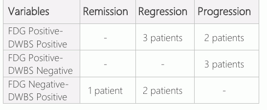
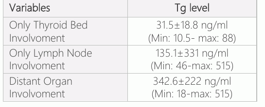
The difference between serum Tg of the two groups was significant (p<0.01). Ten of these patients (30.3%) with a mean Tg level of 135.1±331 ng/ml (min 46-max 515), had head-neck lymph node involvements. The patients with head-neck lymph node involvements were referred to surgery. The mean SUVmax for these patients was 9.1±3.2 (min 2-max 21.2). After neck dissection, histopathological data were compatible with lymph node metastases for patients. Eleven of patients with pathological involvements outside the thyroid bed (33.3%) had distant organ or lymph node involvements on the FDG-PET and/or TWBS. The mean Tg of these patients was 342.6±222 ng/ml (min: 18- max: 515). The difference between serum Tg of the patients with distant organ involvements and only head-neck lymph node involvements was not significant. The features of patients who involvoment, 2 patients (18.2%) had only single bone involvoment and 2 patients (18.2%) had lung and bone involvements.
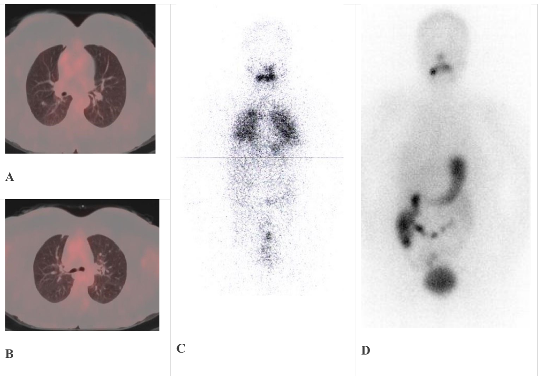
In the patients with negative FDG-PET scan and positive TWBS findings for distant organ involvoment (3 patients), the disease was complete or partial regressed (Fig 1). In the patients with positive FDG-PET and negative TWBS findings for distant organ involvoment (3 patients), the disease was progressed. In the patients with positive FDG-PETscan and positive TWBS findings for distant organ involvoment (5 patients), the disease was progressed in 2 patients and partial regressed in 3 patients. Correlation of FDG PET and TWBS findings for the patients who had organ involvoment, were given on table 2. Also, for the patients with distant organ involvoment who had higher SUVmax levels on FDG PET, the disease tended to progress (Table 2).
Discussion
Tg measurement is one of the main tools in the management and follow-up of patients with DTC after the RIT (7, 14, 15). Stimulated Tg measurement is more sensitive than the basal Tg (16). Although Tg measurement is used in the follow-up DTC patients for detection of residual and recurrence disease, there are some studies that showing the benefit of Tg levels which measured after total/nearly total thyroidectomy and before RIT. Higher Tg level which measured at the staging, is correlated with local/distant metastasis and it can be useful for predicting of prognosis (5–8). Especially Tg levels higher than 300 ng/ml, are suggestive of distant metastasis (8).
In our study, patients with involvoment outside the thyroid bed had higher Tg levels than others. Additionally, the patients with distant organ involvoment had higher Tg levels than patients with only lymph node involvements. The benefit of FDG-PET has been proven in the DTC patients with positive Tg level and negative whole body radioiodine scan (WBS). A negative WBS result with elevated serum Tg level (>10 ng/ml) is the main indication for FDG-PET (17, 18). But the role of FDG-PET in initial staging of DTC patients after surgery is controversial and information on this subject is limited. There are some studies that provide the benefit of FDG-PET in the staging of selected DTC who have suspicious findings such as high Tg, aggressive histopathological subtype (9–13). There is no specific cut-off value of Tg for performing FDG-PET at the time of staging. But our clinical experience has shown that Tg level is usually under 10 ng/ml after a successful thyroidectomy if the patient does not have locally or distant metastases. For this reason, we chose the cut-off value as 10 ng/ml. Liu et al were chose the cut-off value of Tg as 10 ng/ml too for performing FDG-PET to DTC patients at the staging time (9). Iwano et al. concluded that FDG-PET performed concurrently with RIT can detect lymph node metastases in which radioiodine does not accumulate and this situation may influence the management and treatment options for DTC patients (10). In our study 10 patients who had lymph node metastases were referred to resurgery before RIT. Thus, the treatment management has changed in these patients.
In a retrospective study by Nascimento et all, FDG-PET was performed 38 DTC patients with aggressive histology without known persistent disease at the time of postoperative RIT (11). They concluded that postoperative FDG-PET should be performed routinely in DTC with aggressive histology. The FDG-PET scan also provides prognostic information. 18F-FDG-avid metastatic DTC lesions are usually resistant to 131-I treatment. Gartner et al concluded that FDG PET performed at the time of thyroid remnant ablation in high-risk patients with DTC is highly predictive for survival (12). The patients with positive FDG-PET findings showed worse biochemical response and lower survival as compared with patients who had negative FDG-PET scan. And complete remission was more common in patients with negative FDG- PET scan findings. Our study confirmed this status. We found that the disease was complete or partial regressed in the patients with distant organ involvoment who had negative FDG-PET scan and positive TWBS results. On the contrary in the patients with distant organ involvoment who had positive FDG-PET scan and negative TWBS, our findings showed that the disease was progressed. Liu et al. suggested that FDG-PET can play vital role in identifying metastases in postoperative DTC patients with elevated Tg levels (>10 ng/mL) before RIT (9). Also, FDG- PET positive metastatic foci with SUVmax of greater than 4.0 possesses higher probability of non-avidity to radioiodine. In our study, most of the patients with distant metastasis who progressed on the follow-up had higher SUV max than 4.0.
Conclusion
In conclusion our data suggest that FDG-PET scan which performed to the DTC patients with high Tg levels at the time of staging, would be the right approach. This approach will be very useful especially in demonstrating lymph node involvoment and predicting prognosis. Results of FDG-PET may also change the treatment management. Especially in the DTC patients with distant organ metastasis, FDG-PET scan which correlated with WBS, should be used in both staging and follow-up. Thus, radioiodine avid disease and radioiodine-refractory patients can be selected.
Declarations
- Author contributions (CRediT)
- All authors contributed equally to all 14 CRediT roles (Conceptualization, Data curation, Formal analysis, Funding acquisition, Investigation, Methodology, Project administration, Resources, Software, Supervision, Validation, Visualization, Writing – original draft, Writing – review & editing).
- Funding
-
Funding information was not reported in the published version of this article.
- Conflicts of interest
- The authors declare no conflict of interest for the present study.
- Ethics approval
- Approval was obtained from the institutional review board of Gaziantep University. All patients signed an informed consent form that allows the participating institution to use their clinical data (Ethics approval No: 2023/172 and date: 24/05/2023).
- Data availability
- A data availability statement was not reported in the published version of this article.
References (19)
15 of 19 references are linked to their DOI.
- Jemal A, Siegel R, Ward E, et al. Cancer statistics. CA: A Cancer Journal for Clinicians 2006;56:106–130. DOI Crossref PubMed Google Scholar
- Pellegriti G, Frasca F, Regalbuto C, Squatrito S, Vigneri R. Worldwide increasing incidence of thyroid cancer: update on epidemiology and risk factors. J Cancer Epidemiol. 2013;2013:965212. DOI Crossref PubMed Google Scholar
- Hundahl SA, Cady B, Cunningham MP, et al. Initial results from a prospective cohort study of 5583 cases of thyroid carcinoma treated in the United States during 1996. US and German Thyroid Cancer Study Group. An American College of Surgeons Commission on Cancer Patient Care Evaluation study. Cancer. 2000;89:202-17. DOI Crossref PubMed Google Scholar
- Van Herle AJ, Uller RP, Matthews NI, Brown J. Radio immunoassay for measurement of thyroglobulin in human serum. J Clin Invest 1973; 52: 1320-7. PubMed Google Scholar
- Bernier MO, Morel O, Rodien P, et al. Prognostic value of an increase in the serum thyroglobulin level at the time of the first ablative radioiodine treatment in patients with differentiated thyroid cancer. Eur J Nucl Med Mol Imaging 2005; 32: 1418-21. DOI Crossref PubMed Google Scholar
- Lima N, Cavaliere H, Tomimori E, Knobel M, Neto GM. Prognostic value of serial serum thyroglobulin determinations after total thyroidectomy for differentiated thyroid cancer. J Endocrinol Invest 2002; 25: 110-5. DOI Crossref PubMed Google Scholar
- Ronga G, Filesi M, Ventroni G, Vestri AR, Signore A. Value of the first serum thyroglobulin level after total thyroidectomy for the diagnosis of metastases from differentiated thyroid carcinoma. Eur J Nucl Med 1999; 26: 1448-52. DOI Crossref PubMed Google Scholar
- Kalender E, Celen YZ, Elboga U, Demir HD, Karaoglan H, Yilmaz M. The relationship between thyroglobulin (Tg) levels and distant metastases before radioiodine (RAI) treatment in differentiated thyroid carcinoma. Gaziantep Med J 2012;18:11-13. DOI Crossref PubMed Google Scholar
- Liu M, Cheng L, Jin Y, Ruan M, Sheng, S, Chen L. Predicting 131 I-avidity of metastases from differentiated thyroid cancer using 18F-FDG PET/CT in postoperative patients with elevated thyroglobulin. Sci Rep 2018;8:4352. DOI Crossref PubMed Google Scholar
- Iwano S, Kato K, Ito S, Tsuchiya K, Naganawa S. FDG PET performed concurrently with initial I-131 ablation for differentiated thyroid cancer. Ann Nucl Med 2012; 26: 207–13. DOI Crossref PubMed Google Scholar
- Nascimento C, Borget I, Al Ghuzlan A, et al. Postoperative fluorine-18-fluorodeoxyglucose positron emission tomography/computed tomography: an important imaging modality in patients with aggressive histology of differentiated thyroid cancer. Thyroid 2015;25:437–44. DOI Crossref PubMed Google Scholar
- Alzahrani AS, Abouzied ME, Salam SA, et al. The role of F-18-fluorodeoxyglucose positron emission tomography in the postoperative evaluation of differentiated thyroid cancer. Eur J Endocrinol 2008; 158: 683–9. DOI Crossref PubMed Google Scholar
- Gaertner FC, Okamoto S, Shiga T, et al. FDG PET performed at thyroid remnant ablation has a higher predictive value for 184 77 FDG-PET in Differentiated Thyroid Cancer Ebuzer Kalender longg term survival of high-risk patients with well-differentiated thyroid cancer than radioiodine uptake. Clin Nucl Med 2015; 40: 378–83. DOI Crossref PubMed Google Scholar
- Mazzaferri EL: NCCN thyroid carcinoma practice guidelines. Oncology. 1999; 13: 391–442. PubMed Google Scholar
- Schlumberger MJ: Papillary and follicular thyroid carcinoma. N Engl J Med 1998; 338: 297-306. DOI Crossref PubMed Google Scholar
- Mazzaferri EL, Kloos RT. Current approaches to primary therapy for papillary and follicular thyroid cancer. J Clin Endocrinol Metab. 2001; 86: 1447-63. DOI Crossref PubMed Google Scholar
- Schlumberger M, Mancusi F, Baudin E, Pacini F. 131-I therapy for elevated thyroglobulin levels. Thyroid 1997; 7: 273-6. PubMed Google Scholar
- Lind P, Kresnik E, Kumnig G, et al. 18F-FDG-PET in the follow-up of thyroid cancer. Acta Med Austriaca 2003; 30: 17-21. PubMed Google Scholar
- Wang W, Larson SM, Tuttle RM, et al. Resistance of [18f]- fluorodeoxyglucose-avid metastatic thyroid cancer lesions to treatment with high-dose radioactive iodine. Thyroid 2001; 11: 1169-75. DOI Crossref PubMed Google Scholar
Read the full text
Open the PDF here
Loading the PDF…
If the PDF does not show here, use the download button above.
How to cite this article
Kalender E. The Role of the FDG-PET in the Staging of Patients with Differentiated Thyroid Cancer. Ulutas Med J. 2023;9(3):178-184. doi:10.5455/umj.20230901081616
Kalender, E. (2023). The Role of the FDG-PET in the Staging of Patients with Differentiated Thyroid Cancer. The Ulutas Medical Journal, 9(3), 178–184. https://doi.org/10.5455/umj.20230901081616
Kalender, E. (2023) 'The Role of the FDG-PET in the Staging of Patients with Differentiated Thyroid Cancer', The Ulutas Medical Journal, 9(3), pp. 178–184. doi: 10.5455/umj.20230901081616.