The Ulutas Medical Journal · ISSN 2149-0430 (print), 2149-388X (online) · https://www.ulutasmedicaljournal.com/?mno=42850
Case Report · Open access
Median Arcuate Ligament Syndrome as A Rare Cause of Recurrent Chronic Abdominal Pain: Radiological Findings
Also available in: العربية · Türkçe · 中文
- Cumhuriyet University, School of Medicine, Department of Radiology, Sivas, Turkey ROR
- Nazilli State Hospital, Health Ministry, Department of Radiology, Nazilli, Aydın, Turkey ROR
Abstract
- Background
- Median arcuate ligament syndrome (MALS), also known as celiac artery compression syndrome, is characterized by gastrointestinal ischemia secondary to the compression of the median arcuate ligament of the diaphragm to the proximal celiac trunk.
- Case Presentation
- Herein, we aimed to report the Color Doppler Ultrasonography (CDUS) and Computerized Tomography (CT) findings of MALS in a 27-year-old man presenting with complaints of abdominal pain and slimming.
- Conclusion
- We think that MALS should be considered in young patients with persistent abdominal pain who are admitted with chronic epigastric pain. Non-invasive methods such as CDUS and CT should be used for diagnosis.
Keywords
Full text
Introduction
Median arcuate ligament syndrome (MALS) is also called celiac artery compression syndrome or Dunbar syndrome. The most common clinical finding, upper abdominal angina, is due to the compression of the celiac artery trunk with diaphragmatic crura. The typical age is 20- 40 years. Lipshutz first reported it in 1917. MALS was described by Dunbar et al. in 1965 as Dunbar syndrome (1–3). Herein, we aimed to present Multi-Slice Computed Tomography (MSCT) and Colour Doppler Ultrasonography (CDUS) findings in a case with MALS.
Case Presentation
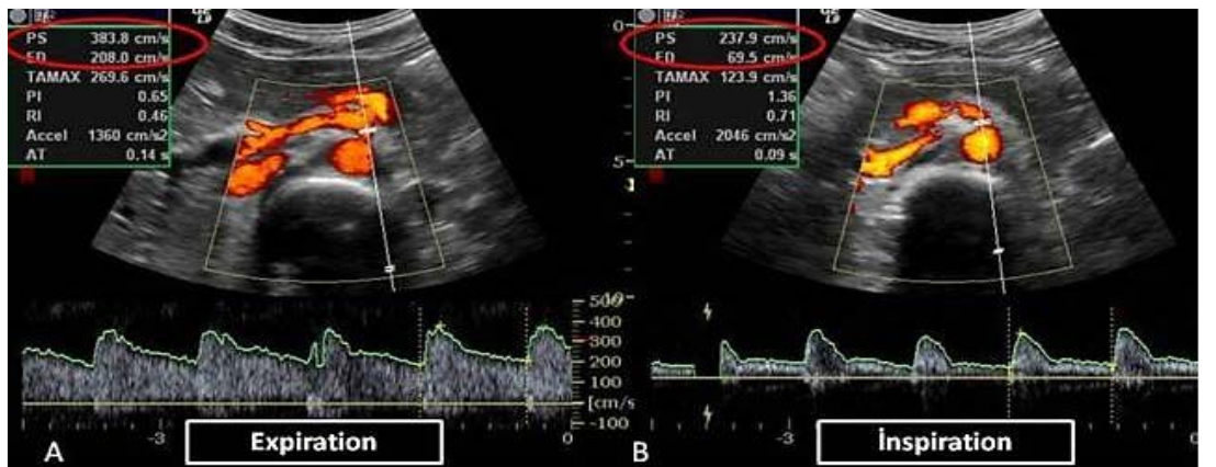
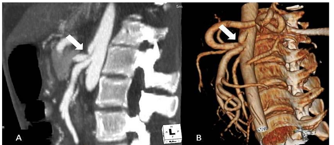
A 27-year-old man presented with worsening postprandial abdominal pain progressively and weight loss for six months. He also had nausea and vomiting. Physical examination was normal except epigastric pain. His history was unremarkable for any former disorder. His electrocardiographic, abdominal, biochemical, and thoracic plain radiographic examinations were all in normal limits. He was examined with CDUS examination of the abdominal aorta and its branches, which revealed stenosis of the celiac artery with left-sided preponderance. In CDUS analysis, the peak systolic and end-diastolic velocities of the celiac artery at end-expiration were 383 cm/sec and 208 cm/sec. In maximum inspiration, the corresponding velocities were 237 cm/sec and 69 cm/sec (Figure’s 1A, B). CDUS findings suggest the diagnosis of MALS. However, to confirm the diagnosis, an angiographic examination with MSCT was performed. 128-slice MSCT (Aquilion, Toshiba Medical Systems Co., Tokyo, Japan) imaging was performed after the administration of intravenous contrast medium. Multi-planar reconstruction and three-dimensional (3D) volume-rendered (VR) images were obtained from CT images. On CT images, the latter revealed a focal narrowing of a typical shape of a hook and post-stenotic dilation (Figure’s 2A, B), which secured the diagnosis of MALS. Conservative treatment with radiological follow-up was commenced with watchful waiting for symptom progression.
Discussion
The median arcuate ligament is a fibrous structure that connects the diaphragmatic crura on both sides of the aortic hiatus and generally passes over the celiac trunk. The median arcuate ligament is localized at the T12-L1 level. In some persons who suffer the condition named the median arcuate ligament syndrome, this ligament inserts at a lower level that is from the proximal part of the celiac trunk, compressing the latter. This condition results in ischemia of the gastrointestinal system. One of the main symptoms of the latter is postprandial abdominal pain. MALS is characterized by nausea, epigastric pain, weight loss, and postprandial pain. Postprandial pain primarily occurs during expiration. MALS is much more common in patients with postural orthostatic tachycardia syndrome. The reason for this is not yet understood (1, 2). In a study that retrospectively examined the aortograms of 1500 patients, symptomatic celiac artery compression was identified in 1% of patients (3). However, the incidence of MALS in general population is not fully known. MALS is more common in women than in men, and most commonly occurs between 20 and 40 years old. Two theories have been advanced for the occurrence of clinical signs and symptoms. The first theory is characterized by mesenteric ischemia due to the compression of the celiac artery. The second theory hypothesizes the irritation of somatic pain fibers secondary to neurological stimulation due to condensation on the celiac ganglion and plexus. (4, 5). The diagnosis of MALS can be examined by CDUS, CT-angiography, catheter angiography and magnetic resonance angiography. CDUS has been reported to confer a high sensitivity for MALS and has been recommended as the diagnostic modality of choice (6). The diagnosis of MALS can be made by Doppler flow measurements of the celiac artery and during inspiration and expiration. In Doppler flow velocity measurements in the compressed segment of the celiac artery, a change in peak systolic velocity of more than 200 cm/s during expiration is considered to be diagnostic (7). Thanks to advances in MSCT technology, MALS can be diagnosed even using routine contrast-enhanced abdominal CT. CT-angiography using MSCT characteristically demonstrates focal narrowing of the proximal celiac artery. The focal narrowing is characteristically seen as a hook. This sign may be beneficial for differentiating the condition from other conditions like atherosclerosis that may cause celiac artery stenosis. Post-stenotic dilation and collateral vessels are the other MSCT findings. The characteristic hook sign and post-stenotic dilation were present in our case. MSCT is a minimally invasive technique which helps in the diagnosis of MALS and may be preferred as an alternative to catheter angiographic procedures (4–6).
In the differential diagnosis of MALS, superior mesenteric artery compression syndrome (SMA syndrome, or Wilkie syndrome) and Nutcracker Syndrome should be considered as a priority. In particularly, SMA syndrome can be easily confused with MALS due to the similarity of clinical findings. The pain in the SMA syndrome decreases in the position of the prone or left lateral decubitus. As in the diagnosis of MALS, CT is the most appropriate diagnostic method in the diagnosis of SMA syndrome (4, 5, 8, 9). The treatment options for MALS include operative resection of the median arcuate ligament, celiac ganglion destruction, and vascular bypass surgery (10).
In conclusion, MALS is a rare and challenging diagnosis that should be considered in patients presenting with non-specific chronic abdominal pain. Doppler US with a dynamic evaluation during inspiration-expiration and in the supine as well as upright position is very useful in the diagnostic workup of suspected MALS. Confirmatory CT and CT-angiography should usually be obtained before treatment.
Declarations
- Author contributions (CRediT)
- All authors contributed equally to all 14 CRediT roles (Conceptualization, Data curation, Formal analysis, Funding acquisition, Investigation, Methodology, Project administration, Resources, Software, Supervision, Validation, Visualization, Writing – original draft, Writing – review & editing).
- Funding
-
Funding information was not reported in the published version of this article.
- Conflicts of interest
- The authors declare that they have no conflict of interest in the current study.
- Ethics approval
- Ethics approval details were not reported in the published version of this article.
- Data availability
- A data availability statement was not reported in the published version of this article.
References (10)
8 of 10 references are linked to their DOI.
- Gürol Ö, Yücel Ü, Mehmet İ, Arif Ö, Ahmet Ö, Burhan K. Celiac artery compression syndrome: Report of a case and literature review. Turkish Journal of Gastroenterology. 1999; 10: 161-3 PubMed Google Scholar
- Güngör Ö, Meral M, Acarbay S, Biberoglu K,Gönen Ö. Çölyak Arter Bası Sendromu: Karın Ağrısının Nadir Bir Nedeni. Turkiye Klinikleri J Cardiovasc Sci 2006; 18: 248-51 PubMed Google Scholar
- Cornell SH. Severe stenosis of the celiac artery. Analysis of patients with and without symptoms. Radiology. 1971; 99: 311-6 DOI Crossref PubMed Google Scholar
- Fong JK, Poh AC, Tan AG, Taneja R. Imaging findings and clinical features of abdominal vascular compression syndromes. AJR Am J Roentgenol. 2014; 203: 29-36 DOI Crossref PubMed Google Scholar
- Srisajjakul S, Prapaisilp P, Bangchokdee S. Imaging features of vascular compression in abdomen: Fantasy, phenomenon, or true syndrome. Indian J Radiol Imaging. 2017; 27: 216-24 DOI Crossref PubMed Google Scholar
- Scholbach T. Celiac artery compression syndrome in children, adolescents, and young adults: clinical and color duplex sonographic features in a series of 59 cases. J Ultrasound Med. 2006; 25: 299-305 DOI Crossref PubMed Google Scholar
- Erden A, Yurdakul M, Cumhur T. Marked increase in flow velocities during deep expiration: A duplex Doppler sign of celiac artery compression syndrome. Cardiovasc Intervent Radiol 1999; 22: 331-2 DOI Crossref PubMed Google Scholar
- Baskan O, Kaya E, Gungoren FZ, Erol C. Compression of the Celiac Artery by the Median Arcuate Ligament: Multidetector Computed Tomography Findings and Characteristics. Can Assoc Radiol J. 2015; 66: 272 -6 DOI Crossref PubMed Google Scholar
- Lamba R, Tanner DT, Sekhon S, McGahan JP, Corwin MT, Lall CG. Multidetector CT of vascular compression syndromes in the abdomen and pelvis. Radiographics. 2014; 34:93-115 DOI Crossref PubMed Google Scholar
- Meyer M, Gharagozloo F, Nguyen D, Tempesta B, Strother E, Margolis M. Robotic-assisted treatment of celiac artery compression syndrome: report of a case and review of the literature. Int J Med Robot. 2012; 8: 379 -83 DOI Crossref PubMed Google Scholar
Respond to this article
Comments on a published article are welcome as a Letter to the Editor.
Get an e-mail when new articles in your fields are published
Questions to the authors
Questions are moderated: an editor approves each question, the authors answer, and an editor approves the answer before both appear here.
No answered questions yet.
Ask the authors a question
Discussion
Comments are moderated: they appear after the writer confirms the e-mail address and an editor approves them. Each comment carries a conflict-of-interest statement.
No comments yet.
Write a comment
Tell me when this article is cited
Leave your e-mail address and confirm it. These notices are not being sent yet; your confirmed address is kept for them and used for nothing else.
Read the full text
Open the PDF here
Loading the PDF…
If the PDF does not show here, use the download button above.
How to cite this article
Başpınar N, Atalar MH. Median Arcuate Ligament Syndrome as A Rare Cause of Recurrent Chronic Abdominal Pain: Radiological Findings. Ulutas Med J. 2019;5(3):221-224. doi:10.5455/umj.20190706071036
Başpınar, N., & Atalar, M. H. (2019). Median Arcuate Ligament Syndrome as A Rare Cause of Recurrent Chronic Abdominal Pain: Radiological Findings. The Ulutas Medical Journal, 5(3), 221–224. https://doi.org/10.5455/umj.20190706071036
Başpınar, N. and Atalar, M.H. (2019) 'Median Arcuate Ligament Syndrome as A Rare Cause of Recurrent Chronic Abdominal Pain: Radiological Findings', The Ulutas Medical Journal, 5(3), pp. 221–224. doi: 10.5455/umj.20190706071036.
@article{umj42850,
  title = {Median Arcuate Ligament Syndrome as A Rare Cause of Recurrent Chronic Abdominal Pain: Radiological Findings},
  author = {Başpınar, Nisa and Atalar, Mehmet H},
  journal = {The Ulutas Medical Journal},
  year = {2019},
  volume = {5},
  number = {3},
  pages = {221--224},
  doi = {10.5455/umj.20190706071036},
  url = {https://www.ulutasmedicaljournal.com/?mno=42850},
  issn = {2149-388X}
}