The Ulutas Medical Journal · ISSN 2149-0430 (print), 2149-388X (online) · https://www.ulutasmedicaljournal.com/?mno=328225
Review Article · Open access
Primary Cardiac Tumors in Children: A State-of-the-Art Review
Also available in: العربية · Türkçe · 中文
- Department of Internal Medicine, University of Nebraska Medical Center, Omaha, NE, USA ROR
- Department of Pharmacy, Campbell University College of Pharmacy & Health Sciences, Buies Creek, NC, USA ROR
- Department of Internal Medicine, Creighton University Medical Center-Bergan Mercy, Omaha, NE, USA ROR
- Department of Pediatrics, University of South Florida College of Medicine, Tampa, FL, USA ROR
Abstract
Cardiac tumors are benign or malignant neoplasms arising from the endocardium, myocardium, or the surrounding pericardium. They can be primary or metastatic. Primary tumors are rare, with a reported autopsy frequency of up to 0.30%. These tumors can occur in the neonatal or fetal period, with an incidence of 0.14% in the latter. Most pediatric cardiac tumors are benign, with approximately 10% being malignant. Rhabdomyoma is the most prevalent benign tumor in children and accounts for more than 50% of all primary cardiac tumors in this population. Multiple cardiac tumors noted during the fetal or neonatal period are commonly associated with tuberous sclerosis. Additional risk factors include family history and other genetic predispositions such as the Carney complex. When clinically suspected, echocardiography, computed tomography, and magnetic resonance imaging help with diagnosis. Treatment includes surgery, chemotherapy, and targeted therapy. This article provides a scoping review of cardiac tumors in children, their epidemiology, histological patterns, diagnosis, and treatment.
Keywords
Full text
Introduction
Cardiac tumors in children are rare, arising most commonly from the endocardium, myocardium, or the surrounding pericardium (1, 2). These can be primary or metastatic, with primary tumors accounting for a prevalence between 0.0017% and 0.28% in autopsy series (3). In the fetal period, the incidence of cardiac tumors has been reported to be up to 0.14% (3, 4). The overwhelming majority of primary cardiac neoplasms in children are benign, with up to 10% being malignant (5). Secondary malignant tumors are much more prevalent than primary malignant neoplasms, and the type and frequency of cardiac tumors differ in adults and children (3–5).
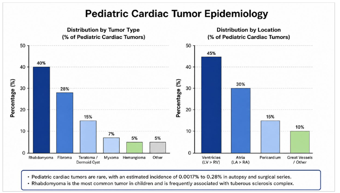
In adults, 75% of tumors are benign, and 25% are malignant (3–6). Myxomas are the most common primary tumor in adults, and sarcomas constitute up to 75% of malignant masses (7–9). Rhabdomyoma is the most common cardiac neoplasm during childhood and fetal life, followed by fibroma and teratoma (Figure 1) (10, 11). Patients with cardiac masses may be asymptomatic or present with heart failure, cardiac murmurs, sudden cardiac death (SCD), or arrhythmias (12). Echocardiography is the primary diagnostic modality, and cardiac magnetic resonance imaging (cMRI) further characterizes the tumor and the extent of adjacent cardiac structure to support treatment planning (13, 14). Management varies from conservative to resection or transplantation. This article provides a scoping review of cardiac tumors in children, including their diagnosis and treatment.
Epidemiology
Primary cardiac tumors (PCTs) are scarce in the pediatric population, with a prevalence of up to 0.28% in various autopsy series (3). In addition, the incidence of cardiovascular neoplasms during fetal life has been reported to be approximately 0.14% in autopsy series (3, 4). Most PCTs are benign, with an estimated 10% malignant (3, 4, 15). Cardiac tumor types and frequencies differ between children and adults. In the adult cohort, benign primary tumors constitute up to 75% of cardiac neoplasms, while malignant tumors represent approximately 25% (16, 17).
Furthermore, myxomas are the most common primary neoplasms in adults, accounting for about 40% of benign tumors, in contrast with neonates and fetuses, where they are exceedingly rare (18). Rhabdomyoma is the most common PCT in childhood and fetal life, accounting for nearly 70% of primary neoplasms, followed by teratoma, fibroma, and hemangioma (19, 20). Similar to myxoma, rhabdomyomas are exceedingly rare in adults, and secondary malignant cardiac tumors are up to 20x more frequent than primary malignant tumors (19–21). Over the past several years, the incidence of PCTs has increased noticeably (22). Part of this trend may be attributed to advances in imaging techniques, with more sophisticated imaging being readily accessible.
Clinical Manifestations
In general, cardiac tumors may appear in fetal or postnatal life. Some tumors may be symptomatic or found incidentally during evaluation for another reason. Symptoms usually relate to the tumor's cardiac location, although some may cause constitutional symptoms. The clinical manifestations of cardiac neoplasms in fetal life may include congestive heart failure (CHF), arrhythmias, and hydrops fetalis (23, 24). In postnatal life, tumors may present with myocardial dysfunction, respiratory distress, murmurs, cyanosis, arrhythmias, valvular insufficiency, and SCD due to in- or outflow obstructions (23–25). Ventricular tumors more commonly may obstruct either the left or right outflow tract, which may present as chest pain or syncope. Large atrial tumors can obstruct atrioventricular (AV) valvular flow and mimic valvular stenosis.
From an arrhythmia standpoint, intracavity or intramyocardial neoplasm can affect cardiac rhythm through myocardial irritation or direct infiltration of conduction tissue, which may result in AV block and ventricular tachycardia. However, presentation may also be direct SCD. The presence of ventricular arrhythmias should always lead to a search for structural heart disease, where cardiac tumors are sometimes incidentally found (26, 27). Patients may also present with pulmonary and/or systemic thromboembolism from the tumor (28). Multiple minuscule emboli may mimic endocarditis or vasculitis, while larger fragments may lead to ischemic strokes when left-sided tumors are involved (28, 29). Right-sided tumors classically mobilize to the lungs, which may produce pleuritic symptoms and possible right heart failure.
Lastly, patients may present with systemic symptoms such as fever, arthralgia, weight loss, and fatigue, as well as paraneoplastic syndromes (30, 31). Hematologic abnormalities, including polycythemia, anemia, leukocytosis, thrombocytosis, and elevated inflammatory markers (i.e., C-reactive protein and erythrocyte sedimentation rate), may also occur (30–32).
Diagnostic Work-up
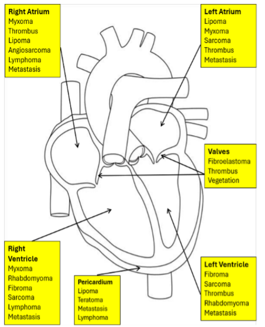
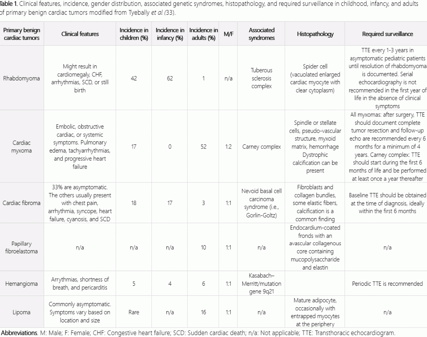
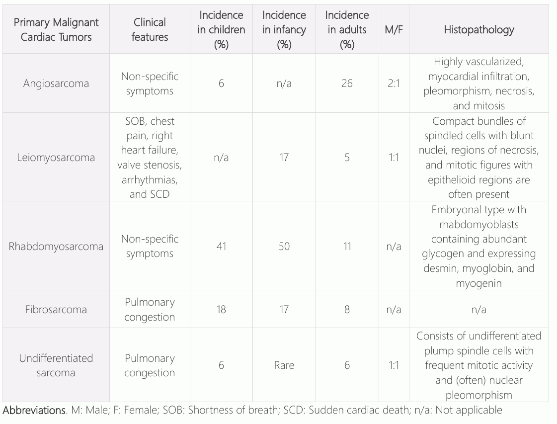
The clinical presentation and prognosis of patients with cardiac tumors vary depending on lesion location and histologic type (Figure 2). Additionally, the setting provides diagnostic clues that help determine the lesion or tumor's etiology. For example, vegetation may be observed in patients with prosthetic or native valve disease who present with bacteremia and elevated inflammatory markers. In contrast, cardiac thrombi are more likely to be associated with arrhythmias, such as atrial fibrillation, or a left ventricular apical infarct with hypokinesis or akinesis. If a cardiac mass is ultimately found, determine its etiology by considering four factors: the patient's age, the tumor's location, tissue characterization based on cMRI, and the epidemiological likelihood or clinical probability of the tumor (Tables 1-2) (33).
Primary Benign Cardiac Tumors
Rhabdomyoma
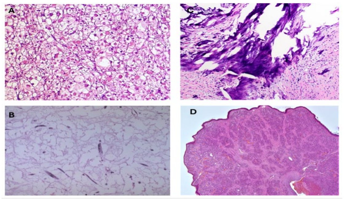
Cardiac rhabdomyoma is the most common primary cardiac tumor in children, accounting for more than 60% of PCTs (10, 11, 34). It is most commonly associated with tuberous sclerosis complex, an autosomal dominant disorder characterized by noncancerous tumors or lesions in the brain and other areas (35). Rhabdomyomas are typically located within the ventricles. On echocardiogram, they appear as homogeneous, well-circumscribed echogenic masses in the ventricular myocardium (36). Computed tomography (CT) or MRI can better delineate the relationship of the masses with the amount of involved myocardium and adjacent structures. These patients should also undergo whole-body imaging to rule out other benign tumors and malignancies, such as renal cell carcinoma and breast and thyroid cancer, among others (37–39). Rhabdomyomas show pathognomonic spider cells with centrally located cytoplasm, with the nucleus and myofibrils radiating to the cell wall (Figure 3A) (40). Clinical presentation varies with the tumor's position, number, and size. Their presence at the AV junction can sometimes enable the tumors to act like an accessory pathway, which can cause tachyarrhythmias (41). Cardiac rhabdomyomas can lead to CHF and infant respiratory distress (10, 11, 40, 41). Given their high rate of spontaneous regression after birth, they can be managed conservatively with echocardiography and electrocardiogram monitoring (42). Surgical intervention should be reserved for only hemodynamic compromise, intractable arrhythmias, or severe obstruction.
Myxoma
Cardiac myxomas are common benign PCTs most often found in the left atrium (Figure 3B) (43). Myxomas are sporadic; however, a familial incidence has been described in up to 7% of all cardiac myxomas, sometimes associated with Carney complex, a rare genetic disorder caused by mutations in the PRKAR1A gene, which causes pigmentation changes, benign tumors, and endocrine abnormalities (33, 44, 45). The tumors are pedunculated with a short, broadbased attachment to the atrial wall with a gelatinous consistency. On echocardiography, myxomas appear as a mobile mass attached to the endocardial surface by a stalk, usually originating from the fossa ovalis (46). More than 50% of patients with left atrial myxomas experience symptoms, which can include dyspnea and orthopnea from mitral valve stenosis or obstruction (47). Fragmentation of tumor may embolize and cause cerebrovascular events. Surgical excision remains the mainstay of treatment and leads to excellent long-term survival, with most patients having a good prognosis (48).
Fibroma
Cardiac fibromas are solitary neoplasms derived from connective tissue fibroblasts, which are mainly located within the ventricular septum (49). They are commonly seen in childhood and range from 1 to 10 cm (49, 50). On the macroscopic level, fibromas appear clearly demarcated from the myocardium. Microscopically, interdigitations surround the myocardium, and calcification of the central portion of the neoplasm is pathognomonic, reflecting poor blood supply to the tumor (Figure 3C) (51). On echocardiography, they appear as a large, well-circumscribed solitary mass in the ventricular free wall or septum (52). A solitary, large, homogeneous myocardial mass centered within the ventricles is typically evident on CT or MRI (52, 53).
Fibromas can invade the ventricular muscle, replace functional myocardium, and potentially cause refractory cyanosis or CHF (49–51). Occasionally, the tumor may extend into the ventricular conduction system, resulting in AV block or arrhythmias (50–54). These neoplasms commonly remain dormant, and spontaneous regression rarely occurs. As a result, surgical resection is usually recommended (55). The progressive loss of functional myocardial fibers and heart failure does not resolve following resection, which may lead to consideration of heart transplantation.
Papillary fibroelastoma
Papillary fibroelastoma is one of the most common PCTs (56). They are now increasingly recognized as imaging quality and techniques improve over the years. On gross inspection, they appear as small intracavity masses that are usually firm, with a short pedicle and multiple papillary fronds (57). These tumors are typically small and rarely present clinically with signs of obstruction. Cases treated with surgery occur in patients with symptoms related to cerebral ischemia (58).
Hemangioma
Angiomas are benign neoplasms composed predominantly of blood vessels (59). They are most frequently found in the right atrium and ventricular septum, and are most often discovered incidentally (59, 60). Multiple skin hemangiomas in neonates should prompt work-up for cardiac hemangiomas (60, 61). On histology, they appear as a proliferation of blood vessels lined by endothelial cells, which may be large, thin-walled vessels (such as cavernous hemangioma, the most common type), small capillaries (capillary hemangioma), or dysplastic malformation of veins and arteries (arteriovenous hemangioma) (Figure 3D) (62). Hemangiomas are benign tumors that typically regress spontaneously and have a good prognosis (60–62).
Lipoma
Lipomas are benign tumors predominantly composed of mature fat cells (63). They can appear at any age and occur with equal frequency in males and females. Lipomas may occur anywhere in the heart and present clinically depending on the tumor's location. MRI is commonly used to diagnose lipomas, which clearly demonstrate fatty tissue (64). These tumors are commonly silent, although they can sometimes cause arrhythmias and AV blocks (63, 64).
Primary Malignant Cardiac Tumors
Angiosarcoma
Angiosarcoma is an aggressive neoplasm that frequently originates in the right atrium and has a high likelihood of metastases at the time of presentation (65). Histopathology commonly shows a tumor that is highly vascularized with myocardial infiltration, pleomorphism, necrosis, and mitosis (66). Echocardiography usually reveals a nodular or lobulated mass in the right atrium near the inferior vena cava (67). CT or MRI shows arterial-phase enhancement, which improves diagnostic accuracy (68, 69). Clinical presentation is often nonspecific and may include symptoms of pericardial involvement or the right-sided heart failure (66, 67). Local pulmonary, pleural, and mediastinal metastases are also common (66–69). Treatment typically involves a combination of surgery and radiation, along with chemotherapy (68–70).
Leiomyosarcoma
Leiomyosarcoma is a primary sarcoma of smooth muscle cells typically originating in two sites (71). It can form in the left atrium, mimicking left atrial myxoma and its symptoms, or the pulmonary infundibulum and artery, mimicking a pulmonary embolism (72, 73). Dyspnea is the main complaint, but patients may sometimes present with pleuritic chest pain, hemoptysis, and palpitations from tachyarrhythmias such as atrial fibrillation. Treatment includes surgical excision (74). Chemoradiation therapy may provide palliation (72–74).
Rhabdomyosarcoma
Cardiac rhabdomyosarcomas are the most common malignant cardiac tumors in children and often affect the valves (75). These are malignant tumors of striated muscle that account for approximately 4% to 7% of cardiac sarcomas, with a male prevalence (75, 76). On gross inspection, they appear mucoid or gelatinous in consistency. Histologically, the embryonal type shows rhabdomyoblasts with abundant glycogen and expression of desmin, myoglobin, and myogenin (75). Patients may present with symptoms of pericardial disease, pleural effusion, or embolic phenomena but could also manifest systemic illness, including fever, weight loss, and malaise. Compared with angiosarcoma, diffuse pericardial involvement is uncommon, and the neoplasm rarely invades beyond the parietal pericardium. The prognosis is generally poor (77).
Fibrosarcoma
Fibrosarcomas are mesenchymal tumors that originate from fibroblasts and are generally seen within the left or right heart chambers (78). Clinical presentation varies based on tumor location and size. Symptoms of mitral stenosis, pulmonary congestion, and pulmonary vein obstruction are common with left chamber involvement (79). The prognosis of cardiac fibrosarcoma is generally poor, with a median survival time of less than a year (80).
Undifferentiated pleomorphic sarcoma
Undifferentiated cardiac sarcomas are invasive and possess no specific histological pattern (81, 82). They are most often seen in the left atrium, and most patients present with pulmonary congestion. The prognosis is generally poor despite surgery and palliative chemotherapy (81–83). Mean postoperative survival ranges from 5 to 18 months (81–84).
Limitations of Review
The available literature on pediatric cardiac tumors is limited by their rarity and small sample sizes, with most published evidence comprising single-center case series, reviews, and multicenter collaborations that still enroll modest cohorts. Primary cardiac tumors occur in approximately 0.0017–0.28% of pediatric autopsy series and represent fewer than 0.1% of pediatric cardiac surgeries (3, 85). The largest multicenter cardiovascular magnetic resonance study to date included 213 confirmed pediatric cardiac masses across 31 international centers, yet even this substantial effort reflects the aggregation of rare cases over many years (86). Single-institution surgical series typically report 16–120 patients accumulated over 2–3 decades, and malignant histotypes such as primary cardiac Ewing sarcoma are documented in fewer than 20 cases worldwide (85, 87–89). This data paucity limits the development of evidence-based management guidelines; the 2022 Society for Cardiovascular Magnetic Resonance/American Society of Echocardiography consensus notes that published cardiovascular magnetic resonance experience consists largely of "case reports and relatively small case series"(90). Consequently, diagnostic algorithms rely on expert consensus rather than prospective validation, surgical strategies are extrapolated from benign tumor experience, and outcomes—particularly for rare malignant variants—remain poorly characterized (86, 91). Tumor heterogeneity (rhabdomyoma, fibroma, myxoma, teratoma, hemangioma, and various sarcomas) further fragments the available evidence, complicating efforts to establish histotype-specific prognostic models or standardized treatment protocols.
Conclusions
In summary, cardiac tumors are rare and can potentially lead to serious complications such as fatal arrhythmias and intracardiac obstruction, even when benign. Imaging techniques such as MRI and CT scans play a pivotal role in diagnosis, helping distinguish between tumor types and ultimately shortening the time from diagnosis to treatment. These neoplasms can be treated surgically with an acceptable incidence of morbidity and mortality. The strongest predictor of mortality is linked to patient comorbidities, hemodynamic status at presentation, and the percentage of myocardial tissue involvement. Evaluation of patients with pediatric cardiac tumors requires multidisciplinary evaluation and individualized management. Overall, benign tumors possess high survival rates, and patients should be monitored for recurrence following resection. Competing interest statement: The authors report no relevant competing interests.
Declarations
- Author contributions (CRediT)
- All authors contributed equally to all 14 CRediT roles (Conceptualization, Data curation, Formal analysis, Funding acquisition, Investigation, Methodology, Project administration, Resources, Software, Supervision, Validation, Visualization, Writing – original draft, Writing – review & editing).
- Funding
-
This article received no specific grant or support from any public or private agencies.
- Conflicts of interest
- The authors report no relevant competing interests.
- Ethics approval
- Ethics committee approval was not required for this article, as it is based on previously published literature and does not involve human participants, patient data or animals.
- Data availability
- Data sharing is not applicable to this article as no new data were created or analysed in this study.
References (91)
82 of 91 references are linked to their DOI.
- Tao TY, Yahyavi-Firouz-Abadi N, Singh GK, Bhalla S. Pediatric cardiac tumors: clinical and imaging features. Radiographics 2014; 34(4): 1031-1046. DOI Crossref PubMed Google Scholar
- Faim D, Francisco A, Pires A. Cardiac tumours in children: a single-centre experience and literature review. Cardiol Young 2022: 1-10. DOI Crossref PubMed Google Scholar
- Uzun O, Wilson DG, Vujanic GM, Parsons JM, De Giovanni JV. Cardiac tumours in children. Orphanet J Rare Dis 2007; 2: 11. DOI Crossref PubMed Google Scholar
- Yadava OP. Cardiac tumours in infancy. Indian Heart J 2012; 64(5): 492-496. DOI Crossref PubMed Google Scholar
- Fresneau B, Oberlin O, Brugières L, Valteau-Couanet D, Patte C. (Malignant primary cardiac tumors in childhood and adolescence). Arch Pediatr 2010; 17(5): 495-501. PubMed Google Scholar
- Hoffmeier A, Sindermann JR, Scheld HH, Martens S. Cardiac tumors--diagnosis and surgical treatment. Dtsch Arztebl Int 2014; 111(12): 205-211. PubMed Google Scholar
- Saad EA, Mukherjee T, Gandour G, Fatayerji N, Rammal A, Samuel P, Abdallah N, Ashok T. Cardiac myxomas: causes, presentations, diagnosis, and management. Ir J Med Sci 2024; 193: 677-688. DOI Crossref PubMed Google Scholar
- Okongwu CC, Olaofe OO. Cardiac myxoma: a comprehensive review. J Cardiothorac Surg 2025; 20(1): 151. DOI Crossref PubMed Google Scholar
- Siontis BL, Leja M, Chugh R. Current clinical management of primary cardiac sarcoma. Expert Rev Anticancer Ther 2020; 20(1): 45-51. DOI Crossref PubMed Google Scholar
- Ng LY, McGuinness J, Prendiville T, Franklin O, Walsh M, Kenny D, Nolke L, McMahon CJ. Cardiac Rhabdomyomas Presenting with Critical Cardiac Obstruction in Neonates and Infants: Treatment Strategies and Outcome, A Single-Center Experience. Pediatr Cardiol 2024; 45(5): 1132 -1141. DOI Crossref PubMed Google Scholar
- Rychik J, Khalek N, Gaynor JW, Johnson MP, Adzick NS, Flake AW, Hedrick HL. Fetal intrapericardial teratoma: natural history and management including successful in utero surgery. Am J Obstet Gynecol 2016; 215(6): 780.e1-780.e7. DOI Crossref PubMed Google Scholar
- Poterucha TJ, Kochav J, O'Connor DS, Rosner GF. Cardiac Tumors: Clinical Presentation, Diagnosis, and Management. Curr Treat Options Oncol 2019;20(8):66. DOI Crossref PubMed Google Scholar
- Mankad R, Herrmann J. Cardiac tumors: echo assessment. Echo Res Pract 2016; 3(4): R65-R77. DOI Crossref PubMed Google Scholar
- Li X, Chen Y, Liu J, Xu L, Li Y, Liu D, Sun Z, Wen Z. Cardiac magnetic resonance imaging of primary cardiac tumors. Quant Imaging Med Surg 2020;10(1): 294-313. DOI Crossref PubMed Google Scholar
- Bussani R, Castrichini M, Restivo L, Fabris E, Porcari A, Ferro F, Pivetta A, Korcova R, Cappelletto C, Manca P, Nuzzi V, Bessi R, Pagura L, Massa L, Sinagra G. Cardiac Tumors: Diagnosis, Prognosis, and Treatment. Curr Cardiol Rep 2020; 22(12): 169. DOI Crossref PubMed Google Scholar
- Salzillo C, Lucà S, Ronchi A, Franco R, Iacobellis G, Leggio A, Marzullo A. Cardiac Tumors Causing Sudden Cardiac Death: A State-of-the-Art Review in Pathology. Cancers (Basel) 2025; 17(4): 669. DOI Crossref PubMed Google Scholar
- Bruckner BA, Reardon MJ. Benign cardiac tumors: a review. Methodist Debakey Cardiovasc J 2010; 6(3): 20-26. DOI Crossref PubMed Google Scholar
- Islam AKMM. Cardiac myxomas: A narrative review. World J Cardiol 2022;1 4(4):206-219. DOI Crossref PubMed Google Scholar
- Shi L, Wu L, Fang H, Han B, Yang J, Ma X, Liu F, Zhang Y, Xiao T, Huang M, Huang M. Identification and clinical course of 166 pediatric cardiac tumors. Eur J Pediatr 2017; 176(2): 253-260. DOI Crossref PubMed Google Scholar
- Mackie AS, Kozakewich HP, Geva T, Perez-Atayde AR, Mulliken JB. Vascular tumors of the heart in infants and children: case series and review of the literature. Pediatr Cardiol 2005; 26(4): 344-349. DOI Crossref PubMed Google Scholar
- Aktoz M, Tatli E, Ege T, Yalçin O, Büyüklü M, Aksu F, Gül C, Ozdemir C. Cardiac rhabdomyoma in an adult patient presenting with right ventricular outflow tract obstruction. Int J Cardiol 2008; 130(3): e105-e107. DOI Crossref PubMed Google Scholar
- He S, Cao Y, Qin W, Chen W, Yin L, Chai H, Tao Z, Tang S, Qiu Z, Chen X. Prevalence of primary cardiac tumor malignancies in retrospective studies over six decades: a systematic review and meta-analysis. Oncotarget 2017; 8(26): 43284-43294. 38 4-Pediatric Cardiac Tumors English et al. DOI Crossref PubMed Google Scholar
- Leja MJ, Shah DJ, Reardon MJ. Primary cardiac tumors. Tex Heart Inst J 2011; 38(3): 261-262. PubMed Google Scholar
- Campisi A, Ciarrocchi AP, Asadi N, Dell'Amore A. Primary and secondary cardiac tumors: clinical presentation, diagnosis, surgical treatment, and results. Gen Thorac Cardiovasc Surg 2022; 70(2): 107-115. DOI Crossref PubMed Google Scholar
- Butany J, Nair V, Naseemuddin A, Nair GM, Catton C, Yau T. Cardiac tumours: diagnosis and management. Lancet Oncol 2005; 6(4): 219-228. DOI Crossref PubMed Google Scholar
- Gillespie HS, Lin CC, Prutkin JM. Arrhythmias in structural heart disease. Curr Cardiol Rep 2014;16(8): 510. DOI Crossref PubMed Google Scholar
- Lopez EM, Malhotra R. Ventricular Tachycardia in Structural Heart Disease. J Innov Card Rhythm Manag 2019; 10(8): 3762-3773. PubMed Google Scholar
- He DK, Zhang YF, Liang Y, Ye SX, Wang C, Kang B, Wang ZN. Risk factors for embolism in cardiac myxoma: a retrospective analysis. Med Sci Monit 2015; 21: 1146-1154. DOI Crossref PubMed Google Scholar
- Adams HP Jr. Cerebrovascular manifestations of tumors of the heart. Handb Clin Neurol 2021;177:275-282. DOI Crossref PubMed Google Scholar
- Joshi M, Kumar S, Noshirwani A, Harky A. The Current Management of Cardiac Tumours: a Comprehensive Literature Review. Braz J Cardiovasc Surg 2020; 35(5): 770-780. DOI Crossref PubMed Google Scholar
- Arakelyanz AA, Morozova TE, Vlasova AV, Lischke R. Cardiac tumours - a brief review. Br J Cardiol 2020; 27(1): 03. DOI Crossref PubMed Google Scholar
- De Martino A, Pattuzzi C, Garis S, Bosco F, Virgone VM, Salsano A, Santini F, Pucci A. A Comprehensive Review of Cardiac Tumors: Imaging, Pathology, Treatment, and Challenges in the Third Millennium. Diagnostics (Basel) 2025; 15(11): 1390. DOI Crossref PubMed Google Scholar
- Tyebally S, Chen D, Bhattacharyya S, Mughrabi A, Hussain Z, Manisty C, Westwood M, Ghosh AK, Guha A. Cardiac Tumors: JACC CardioOncology State-of-the-Art Review. JACC CardioOncol 2020; 2(2): 293-311. PubMed Google Scholar
- Kutluk T, Demir HA, Büyükpamukçu M, Ozkutlu S, Akyüz C, Varan A, Yalçın B. Cardiac rhabdomyomas in childhood: six cases from a single institution. Turk J Pediatr 2013; 55(1): 69-73. PubMed Google Scholar
- Kocabaş A, Ekici F, Cetin Iİ, Emir S, Demir HA, Arı ME, Değerliyurt A, Güven A. Cardiac rhabdomyomas associated with tuberous sclerosis complex in 11 children: presentation to outcome. Pediatr Hematol Oncol 2013; 30(2): 71-79. DOI Crossref PubMed Google Scholar
- Di Liang C, Ko SF, Huang SC. Echocardiographic evaluation of cardiac rhabdomyoma in infants and children. J Clin Ultrasound 2000; 28(8): 381-386. DOI Crossref PubMed Google Scholar
- Wang MX, Segaran N, Bhalla S, Pickhardt PJ, Lubner MG, Katabathina VS, Ganeshan D. Tuberous Sclerosis: Current Update. Radiographics 2021; 41(7): 1992-2010. DOI Crossref PubMed Google Scholar
- Henske EP, Cornejo KM, Wu CL. Renal Cell Carcinoma in Tuberous Sclerosis Complex. Genes (Basel) 2021; 12(10): 1585. DOI Crossref PubMed Google Scholar
- Zhu QY, He ZM, Cao WM, Li B. The role of TSC2 in breast cancer: a literature review. Front Oncol 2023; 13: 1188371. DOI Crossref PubMed Google Scholar
- Fenoglio JJ Jr, MCAllister HA Jr, Ferrans VJ. Cardiac rhabdomyoma: a clinicopathologic and electron microscopic study. Am J Cardiol 1976; 38(2): 241-251. DOI Crossref PubMed Google Scholar
- Wacker-Gussmann A, Strasburger JF, Cuneo BF, Wiggins DL, Gotteiner NL, Wakai RT. Fetal arrhythmias associated with cardiac rhabdomyomas. Heart Rhythm 2014; 11(4): 677-683. DOI Crossref PubMed Google Scholar
- Wu SS, Collins MH, de Chadarévian JP. Study of the regression process in cardiac rhabdomyomas. Pediatr Dev Pathol 2002; 5(1): 29-36. DOI Crossref PubMed Google Scholar
- Griborio-Guzman AG, Aseyev OI, Shah H, Sadreddini M. Cardiac myxomas: clinical presentation, diagnosis and management. Heart 2022; 108(11): 827-833. DOI Crossref PubMed Google Scholar
- Ergi DG, Klarich KW, Maleszewski JJ, Carney JA, Rowse PG, Crestanello JA, Schaff HV, Todd A, Dearani JA, Daly RC, Arghami A. Cardiac Myxomas in Carney Complex: Single Institution Multidecade Experience. Ann Thorac Surg 2024; 118(6): 1216-1223. DOI Crossref PubMed Google Scholar
- Wilkes D, Charitakis K, Basson CT. Inherited disposition to cardiac myxoma development. Nat Rev Cancer 2006; 6(2): 157-165. DOI Crossref PubMed Google Scholar
- Hrabak-Paar M, Muršić M, Balaško-Josipović T, Dilber D, Bulj N. Multimodality Imaging of Cardiac Myxomas. Rev Cardiovasc Med 2024; 25(6): 204. DOI Crossref PubMed Google Scholar
- Goswami KC, Shrivastava S, Bahl VK, Saxena A, Manchanda SC, Wasir HS. Cardiac myxomas: clinical and echocardiographic profile. Int J Cardiol 1998; 63(3): 251-259. DOI Crossref PubMed Google Scholar
- Garatti A, Nano G, Canziani A, Gagliardotto P, Mossuto E, Frigiola A, Menicanti L. Surgical excision of cardiac myxomas: twenty years experience at a single institution. Ann Thorac Surg 2012; 93(3): 825-831. DOI Crossref PubMed Google Scholar
- Covington MK, Young PM, Bois MC, Maleszewski JJ, Anand V, Dearani JA, Klarich KW. Clinical Impact of Cardiac Fibromas. Am J Cardiol 2022; 182: 95-103. DOI Crossref PubMed Google Scholar
- Zhou C, Liu J, Li S, Wang H, Jin Y, Liu J. Left ventricular fibromas in pediatric patients: a case series and review of the literature. J Cardiothorac Surg 2025; 20(1): 180. DOI Crossref PubMed Google Scholar
- Teng F, Yang S, Chen D, Fang W, Shang J, Dong S, Cui Y, Fu W, Zheng M, Li Y, Lian G. Cardiac fibroma: A clinicopathologic study of a series of 12 cases. Cardiovasc Pathol 2022; 56: 107381. DOI Crossref PubMed Google Scholar
- Lin Y, Xie M, Qian M, Gao L, Ji MM, Li Y. Cardiac fibroma: characteristics on echocardiography and cardiac magnetic resonance imaging. QJM 2022; 115(6): 412-414. DOI Crossref PubMed Google Scholar
- Araoz PA, Mulvagh SL, Tazelaar HD, Julsrud PR, Breen JF. CT and MR imaging of benign primary cardiac neoplasms with echocardiographic correlation. Radiographics 2000; 20(5): 1303-1319. DOI Crossref PubMed Google Scholar
- Finke K, Gietzen T, Steven D, Baldus S, Ten Freyhaus H, Maintz D, Pennig L, Gietzen C. Cardiac fibromas in adult patients: a case series focusing on rhythmology and radiographic features. Eur Heart J Case Rep 2024; 8(8): ytae410. DOI Crossref PubMed Google Scholar
- Cho JM, Danielson GK, Puga FJ, Dearani JA, McGregor CG, Tazelaar HD, Hagler DJ. Surgical resection of ventricular cardiac fibromas: early and late results. Ann Thorac Surg 2003; 76(6): 1929-1934. DOI Crossref PubMed Google Scholar
- Gowda RM, Khan IA, Nair CK, Mehta NJ, Vasavada BC, Sacchi TJ. Cardiac papillary fibroelastoma: a comprehensive analysis of 725 cases. Am Heart J 2003; 146(3): 404-410. DOI Crossref PubMed Google Scholar
- Khair T, Mazidi P, Laos LF. Cardiac Papillary Fibroelastoma: case report and review of the literature. Int J Cardiol 2010; 139(1): 102-104. DOI Crossref PubMed Google Scholar
- Mazur P, Kurmann R, Klarich KW, Dearani JA, Arghami A, Daly RC, Greason K, Schaff HV, Ahmad A, El-Am E, Sorour A, Bois MC, Viehman J, King KS, Maleszewski JJ, Crestanello JA. 39 4-Pediatric Cardiac Tumors English et al. Operative management of cardiac papillary fibroelastomas. J Thorac Cardiovasc Surg 2024; 167(3): 1088-1097.e2. DOI Crossref PubMed Google Scholar
- Munteanu IR, Novaconi RC, Merce AP, Dima CN, Falnita LS, Manzur AR, Streian CG, Feier HB. Cardiac Hemangiomas: A Five-Year Systematic Review of Diagnosis, Treatment, and Outcomes. Cancers (Basel) 2025; 17(9): 1532. DOI Crossref PubMed Google Scholar
- Chao JC, Reyes CV, Hwang MH. Cardiac hemangioma. South Med J 1990; 83(1): 44-47. PubMed Google Scholar
- Tal R, Dotan M, Lorber A. Approach to haemangio-matosis causing congestive heart failure. Acta Paediatr 2016; 105(6): 600-604. DOI Crossref PubMed Google Scholar
- Eftychiou C, Antoniades L. Cardiac hemangioma in the left ventricle and brief review of the literature. J Cardiovasc Med (Hagerstown) 2009; 10(7): 565-567. DOI Crossref PubMed Google Scholar
- Shu S, Wang J, Zheng C. From pathogenesis to treatment, a systemic review of cardiac lipoma. J Cardiothorac Surg 2021; 16(1): 1. DOI Crossref PubMed Google Scholar
- D'Souza J, Shah R, Abbass A, Burt JR, Goud A, Dahagam C. Invasive Cardiac Lipoma: a case report and review of literature. BMC Cardiovasc Disord 2017; 17(1): 28. DOI Crossref PubMed Google Scholar
- Kumari N, Bhandari S, Ishfaq A, Butt SRR, Ekhator C, Karski A, Kadel B, Altayb Ismail MA, Sherpa TN, Al Khalifa A, Khalifah B, Nguyen N, Lazarevic S, Zaman MU, Ullah A, Yadav V. Primary Cardiac Angiosarcoma: A Review. Cureus 2023; 15(7): e41947. DOI Crossref PubMed Google Scholar
- Leduc C, Jenkins SM, Sukov WR, Rustin JG, Maleszewski JJ. Cardiac angiosarcoma: histopathologic, immuno-histochemical, and cytogenetic analysis of 10 cases. Hum Pathol 2017; 60: 199-207. DOI Crossref PubMed Google Scholar
- Kupsky DF, Newman DB, Kumar G, Maleszewski JJ, Edwards WD, Klarich KW. Echocardiographic Features of Cardiac Angiosarcomas: The Mayo Clinic Experience (1976-2013). Echocardiography 2016; 33(2): 186-192. DOI Crossref PubMed Google Scholar
- Chen Y, Li Y, Zhang N, Shang J, Li X, Liu J, Xu L, Liu D, Sun Z, Wen Z. Clinical and Imaging Features of Primary Cardiac Angiosarcoma. Diagnostics (Basel) 2020; 10(10): 776. DOI Crossref PubMed Google Scholar
- Look Hong NJ, Pandalai PK, Hornick JL, Shekar PS, Harmon DC, Chen YL, Butrynski JE, Baldini EH, Raut CP. Cardiac angiosarcoma management and outcomes: 20-year single-institution experience. Ann Surg Oncol 2012; 19(8): 2707-2715. DOI Crossref PubMed Google Scholar
- Krishnan T, Pettersson G, Mukherjee R, Singhal N. Cardiac angiosarcoma: A diagnostic and therapeutic challenge. J Cardiol Cases 2020; 22(2): 90-93. DOI Crossref PubMed Google Scholar
- Wang JG, Cui L, Jiang T, Li YJ, Wei ZM. Primary cardiac leiomyosarcoma: an analysis of clinical characteristics and outcome patterns. Asian Cardiovasc Thorac Ann 2015; 23(5): 623-630. DOI Crossref PubMed Google Scholar
- Morin JE, Rahal DP, Hüttner I. Myxoid leiomyosarcoma of the left atrium: a rare malignancy of the heart and its comparison with atrial myxoma. Can J Cardiol 2001; 17(3): 331-336. PubMed Google Scholar
- Leiva O, Hollingsworth P, Singh V, Qian X, Blankstein R, Vaduganathan M. What Looks Like a Clot But Is Not a Clot?: Cardiac Leiomyosarcoma Mimicking Pulmonary Embolism. JACC Case Rep 2020; 2(12): 1966-1968. DOI Crossref PubMed Google Scholar
- Ramlawi B, Leja MJ, Abu Saleh WK, Al Jabbari O, Benjamin R, Ravi V, Shapira OM, Blackmon SH, Bruckner BA, Reardon MJ. Surgical Treatment of Primary Cardiac Sarcomas: Review of a Single-Institution Experience. Ann Thorac Surg 2016; 101(2): 698-702. DOI Crossref PubMed Google Scholar
- Castorino F, Masiello P, Quattrocchi E, Di Benedetto G. Primary cardiac rhabdomyosarcoma of the left atrium: an unusual presentation. Tex Heart Inst J 2000; 27(2): 206-208. PubMed Google Scholar
- Kimura A, Tsuji M, Isogai T, Nagata K, Kato K, Hisagi M, Nonaka T, Ninomiya M, Kiriu T, Tanaka H, Tejima T. A Mass Filling the Right Atrium: Primary Cardiac Rhabdomyo sarcoma. Intern Med 2018; 57(24): 3575-3580. DOI Crossref PubMed Google Scholar
- Panagiotopoulos I, Katinioti A, Mousafeiris V, Leivaditis V, Skevis K, Tasios K, Antzoulas A, Pitros C, Verras GI, Mulita F, Prapas S. Multifocal, biatrial, primary cardiac embryonal rhabdomyosarcoma. Prz Menopauzalny 2023; 22(3): 173-176. DOI Crossref PubMed Google Scholar
- Chan EY, Ali A, Zubair MM, Nguyen DT, Ibarra-Cortez SH, Graviss EA, Shapira OM, Ravi V, MacGillivray TE, Reardon MJ. Primary cardiac sarcomas: Treatment strategies. J Thorac Cardiovasc Surg 2023; 166(3): 828-838.e2. DOI Crossref PubMed Google Scholar
- Paruchuru PK, Brann SH, Patel RL. Primary left atrial fibrosarcoma. Cardiovasc Pathol 2001; 10(6): 317-319. DOI Crossref PubMed Google Scholar
- Kabashi S, Hoxha N, Gashi S, Ahmegjekaj I, Bejta I, Sadiku M, Ymeri H, Kabashi A, Bicaj X, Mucaj S. Primary fibro sarcoma of the heart. Acta Inform Med 2013; 21(3): 213-215. DOI Crossref PubMed Google Scholar
- Gandhi R, King S, Shi B, Sasse A. Undifferentiated Pleomorphic Cardiac Sarcoma: A Rare and Often Fatal Diagnosis. JACC Case Rep 2024; 29(24): 102844. DOI Crossref PubMed Google Scholar
- Stoica AI, Harpa MM, Banceu CM, Ghiragosian C, Opris CE, Al-Hussein H, Al-Hussein H, Flamind Oltean S, Mezei T, Mares RG, Suciu H. A Rare Case of Undifferentiated Pleomorphic Cardiac Sarcoma with Inflammatory Pattern. Medicina (Kaunas) 2022;58(8): 1009. DOI Crossref PubMed Google Scholar
- Guerra-Raygada M, Saavedra-Sanchez AJ, Hidalgo-Avendaño D, Bermudez-Pelaez MF, Guevara-Lazo D, Nombera-Aznaran N. From dyspnea to diagnosis, unmasking undifferentiated cardiac sarcoma: a case report. Egypt Heart J 2024; 76(1): 86. DOI Crossref PubMed Google Scholar
- Vaitiekiene A, Vaitiekus D, Urbonaite L, Jankauskas A, Portacenko J, Lapinskas T, Benetis R, Siudikas A, Veikutiene A, Poskiene L, Kavoliuniene A, Janciauskiene R, Jarusevicius L, Juozaityte E, Zaliunas R, Ereminiene E. Multidisciplinary approach to rare primary cardiac sarcoma: a case report and review. BMC Cancer 2019; 19(1): 529. DOI Crossref PubMed Google Scholar
- Bielefeld KJ, Moller JH. Cardiac tumors in infants and children: study of 120 operated patients. Pediatr Cardiol 2013; 34(1):125-8. DOI Crossref PubMed Google Scholar
- Beroukhim RS, Ghelani S, Ashwath R, Balasubramanian S, Biko DM, Buddhe S, Campbell MJ, Cross R, Festa P, Griffin L, Grotenhuis H, Hasbani K, Hashemi S, Hegde S, Hussain T, Jain S, Kiaffas M, Kutty S, Lam CZ, Liberato G, Merlocco A, Misra N, Mowers KL, Muniz JC, Nutting A, Parra DA, Patel JK, Perez-Atayde AR, Prasad D, Rosental CF, Shah A, Samyn MM, Sleeper LA, Slesnick T, Valsangiacomo E, Geva T. Accuracy of Cardiac Magnetic Resonance Imaging in Diagnosing Pediatric Cardiac Masses: A Multicenter Study. JACC Cardiovasc Imaging 2022; 15(8):1391-1405. DOI Crossref PubMed Google Scholar
- Delmo Walter EM, Javier MF, Sander F, Hartmann B, Ekkernkamp A, Hetzer R. Primary Cardiac Tumors in Infants and Children: Surgical Strategy and Long-Term Outcome. Ann Thorac Surg 2016; 102(6):2062-2069. DOI Crossref PubMed Google Scholar
- Liu X, Hong H, Zhang H, Xu Z, Liu J, Qiu L. Treatment Strategies for Primary Tumors of the Heart in Children: A 10-Year Experience. Ann Thorac Surg. 2015; 100(5):1744-9. 40 4-Pediatric Cardiac Tumors English et al. DOI Crossref PubMed Google Scholar
- Khajeh-Mehrizi A, Karimi M, Mortezazadeh M, Amiri B, Zarei H, Saffar H. Primary cardiac ewing sarcoma presenting as an infiltrative myocardial mass: a rare case with long-term survival. J Cardiothorac Surg 2025; 21(1):48. DOI Crossref PubMed Google Scholar
- Fogel MA, Anwar S, Broberg C, Browne L, Chung T, Johnson T, Muthurangu V, Taylor M, Valsangiacomo-Buechel E, Wilhelm C. Society for Cardiovascular Magnetic Resonance/European Society of Cardiovascular Imaging/American Society of Echocardiography/Society for Pediatric Radiology/North American Society for Cardiovascular Imaging Guidelines for the Use of Cardiac Magnetic Resonance in Pediatric Congenital and Acquired Heart Disease: Endorsed by The American Heart Association. Circ Cardiovasc Imaging 2022; 15(6):e014415. DOI Crossref PubMed Google Scholar
- Padalino MA, Vida VL, Boccuzzo G, Tonello M, Sarris GE, Berggren H, Comas JV, Di Carlo D, Di Donato RM, Ebels T, Hraska V, Jacobs JP, Gaynor JW, Metras D, Pretre R, Pozzi M, Rubay J, Sairanen H, Schreiber C, Maruszewski B, Basso C, Stellin G. Surgery for primary cardiac tumors in children: early and late results in a multicenter European Congenital Heart Surgeons Association study. Circulation 2012; 126(1):22-30. DOI Crossref PubMed Google Scholar
Respond to this article
Comments on a published article are welcome as a Letter to the Editor.
Get an e-mail when new articles in your fields are published
Questions to the authors
Questions are moderated: an editor approves each question, the authors answer, and an editor approves the answer before both appear here.
No answered questions yet.
Ask the authors a question
Discussion
Comments are moderated: they appear after the writer confirms the e-mail address and an editor approves them. Each comment carries a conflict-of-interest statement.
No comments yet.
Write a comment
Tell me when this article is cited
Leave your e-mail address and confirm it. These notices are not being sent yet; your confirmed address is kept for them and used for nothing else.
Read the full text
Open the PDF here
Loading the PDF…
If the PDF does not show here, use the download button above.
How to cite this article
English K, Dzukey E, Garcia R, Kumar KA. Primary Cardiac Tumors in Children: A State-of-the-Art Review. Ulutas Med J. 2026;12(3):29-40. doi:10.5455/umj.20260718040933
English, K., Dzukey, E., Garcia, R., & Kumar, K. A. (2026). Primary Cardiac Tumors in Children: A State-of-the-Art Review. The Ulutas Medical Journal, 12(3), 29–40. https://doi.org/10.5455/umj.20260718040933
English, K. et al. (2026) 'Primary Cardiac Tumors in Children: A State-of-the-Art Review', The Ulutas Medical Journal, 12(3), pp. 29–40. doi: 10.5455/umj.20260718040933.
@article{umj328225,
  title = {Primary Cardiac Tumors in Children: A State-of-the-Art Review},
  author = {English, Kevan and Dzukey, Elfreda and Garcia, Reicelle and Kumar, Kritikka Ajit},
  journal = {The Ulutas Medical Journal},
  year = {2026},
  volume = {12},
  number = {3},
  pages = {29--40},
  doi = {10.5455/umj.20260718040933},
  url = {https://www.ulutasmedicaljournal.com/?mno=328225},
  issn = {2149-388X}
}