The Ulutas Medical Journal · ISSN 2149-0430 (印刷版), 2149-388X (在线版) · https://www.ulutasmedicaljournal.com/?mno=268878
综述 · 开放获取
肝细胞癌中肝脏内皮生长因子和转化生长因子β的作用
标题、摘要和全文为机器翻译。 英文原文
- Biochemistry Lab, Department of Zoology, Dr. Harisingh Gour Central University, Sagar - 470003, Madhya Pradesh, India ROR
- 开放获取
- CC BY-NC 4.0
- ORCID
- 资助
- 伦理批准
- 数据可用性
摘要
肝细胞癌是全世界最常见的恶性肿瘤之一,也是导致死亡的主要原因。 据报道,肿瘤基因、肿瘤血栓的信号、诱导的炎性细胞和缺氧性血管疾病可切换血管。 血管内皮生长因子是调节细胞增殖,血管渗透的多肽因子,并通过其不同的血管受体在肿瘤血管增生中发挥重要作用. 多功能调控细胞基质转化生长因子-β也控制细胞增殖,分化,细胞外基质生产以及许多类型细胞的生存/或吞噬. 通过提高上位细胞-中位细胞过渡水平,转化生长因子-β为肿瘤细胞生长创造了有利的微观环境. 视癌症发展阶段而定,它可以引发肿瘤发育、进化和元化,也可以起到抑制肿瘤的作用。 在本次审查中,已试图全面界定肝细胞癌的发育和进展中,肝细胞内皮生长因子的作用以及改变增长因子β的作用.
关键词
全文
全文为机器翻译;以英文原文为准。 英文原文
导言
据报告,肝细胞癌是全世界第七大常见癌症,也是第三大癌症致死原因。 (1)由于缺乏有效的治疗方案,特别是在后期阶段,已观察到高死亡率。 虽然外科手术和外科外科外科外科外科外科外科外科外科外科外科外科外科外科外科外科外科外科外科外科外科外科外科外科外科外科外科外科外科外科外科外科外科外科外科外科外科外科外科外科外科外科外科外科外科外科外科外科外科外科外科外科外科外科外科外科外科外科外科外科外科外科外科外科外科外科外科外科外科外科外科外科外科外科外科外科外科外科外科外科外科外科外科外科外科外科外科外科外科外科外科外科外科外科外科外科外科外科外科外科外科外科外科外科外科外科外科外科外科外科外科外科外科外科外科外科外科外科外科外科外科外科外科外科外科外(2)此外,由于早期总部合同委员会的无症状性质、缺乏认识和筛查战略不明确,大多数患者(约80%)患有高级或无法治愈的疾病。 (3)。 。 。
与HCC有关的肿瘤因素包括慢性肝病,肝炎病毒(HBV & HCV)和酗酒. 不论肝硬化是肿瘤发展的主要风险因素之一,据观察,近80%的HCC患者在肿瘤发作前经过这一阶段。 (4)此外,据报告,黄道毒素对丙型肝炎病毒引起的肝癌具有协同效应。 (5)从流行病学研究中可以明显看出,食物受AB1污染是人类肝癌的主要风险因素。 (6, 7)。 。 。
HCC被认为是血管肿瘤生长的极端类型,依赖血管增生,即新血管增生. 在癌症从入侵前和休眠状态到入侵和元静态阶段的病原和演变过程中,人们观察到血管疾病通过产生肿瘤生长、入侵和元静态所必需的血管疾病因素而发挥关键作用。 多项研究表明,肿瘤血管病的程度与疾病阶段、元病可能性和癌症复发有着积极的联系。 (8)。 。 。
血管内皮生长因子(VEGF)是一个众所周知的强效血管致病因子,据报道,该因子可增强血管通透性,促进血管新血管化和内皮细胞在HCC中由于其内皮细胞的线粒体特性而扩散. (9)除了VEGF外,在肝脏疾病发作期间,还报告转化生长系数b(TGF-b)在损伤纤维化方面发挥着重要作用。 (10)虽然在正常的肝脏中TGF-β抑制肝细胞生长,但观察到肝脏受伤者可激活肝细胞,促进肝细胞癌的发展。 (11). 在癌症发作期间,激活TGF-β信号,导致TGF-β粘合在细胞膜上的受体上 (12)。 。 。
VEGF 作为肝细胞癌的生物标志
1989年首次描述 (13),VEGF蛋白家族目前由几个成员组成:VEGF(或VEGF-A),VEGF-B,VEGF-C和VEGF-D,VEGF-F,胎盘生长因子(PlGF)及其受体VEGFR-1,VEGFR-2和VEGFR-3. (14)VEGF是VEGF tyrosine kinase受体超家族的支架,包括VEGF-A,-B,-C和-D,其支架变体VEGF-A导致几种异构. (15, 16)。 。 。
VEGF家族的韧带与tyrosine kinase受体结合 VEGFR1, VEGFR2和VEGFR3 (图1); 每个受体包含一个大约750个氨基酸残基的细胞外域,在七个免疫球蛋白像折叠一样排列. (17)此外,肝素硫酸甲酯蛋白糖以及神经肽(NRP-1和NRP-2)可作为VEGF的核心受体,并促进VEGFR激活。 (18, 19)· VEGF/VEGFR路径受到许多因素的影响和调节,例如当地环境缺氧、各种激素、生长因素和细胞基。 激素,如高血压细胞和恶性细胞VEGF生产和血管增生,促进前列腺癌生长 (20, 21)。 。 。
VEGF被描述为主要的血管生长因子,其表达水平被作为生物标志用于识别血管疾病. 据报道,VEGF的表达水平随着合同委员会的发展和病因的产生而增加,并且据报告在癌症晚期表现水平很高。 在由于新血管化而形成的固体肿瘤中,VEGF通过提供持续的养分流动及其与肿瘤进化相关的表达,促进肿瘤生长和元化. (22)它已被确定为合同委员会的重要血管增生因素之一。 据报道,其表达方式从低级阻滞结核逐渐增加,到高级阻滞结核逐渐增加至早期合同委员会 (23)在合同委员会早期阶段,观察到合同委员会较高的膜与微血管密度、未发作动脉(即动脉不伴有毛细管,表明血管发作)和CD34染色物在合同委员会中作为鼻索毛细管化标记有关联。 (23). VEGF-C基因多态化导致其过度表达的情况是新angiogenics和细胞在HCC中经常出现,与高级HCC相比,已经观察到扩散活动 (23). 有人建议,开发过程中VEGF的表达程度是合同委员会后期的预测因素 (24)。 。 。
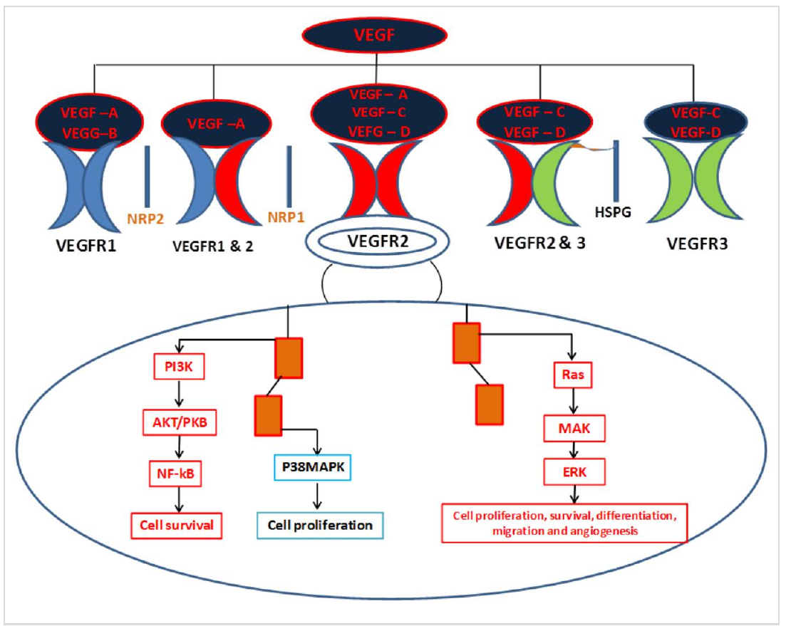
VEGF被核算为主要血管因子,负责HCC肿瘤的新生、发育和/或进展/传播 (25) 血清和组织中VEGF过量表达的HCC患者与存活率低有关 (26)· VEGF 诱导形成新的微管,对各种人类固体肿瘤的生长和进展至关重要,因为它能够向迅速生长的肿瘤提供氧气和营养物质 (27)其中一些快速生长的肿瘤还同时分泌了血管瘤 (28)。 。 。
乙型六氯环己烷的肿瘤和肿瘤功能
TGF-β是一种细胞基质,具有多种功能,如调节细胞增殖,分化和细胞外基质生产. (29)解除对TGF-β表达或信号的调控与癌症和纤维化等各种疾病的病因有关。 TGF-β的表达方式被修正到非常低,在癌症发育的后期会急剧增加,因此被肿瘤细胞积极分泌,因此它不仅起到旁观者的作用,反而在晚期促进细胞生长、入侵、元化和减少宿主-肿瘤免疫反应 (30). TGF-β是大量分泌蛋白质家族的原生成员,根据它们激活的信号途径,可进一步分为两个子家族——TGF-β/Activin/Nodal子家族和Bone Morphogenetic蛋白(BMP)子家族. (31)。 。 。
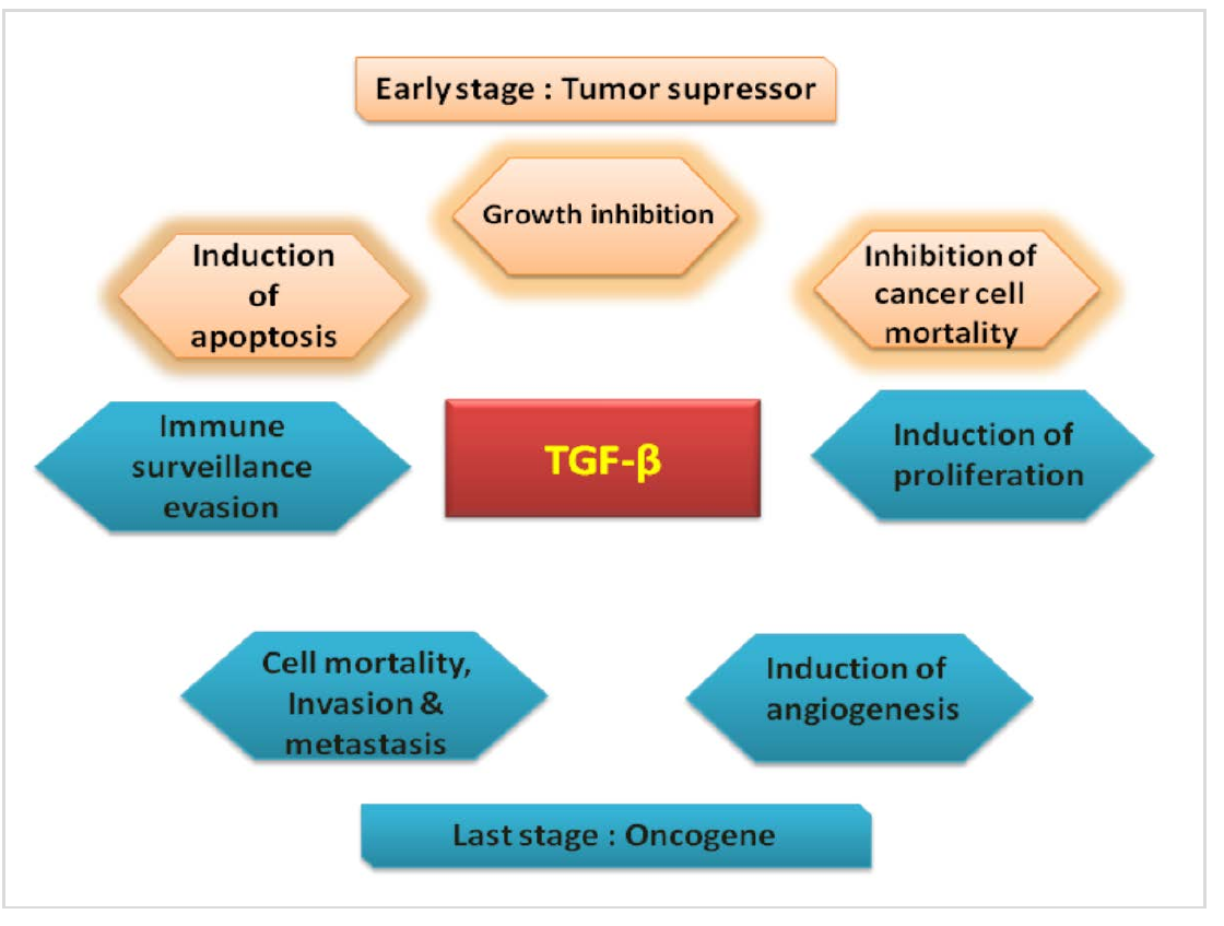
TGF-β能够调节肿瘤的启动、进化和元静态发育。 人们广泛认为,TGF-βs既是肿瘤抑制剂又是肿瘤促进剂,这取决于癌症类型和肿瘤发育时间。 (32, 33). 在肿瘤发育的早期阶段,TGF-β1被报告为肿瘤抑制剂,主要是因为其能够抑制上皮细胞的扩散. 此外,它还可以通过降低C-Myc和ID等肿瘤的表达和/或诱导CDK抑制剂的表达,如p21Cip1和p15lnk4b,导致G1细胞循环停止. (34, 35)据报道,TGF-β具有肿瘤抑制剂和肿瘤促进剂的双重作用(图2),但迄今为止,这一机制尚不清楚。 据报道,HCC中TGF-β的表达值随着烯烃沉积和蛋白质降解减少而增加。 (36)。 。 。
在胃癌、结肠癌、前列腺癌、乳房癌、肺癌、肝癌和胰腺癌等许多恶性肿瘤中观察到的Smad4、TβRI或TβRII(TGF-β II型受体)的频繁突变缺陷表明TGF-β的肿瘤抑制功能。 (37, 38)在合同委员会发展和进展期间,在肝脏上皮细胞中观察到TGF-β的双重作用。 在初始阶段,由于肝细胞中可溶性TβRII的异位表达,TGF-β II型受体(TβRII)的可用性降低,对HCC的易感性明显增强。 (39) 或减少异羟基小鼠的TβRII表达已经证实TGF-β信号通路的肿瘤抑制功能 (40)。 。 。
在大多数由上皮细胞引起的癌症中,TGF-β通过对循环依赖蛋白质激酶(CDK)抑制剂如p15和p21的调节,诱导蛋白质诱导如死亡相关蛋白质激酶(DAP),从而抑制恶性细胞生长. (41)与前列腺癌和子宫颈癌的这一表述相反,人们发现癌症细胞中TGF-β1的表达与进化和元化密切相关。 (42–44)癌症中最常见的变异TGF-β途径基因是TGFBR2,TGFBR1,SMAD4和SMAD2.
结论
HCC表现出许多分子异常,可能涉及癌症的发育和进化过程. 因此,必须确定可能参与癌症致病和发病的关键参与者,以便能够作出准确的预后,并对高血压患者适用合理的选择标准,特别是在早期阶段。 据观察,在包括HCC在内的各种肿瘤中,每当局部入侵癌细胞或变异性分裂部位时,通过血管增生,血液中的VEGF水平就会上升. TGF-β的进一步肿瘤抑制功能发挥着重要作用. 因此,VEGF和TGF-β都通过不同机制对细胞外基质的血管生长、肿瘤增殖和产生以及HCC的免疫抑制进行监管。 在这种情况下,对VEGF和TGF-β途径的更深入了解,将可导致制定模式,将目标对准合同委员会中的癌细胞。 迄今,TGF-β的恶性转化所涉及的确切机制尚不明确,需要从治疗的角度来研究。
声明
- 作者贡献(CRediT)
- 所有作者对全部 14 项 CRediT 角色贡献均等(Conceptualization, Data curation, Formal analysis, Funding acquisition, Investigation, Methodology, Project administration, Resources, Software, Supervision, Validation, Visualization, Writing – original draft, Writing – review & editing)。
- 资助
-
Funding information was not reported in the published version of this article.
- 利益冲突
- The authors declare that there is no conflict of interests.
- 伦理批准
- Ethics committee approval was not required for this article, as it is based on previously published literature and does not involve human participants, patient data or animals.
- 数据可用性
- Data sharing is not applicable to this article as no new data were created or analysed in this study.
参考文献 (44)
44 条参考文献中有 36 条已链接 DOI。
- Simbaqueba Ferlay J, Shin HR, Bray Forman FD, Mathers C, Parkin DM. Estimates of worldwide burden of cancer in 2008: GLOBOCAN 2008, Int J Cancer 2010;127: 2893– 2917. DOI Crossref PubMed Google Scholar
- Mulcahy MF. Management of hepatocellular cancer. Curr Treat Options Oncol 2005;6: 423–435. DOI Crossref PubMed Google Scholar
- Thomas MB, Abbruzzese JL. Opportunities for targeted therapies in hepatocellular carcinoma. J Clin Oncol 2005;23: 8093– 8108. DOI Crossref PubMed Google Scholar
- Minguez B, Tovar V, Chiang D, Villanueva A, Llovet JM. Pathogenesis of hepatocellular carcinoma and molecular therapies. Curr Opin Gastroenterol 2009;25: 186-194. DOI Crossref PubMed Google Scholar
- 5. Kirk GD, Lesi OA, M Mendy et al. 249 (ser) P53 mutation in plasmid DNA, hepatitis B viral infection, and risk of hepatocellular carcinoma. Oncogene 2005;24:5858 5867. PubMed Google Scholar
- Li Y, SU JJ, Qin LL, Yang C, Ban KC, Yan RQ. Synergistic effect of hepatitis B virus and aflatoxin B1 in hepato carcinogenesis in tree shrews. Ann Acad Med Singapore 1999;28: 67-71. DOI Crossref PubMed Google Scholar
- Madden CR, Finegold MJ, Slagle BL. Altered DNA mutation spectrum in aflatoxin b1-treated transgenic mice that express the hepatitis B virus x protein. J Virol 2002;76: 11770-11774. DOI Crossref PubMed Google Scholar
- Weidner N. Angiogenesis as a predictor of clinical outcome in cancer patients. Hum Pathol 2000;31: 403– 405. PubMed Google Scholar
- Dvorak HF, Brown IF, Detmar MA, Dvorak M. Vascular permeability factor/vascular endothelial growth factor, microvascular hypermeability, and angiogenesis. Am J Pathol 1995;146:1029–1039. PubMed Google Scholar
- Gressner AM, Weiskirchen R, Breitkopf K, Dooley S. Roles of TGF-beta in hepatic fibrosis. Front Biosci 2002;7:d793– d807. DOI Crossref PubMed Google Scholar
- Carr BI, Hayashi I, Branum EL, et al. Inhibition of DNA synthesis in rat hepatocytes by platelet-derived type beta transforming growth factor. Cancer Res 1986;46: 2330– 2334. PubMed Google Scholar
- Nakamura T, Tomita Y, Hirai R et al. Inhibitory effect of transforming growth factor-beta on DNA synthesis of adult rat hepatocytes in primary culture. Biochem Biophys Res Commun 1985;133:1042– 1050. DOI Crossref PubMed Google Scholar
- Tischer E, Gospodarowicz D, Mitchell Silva RM, Schilling Lau JK, Crisp T, Fiddes JC, Abraha JA. Vascular endothelial growth factor: a new member of the platelet-derived growth factor gene family. Biochem Biophys Res Commun 1989;165:1198-1206. DOI Crossref PubMed Google Scholar
- Tammela TM, Enholm B, Alitalo K, Paavonen K. The biology of vascular endothelial growth factors. Cardiovas Res 2005;65: 550-563. DOI Crossref PubMed Google Scholar
- Clifford RL, Deacon K, Knox AJ. Novel regulation of vascular endothelial growth factor-A (VEGF-A) by transforming growth factor β1: requirement for Smads, β - catenin, and GSK3 β. J Biol Chem 2008;283:35337– 35353. DOI Crossref PubMed Google Scholar
- Fan F, Wey JS, McCarty MF et al. Expression and function of vascular endothelial growth factor receptor-1 on human colorectal cancer cells. Oncogene 2005;24:2647– 2653. DOI Crossref PubMed Google Scholar
- Olsson A, Dimber KA, Kreuger J, et al. VEGF receptor signalling—in control of vascular function. Nat Rev Mol Cell Biol 2006;7:359– 371. DOI Crossref PubMed Google Scholar
- Ferrara N, Gerber HP, LeCoute JR. The biology of VEGF and its receptors. Nature Medicine 2003;9: 669–676. DOI Crossref PubMed Google Scholar
- Takahashi H, Shibuya M. The vascular endothelial growth factor (VEGF)/VEGF receptor system and its role under physiological and pathological conditions. Clin Sci 2005;109:227–241. DOI Crossref PubMed Google Scholar
- Levine A, Liu C, Greenberg XH PD, et al. Androgen induce the expression of vascular endothelial growth factor in human fetal prostatic fibroblasts. Endocrinology 1998; 139:4672– 4678. PubMed Google Scholar
- Jackson MW. A potential autocrine role for vascular endothelial growth factor in prostate cancer. Cancer Res 2002;62:854– 859. PubMed Google Scholar
- Fukumura D, Xavier R, Suguira T, et al. Tumor induction of VEGF promoter activity in stromal cells. Cell 1998;94:715-725. DOI Crossref PubMed Google Scholar
- Park YN, Kim YB, Yang KM, Park C. Increased expression of vascular endothelial growth factor and angiogenesis in the early stage of multistep hepatocarcinogenesis. Arch Pathol Lab Med 2000;124:1061– 1065. DOI Crossref PubMed Google Scholar
- Hsieh MC, Hsu HT, Hsiao PC, et al. Role of VEGF-C gene polymorphisms in susceptibility to hepatocellular carcinoma and its pathological development. J Clin Lab Anal 2014;28:237-244. DOI Crossref PubMed Google Scholar
- Spinzi G, Paggi S. Sorafenib in advanced hepatocellular carcinoma. N Engl J Med 2008;359:2497-2498. VEGF and TGF-β in Hepatocellular Carcinoma Saurabh Kumar Chhonker, et al. DOI Crossref PubMed Google Scholar
- Zhou L, Liu J, Luo F. Serum tumor markers for detection of hepatocellular carcinoma. World J Gastroenterol 2006;12: 1175-1181. DOI Crossref PubMed Google Scholar
- Leung DW, Carchianes G, Kuang WJ, Goeddel DV, Ferrara N. Vascular endothelial growth factor is a secreted angiogenic mitogen. Science 1989;246:1306– 1309. DOI Crossref PubMed Google Scholar
- Fox SB, Gatter KC, Harris AL. Tumor angiogenesis. J Pathol 1996;179:232–237. PubMed Google Scholar
- Jennings MT, Pietenpol JA. The role of transforming growth factor in glioma progression. J Neurooncol 1998;36:123-140. DOI Crossref PubMed Google Scholar
- Lindholm D, Castren E, Kiefer R, Zafra F, Thoenen H. Transforming growth factor-beta1 in rat brain: increase after injury and inhibition of astrocyte proliferation. J Cell Biol 1992;117: 395-400. DOI Crossref PubMed Google Scholar
- Heldin CH, Miyazono K, Dijke PT. TGF-beta signaling from cell membrane to nucleus through SMAD proteins. Nature 1997;390:465-471. DOI Crossref PubMed Google Scholar
- Roberts AB, Wakefield LM. The two faces of transforming growth factor beta in carcinogenesis. Proc Natl Acad Sci USA 2003;100:8621–8623. DOI Crossref PubMed Google Scholar
- Wang J, Shao N, Ding X, Tan B, Song Wang QN, Jia Y, Ling H, Cheng Y. Crosstalk between transforming growth factor-β signaling pathway and long non-coding RNAs in cancer. Cancer Lett 2016;370:296– 301. DOI Crossref PubMed Google Scholar
- Pardali K, Kurisaki A, Morén A, Ten Dijke P, Kardassis D, Moustakas A. Role of Smad proteins and transcription factor Sp1 in p21(Waf1/Cip1) regulation by transforming growth factor-beta. J Biol Chem 2000;275:29244–29256. DOI Crossref PubMed Google Scholar
- Seoane J, Le HV, Shen L, Anderson SA, Massagué J. Integration of Smad and forkhead pathways in the control of neuroepithelial and glioblastoma cell proliferation. Cell 2004;117:211–223. DOI Crossref PubMed Google Scholar
- Murawaki Y, Ikuta Y, Nishimura Y Koda, M, Kawasaki H. Serum markers for fibrosis and plasma transforming growth factor-beta 1 in patients with hepatocellular carcinoma in comparison with patients with liver cirrhosis, J Gastroenterol Hepatol 1996;11:443–450. DOI Crossref PubMed Google Scholar
- Elliott RL, Blobe GC. Role of transforming growth factor Beta in human. J Oncol Clin 2005;23:2078-2093. DOI Crossref PubMed Google Scholar
- Levy L, Hill CS. Alterations in components of the TGF-beta superfamily signaling pathways in human cancer. Cytokine Growth Factor Rev 2006;17: 41-58. DOI Crossref PubMed Google Scholar
- Kanzler S, Meyer E, Lohse AW, Schirmacher P, Henninger J, Galle PR, Blessing M. Hepatocellular expression of a dominant negative mutant TGF-β type II receptor accelerates chemically induced hepatocarcinogenesis. Oncogene 2001;20:5015–5024. DOI Crossref PubMed Google Scholar
- Im YH, Kim HT, Kim IY, Factor VM, Hahm KB, Anzano M, Jang JJ, Flanders K, Haines DC, Thorgeirsson SS, Sizeland A, Kim SJ. Heterozygous mice for the transforming growth factor-β type II receptor gene have increased susceptibility to hepatocellular carcinogenesis. Cancer Res 2001;61: 6665–6668. PubMed Google Scholar
- Ohgushi M, Kuroki S, Fukamachi H, O'Reilly LA, Kuida K, Strasser A, et al. Transforming growth factor beta-dependent sequential activation of Smad, Bim, and caspase-9 mediates physiological apoptosis in gastric epithelial cells. Mol Cell BioL 2005;25:10017–10028. DOI Crossref PubMed Google Scholar
- Tsushima H, Kawata S, Tamura S, Ito N, Shirai Y, Kiso S, Imai Y, Shimomukai H, Nomura Y, Matsuda Y, et al. High levels of transforming growth factor beta 1 in patients with colorectal cancer: association with disease progression. Gastroenterology 1996;110:375-382. DOI Crossref PubMed Google Scholar
- Wikstrom P, Stattin P, Franck-Lissbrant I, Damber JE, Bergh A. Transforming growth factor beta1 is associated with angiogenesis, metastasis, and poor clinical outcome in prostate cancer. Prostate 1998;37:19-29. DOI Crossref PubMed Google Scholar
- Friedman E, Gold LI, Klimstra D, Zeng ZS, Winawer S, Cohen A. High levels of transforming growth factor beta 1 correlate with disease progression in human colon cancer. Cancer Epidemiol Biomarkers Prev 1995;4:549-554. DOI Crossref PubMed Google Scholar
阅读全文
在此打开 PDF
正在加载 PDF…
若 PDF 未在此显示,请使用上方的下载按钮。
如何引用本文
Chhonker SK, Rawat D, Koiri RK. Role of Vascular Endothelial Growth Factor and Transforming Growth Factor-β in Hepatocellular Carcinoma. Ulutas Med J. 2017;3(2):25-31. doi:10.5455/umj.20170608022309
Chhonker, S. K., Rawat, D., & Koiri, R. K. (2017). Role of Vascular Endothelial Growth Factor and Transforming Growth Factor-β in Hepatocellular Carcinoma. The Ulutas Medical Journal, 3(2), 25–31. https://doi.org/10.5455/umj.20170608022309
Chhonker, S.K., Rawat, D. and Koiri, R.K. (2017) 'Role of Vascular Endothelial Growth Factor and Transforming Growth Factor-β in Hepatocellular Carcinoma', The Ulutas Medical Journal, 3(2), pp. 25–31. doi: 10.5455/umj.20170608022309.