The Ulutas Medical Journal · ISSN 2149-0430 (print), 2149-388X (online) · https://www.ulutasmedicaljournal.com/?mno=137221
Original Article · Open access
Serum Kallistatin Levels are Associated with the Pathogenesis of Sickle Cell Disease
Also available in: العربية · Türkçe · 中文
- Department of Medical Biochemistry, Hatay Mustafa Kemal University, Faculty of Medicine, Hatay, Turkey ROR
- Department of Physiotherapy and Rehabilitation, Biomedical Engineering, Sakarya University, Sakarya, Turkey ROR
- Department of Hematology, Hatay Mustafa Kemal University, Faculty of Medicine, Hatay, Turkey ROR
Abstract
- Introduction
- Sickle cell disease (SCD) is a prevalent monogenic hemoglobin disorder that presents a global burden with life-threatening complications. More studies are needed to understand the complex pathophysiology that causes multi-organ damage and develop effective approaches that ameliorate the clinical complications of patients with SCD. In the present study, we aimed to explore the relationship of Kallistatin with VEGF-A and TNF-α in patients with SCD.
- Materials and methods
- Thirty patients with SCD and 28 age- and sex-matched healthy subjects were included in the study. Serum Kallistatin, VEGF-A and TNF-α levels were measured by Enzyme-Linked Absorbent Assay (ELISA) and biochemical parameters were analyzed with routine techniques.
- Results
- The levels of Kallistatin, VEGF-A, leukocyte, AST, ALT, total bilirubin, direct bilirubin, LDH, and CRP were significantly higher in the patient group than in the control group whereas RBC and hemoglobin levels were significantly lower in patients than healthy individuals. Kallistatin levels were positively correlated with AST, total bilirubin, direct bilirubin, LDH, CRP, VEGF-A. VEGF-A levels were positively correlated with WBC, ALT, total bilirubin, while VEGF-A negatively correlated with RBC and hemoglobin. The areas under the receiver operating characteristic for Kallistatin and VEGF-A were 0.8536 and 0.7488, respectively.
- Conclusion
- We report for the first time that Kallistatin could be a novel marker associated with the pathogenesis of SCD.
Keywords
Full text
Introduction
Sickle Cell Disease (SCD) is a disorder of prevalent monogenic hemoglobin presenting life-threatening complications and abnormal hemoglobin S production play an important role in the molecular pathophysiology of SCD (1). Asingle gene substitution in the hemoglobin gene leads to the formation of Hemoglobin S by substation of valine with glutamic acid in the β-globin chain. HbS polymerizes under low oxygen conditions, resulting in the formation of sickle-shaped red blood cells that increase the viscosity of the circulation, thus slowing down the circulation, leading to a hypoxic environment, especially in small vessels (2). Sickle cell disease is mainly featured by hemolytic anemia and vascular occlusion, ultimately leading to tissue hypoxia and acute and chronic tissue damage. Therefore, more studies are needed to understand the complex pathophysiology that causes multi-organ damage and develop effective approaches that ameliorate the clinical complications of patients with SCD (3).
Tissue hypoxia contributes to angiogenic processes involved in capillary formation by interacting with various cell types, and the proliferation and invasion of endothelial cells stimulated by angiogenic factors and changes in oxygen tension are critical mechanisms. Angiogenic mechanisms play a crucial role in some complications such as proliferative retinopathy and vasculopathy in patients with SCD (4). Vascular endothelial growth factor (VEGF) is the main pro-angiogenic factor that acts as a powerful stimulator of endothelial cell proliferation, and elevated VEGF expressions in SCD patients have been reported (5).
Kallistatin, an endogen measurable protein in human serum, is a unique serine proteinase inhibitor (serpin) involved in some physiological events. Kallistatin is synthesized in the kidney, heart, and blood vessels but is predominantly expressed in the liver tissue (6, 7). Kallistatin is composed of two major functional parts: active site and heparin-binding site that regulate physiological mechanisms such as apoptosis, oxidative stress and angiogenesis. Recent studies reported that Kallistatin suppresses angiogenesis by hindering VEGF-induced growth and migration of endothelial cells, blocks apoptotic activity in endothelial cells and antagonizes tumor necrosis factor-alpha (TNF-α)- triggered oxidative stress. Furthermore, Kallistatin encourages neovascularization by enhancing VEGF concentrations in endothelial progenitor cells (EPCs) (6).
Kallistatin has been studied in animal models of various diseases such as cancer, diabetes mellitus, hypertension, cardiovascular, renal, and lung dysfunction (6, 7). Furthermore, the presence of kallistatin has been reported in patients with diabetes mellitus, liver disease, cancer, pneumonia, and septic syndrome (6, 7). However, there is no study in the literature evaluating serum Kallistatin levels in patients with SCD. Considering the papers mentioned above on the role of kallistatin in angiogenic mechanisms, we speculated that kallistatin could be a critical factor involved in the pathogenesis of SCD. Here, we aimed to explore the relationship of Kallistatin with VEGF- A and TNF-α in patients with SCD.
Materials and Methods
Selection of Patient and Control Groups
Thirty patients with SCD who were admitted to Hatay Mustafa Kemal University Hospital Department of Hematology were enrolled in this study as a patient group. The control group consisted of 28 age- and sex-matched healthy subjects. All of the patients had homozygous sickle cell anemia (HbSS). Patients with kidney or liver failure, coronary heart disease, diabetes mellitus, obesity, any hematologic disorder other than SCD, malignancy and patients with a recent history of transfusion in the last three months were excluded from the study. Demographic data were collected from the hospital information system. All SCD patients and healthy individuals provided informed consent forms before participation in the present study.
Ethical Statement
The current study was approved by the Institutional Ethics Committee of Hatay Mustafa Kemal University and was conducted following the Declaration of Helsinki (2020/101).
Specimen Collection
Fasting venous blood samples were collected by venipuncture from SCD patients and healthy controls into biochemistry tubes coated with gel for serum separation. The blood tubes were centrifuged at 1500×g /10 min, at 4⁰C within 1h after sampling to obtain serum which was then aliquoted to store at -80°C until the assays.
Measurement of Biochemical Parameters
Hemoglobin concentrations, leukocyte and erythrocyte counts in patients with SCD, and healthy controls were detected by Mindray BC6800 Hematology (MINDRAY Medical Int. Co., Shenzhen, China). Total bilirubin, direct bilirubin, Alanine aminotransferase (ALT), AST: Aspartate aminotransferase (AST), and lactate dehydrogenase (LDH) were measured on Advia 1800 Chemistry System (Siemens, Germany). CRP concentrations were determined by Dade Behring BN II System (Siemens Germany) by the nephelometric method.
Enzyme-linked Immunosorbent Assay (ELISA) Measurements Measurement of serum kallistatin (Elabscience, catalog no: E-EL-H5550), VEGF-A (Elabscience, catalog no:E-EL-H0111) and TNF-α (Elabscience, catalog no: E-EL-H0109) from the patients and healthy controls were performed using commercial ELISA kits. The assay ranges for the kallistatin kit were 15.63-1000 ng/mL, sensitivity was 9.38 ng/mL and the intra- and interassay coefficients of variance (CV%) were 5.30% and 6.04%, respectively. The assay ranges for the VEGF-A kit were 31.25-2000 pg/mL, sensitivity was 18.75 pg/mL, and the intra- and interassay CV% of VEGF-Awere 5.84% and 5.19%, respectively. The assay ranges for the TNF-α kit were 7.81-500 pg/mL, sensitivity was 4.69 pg/mL and the intra- and interassay CV% of TNF-a were 6.22% and 5.29%, respectively.
Statistical Analysis
Kolmogorov-Smirnov test was utilized to test the normality of all data, followed by either t-test or Mann-Whitney U test was used for two groups comparisons, continuous variables were demonstrated as mean ± standard deviation. Categorical data were analyzed using the Chi-square test and showed as numbers and percentages. The association of Kallistatin with other parameters was assessed using the Spearman correlation test. Receiver operating characteristic (ROC) analyses were utilized to detect the potential diagnostic value of Kallistatin and VEGF-A in patients with SCD. SPSS version 24.0 and Grappad prism software were used for statistical analyses, and p<0.05 values were considered statistically significant.
Results
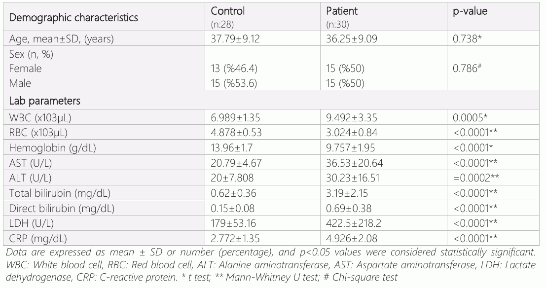
Demographic Characteristics and Laboratory Findings of SCD and Healthy Individuals The demographics and laboratory findings of healthy subjects and patients with SCD who participated in the study are given in Table 1. Of the 30 patients included in the study comprised 15 (50%) females and 15 (50%) males with a mean age of 36.25±9.09 years while 28 healthy individuals who matched the patients in terms of age and gender enrolled in the control group, and no significant difference in age and gender were determined between these groups (p>0.05). Our laboratory results showed that levels of leukocyte, AST, ALT, total bilirubin, direct bilirubin, LDH, and CRP were significantly higher in the patient group compared to the control group (p<0.05), whereas erythrocyte count and hemoglobin concentrations were significantly decreased in patients compared with healthy individuals.
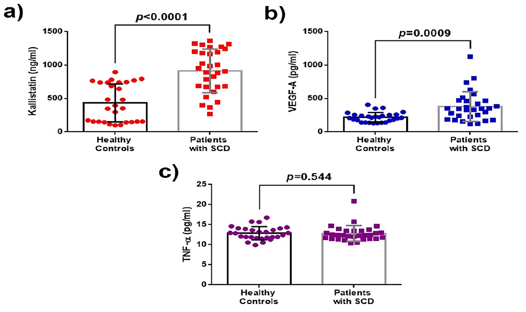
Kallistatin, VEGF-A and TNF-α Levels in Patients with SCD and Healthy Subjects We presented the results of the ELISA parameters in Figure 1. The measurements of Kallistatin, VEGF-A, and TNF-α from the serum samples of patients with SCD and healthy individuals demonstrated that the levels of Kallistatin and VEGF-A were increased in the patients with SCD (Figure 1a, 1b, p<0.05) while there was significantly no difference in TNF-α levels between the groups (Figure 1c, p>0.05). The parameters of ELISA are demonstrated in table 3. Serum levels of kallistatin and VEGF-A were significantly increased in SCD patients compared to healthy controls.
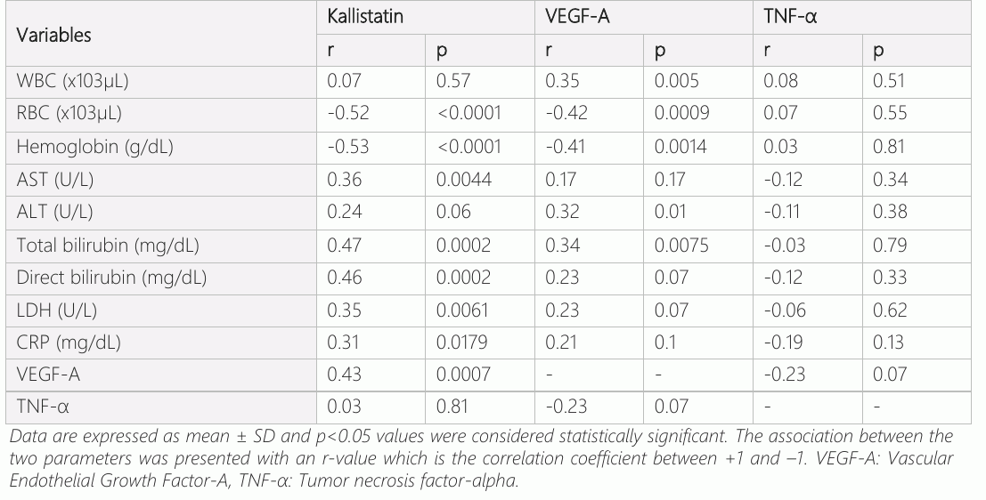
Correlations of Kallistatin, VEGF-A, and TNF-α with Routine Laboratory Parameters in Controls and Patients with SCD We performed correlation analyses to detect the association of Kallistatin, VEGF-A, and TNF-α with routine laboratory parameters in controls and patients with SCD and presented the results in Table 2. Our analyses revealed that serum Kallistatin levels were positively correlated with AST, total bilirubin, direct bilirubin, LDH, CRP, VEGF-A, but it was negatively correlated with RBC and hemoglobin (p<0.05). In addition, correlation analyses of VEGF-A unveiled that VEGF-A levels were positively correlated with WBC, ALT, total bilirubin, while correlated with RBC and hemoglobin negatively. Our results revealed that TNF-α levels were not significantly associated with any laboratory parameters.

ROC Analyses of Kallistatin and VEGF-A
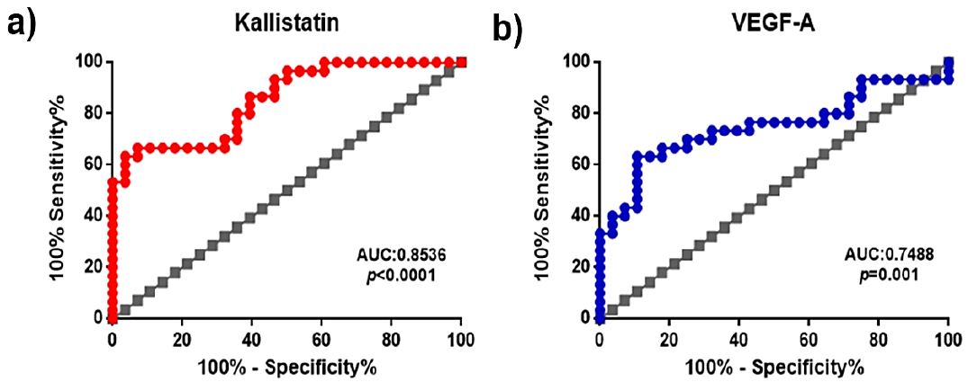
We performed ROC to detect the diagnostic value of serum Kallistatin, VEGF-A, and TNF-a in SCD and showed the results in Table 3 and Figure 2. Our results revealed that the areas under the receiver operating characteristic for Kallistatin and VEGF-A were 0.8536 and 0.7488, respectively (p<0.05). These data suggest that Kallistatin and VEGF-A might have diagnostic importance for patients with SCD.
Discussion
Here, we showed that Kallistatin elevated in SCD, and increased Kallistatin levels were positively correlated with VEGF-A. Angiogenesis involves new blood vessel growth or neo vascularization and plays a key role in vascular-mediated tissue damage in SCD (5, 8). There are some inconsistent data about VEGF levels in patients with SCD. Although increased levels of VEGF were found in previous clinical studies in steady-state SCD patients (5, 9, 10), some studies reported no significant difference in levels of VEGF in SCD patients versus healthy subjects (11, 12). Our results revealed in the current study that VEGF concentrations were significantly elevated in steady-state SCD patients.
Recent studies revealed that Kallistatin levels are elevated in some diseases. For instance; Serum Kallistatin concentrations have been reported to be elevated in patients with type 1 diabetes (13). In another study, McBride et al. (14) indicated elevated serum Kallistatin concentrations in type 2 diabetic patients. Moreover, it has been shown that Kallistatin concentrations are elevated in patients with obesity and prediabetes (15) and patients with rheumatoid arthritis (16). In the current study, significantly higher levels of kallistatin in asymptomatic SCD patients versus healthy individuals were observed.
Endothelial progenitor cells (EPCs), which can differentiate into endothelial cells play a central role in vascular repair by involving in endothelial regeneration and vessel formation (17). Studies have inconsistent findings regarding changes in circulating EPC in SCD patients. van Beem et al. reported that there is no difference in circulating EPCs between asymptomatic state patients and healthy subjects, but EPCs were significantly increased in SCD with painful crises (18). On the other hand, Anjum et al. (19) found significantly lower subsets of circulating EPCs in SCD with pulmonary arterial hypertension (PAH) than in those with no PAH. Moreover, Rodrigo et al. (20) reported that the number of circulating EPCs trended lower in adults with SCD than healthy African-American controls. Kallistatin encourages neovascularization by enhancing VEGF in EPCs (6, 7, 21). Gao et al. examined the role of kallistatin on blood EPCs in a vascular injury model of rats, and their results revealed that Kallistatin improves the vasculogenic capacity, mobility, and viability of EPCs through increasing VEGF, suggesting that Kallistatin may have a protective role on vascular injury (22).
In our study, Kallistatin and VEGF-A raised significantly in steady-state SCD patients, and a positive correlation was observed between Kallistatin and VEGF-A. We speculated that kallistatin via its active site increased VEGF levels in steady-state SCD patients, thus suggesting it could be involved in angiogenesis by affecting EPCs. We also tested the relationship of Kallistatin and VEGF-A with routine laboratory parameters in SCD using correlation analyses. Our findings unveiled that Kallistatin and VEGF- A concentrations were correlated with some biochemical parameters in patients with SCD, suggesting that Kallistatin and VEGF-A could be associated with the pathogenesis of SCD. Moreover, as shown in Figure 2 and Table 3, we reveal that Kallistatin and VEGF-A have diagnostic values that may improve the clinical therapy of patients with SCD.
Biochemical and hemogram parameters are routine tests used in the diagnosis and management of SCD (23–25). Previous studies unveiled that WBC, AST, ALT, total and direct bilirubin, and LDH levels were significantly elevated while hemoglobin and RBC were diminished considerably in SCD compared with healthy controls (23–25). Furthermore, recent studies reported that CRP concentrations were higher in SCD patients than healthy controls (24, 26). In our study, we detected that the levels of WBC, AST, ALT, total and direct bilirubin, LDH, and CRP levels were significantly increased while RBC and hemoglobin levels were significantly diminished in SCD versus controls. Our laboratory findings provided here are consistent with previous studies in the literature (23–26).
TNF-α, as an inflammatory marker, has been investigated in some studies comparing steady-state SCD patients compared with healthy subjects. Some studies reported that there is no significant difference in TNF-α levels between steady-state SCD patients and controls (27–29). In contrast, Other studies revealed that the levels of TNF-α in steady-state SCD patients are higher than in healthy individuals (9, 24, 30, 31). In our study, we did not find any difference in TNF-α levels in SCD patients compared with healthy individuals.
The current study has some limitations. Firstly, the sample size of the study was small because of the single-center design of the study. Secondly, no patients were experiencing a crisis and complications to compare the results.
Conclusion
Our results presented here show that Kallistatin could be a novel marker associated with the pathogenesis of SCD, and there are also many fields to explore about Kallistatin in SCD.
Declarations
- Author contributions (CRediT)
- All authors contributed equally to all 14 CRediT roles (Conceptualization, Data curation, Formal analysis, Funding acquisition, Investigation, Methodology, Project administration, Resources, Software, Supervision, Validation, Visualization, Writing – original draft, Writing – review & editing).
- Funding
-
Funding information was not reported in the published version of this article.
- Conflicts of interest
- The authors report no conflict of interests.
- Ethics approval
- The current study was approved by the Institutional Ethics Committee of Hatay Mustafa Kemal University and was conducted following the Declaration of Helsinki (2020/101).
- Data availability
- A data availability statement was not reported in the published version of this article.
References (31)
30 of 31 references are linked to their DOI.
- Lu M, Rab MA, Shevkoplyas SS, Sheehan VA. Blood rheology biomarkers in sickle cell disease. Experimental biology and medicine. 2020;245(2):155-65 DOI Crossref PubMed Google Scholar
- Kato GJ, Piel FB, Reid CD, Gaston MH, Ohene-Frempong K, Krishnamurti L, et al. Sickle cell disease. Nature reviews Disease primers. 2018;4:18010 DOI Crossref PubMed Google Scholar
- Stuart MJ, Nagel RL. Sickle-cell disease. Lancet. 2004;364 (9442):1343-60 DOI Crossref PubMed Google Scholar
- Conran N, Belcher JD. Inflammation in sickle cell disease. Clinical hemorheology and microcirculation. 2018;68 (2-3): 263-99 DOI Crossref PubMed Google Scholar
- Antwi-Boasiako C, Frimpong E, Gyan B, Kyei-Baafour E, Sey F, Dzudzor B, et al. Elevated Proangiogenic Markers are Associated with Vascular Complications within Ghanaian Sickle Cell Disease Patients. Medical sciences. 2018;6(3) DOI Crossref PubMed Google Scholar
- Chao J, Li P, Chao L. Kallistatin: double-edged role in angiogenesis, apoptosis and oxidative stress. Biological chemistry. 2017;398(12):1309-17 DOI Crossref PubMed Google Scholar
- Chao J, Bledsoe G, Chao L. Protective Role of Kallistatin in Vascular and Organ Injury. Hypertension. 2016;68(3):533-41 DOI Crossref PubMed Google Scholar
- Al-Habboubi HH, Mahdi N, Abu-Hijleh TM, Abu-Hijleh FM, Sater MS, Almawi WY. The relation of vascular endothelial growth factor (VEGF) gene polymorphisms on VEGF levels and the risk of vasoocclusive crisis in sickle cell disease. European journal of haematology. 2012;89(5):403-9 DOI Crossref PubMed Google Scholar
- Niu X, Nouraie M, Campbell A, Rana S, Minniti CP, Sable C, et al. Angiogenic and inflammatory markers of cardiopulmonary changes in children and adolescents with sickle cell disease. PloS one. 2009;4(11):e7956 DOI Crossref PubMed Google Scholar
- Mohan JS, Lip PL, Blann AD, Bareford D, Lip GY. The angiopoietin/Tie-2 system in proliferative sickle retinopathy: relation to vascular endothelial growth factor, its soluble receptor Flt-1 and von Willebrand factor, and to the effects of laser treatment. The British journal of ophthalmology. 2005;89(7):815-9 DOI Crossref PubMed Google Scholar
- Gurkan E, Tanriverdi K, Baslamisli F. Clinical relevance of vascular endothelial growth factor levels in sickle cell disease. Annals of hematology. 2005;84(2):71-5 DOI Crossref PubMed Google Scholar
- Duits AJ, Rodriguez T, Schnog JJ, Group CS. Serum levels of angiogenic factors indicate a pro-angiogenic state in adults with sickle cell disease. British journal of haematology. 2006;134(1):116-9 DOI Crossref PubMed Google Scholar
- Jenkins AJ, McBride JD, Januszewski AS, Karschimkus CS, Zhang B, O'Neal DN, et al. Increased serum kallistatin levels in type 1 diabetes patients with vascular complications. Journal of angiogenesis research. 2010;2:19 227 77 Kallistatin Levels at the Pathogenesis of SCD Doğan et al. DOI Crossref PubMed Google Scholar
- McBride JD, Jenkins AJ, Liu X, Zhang B, Lee K, Berry WL, et al. Elevated circulation levels of an antiangiogenic SERPIN in patients with diabetic microvascular complications impair wound healing through suppression of Wnt signaling. The Journal of investigative dermatology. 2014;134(6):1725-34 DOI Crossref PubMed Google Scholar
- Gateva A, Assyov Y, Velikova T, Kamenov Z. Increased kallistatin levels in patients with obesity and prediabetes compared to normal glucose tolerance. Endocrine research. 2017;42(2):163-8 DOI Crossref PubMed Google Scholar
- Wang CR, Chen SY, Shiau AL, Wu CL, Jou IM, Chao L, et al. Upregulation of kallistatin expression in rheumatoid joints. The Journal of rheumatology. 2007;34(11):2171-6 PubMed Google Scholar
- Umemura T, Higashi Y. Endothelial progenitor cells: therapeutic target for cardiovascular diseases. Journal of pharmacological sciences. 2008;108(1):1-6 DOI Crossref PubMed Google Scholar
- van Beem RT, Nur E, Zwaginga JJ, Landburg PP, van Beers EJ, Duits AJ, et al. Elevated endothelial progenitor cells during painful sickle cell crisis. Experimental hematology. 2009;37(9): 1054-9 DOI Crossref PubMed Google Scholar
- Anjum F, Lazar J, Zein J, Jamaleddine G, Demetis S, Wadgaonkar R. Characterization of altered patterns of endothelial progenitor cells in sickle cell disease related pulmonary arterial hypertension. Pulmonary circulation. 2012;2(1):54-60 DOI Crossref PubMed Google Scholar
- Rodrigo M, Mendelsohn L, Bereal-Williams C, Hunter L, Dalby CK, McGowan V, 2nd, et al. Circulating endothelial progenitor cells in adults with sickle cell disease. Pulmonary circulation. 2013;3(2):448-9 DOI Crossref PubMed Google Scholar
- Shen B, Smith RS, Jr., Hsu YT, Chao L, Chao J. Kruppel-like factor 4 is a novel mediator of Kallistatin in inhibiting endothelial inflammation via increased endothelial nitric-oxide synthase expression. The Journal of biological chemistry. 2009;284(51):35471-8 DOI Crossref PubMed Google Scholar
- Gao L, Li P, Zhang J, Hagiwara M, Shen B, Bledsoe G, et al. Novel role of kallistatin in vascular repair by promoting mobility, viability, and function of endothelial progenitor cells. Journal of the American Heart Association. 2014;3(5):e001194 DOI Crossref PubMed Google Scholar
- Meher S, Patel S, Das K, Dehury S, Jit BP, Maske MM, et al. Association of plasma homocysteine level with vaso-occlusive crisis in sickle cell anemia patients of Odisha, India. Annals of hematology. 2019;98(10): 2257-65 DOI Crossref PubMed Google Scholar
- Iwalokun BA, Iwalokun SO, Hodonu SO, Aina OA, Omilabu S. A study on the association between parvovirus B19 infection, serum tumour necrosis factor and C-reactive protein levels among Nigerian patients with sickle cell anaemia. Singapore medical journal. 2012;53(11):726-31 DOI Crossref PubMed Google Scholar
- Keikhaei B, Mohseni AR, Norouzirad R, Alinejadi M, Ghanbari S, Shiravi F, et al. Altered levels of pro-inflammatory cytokines in sickle cell disease patients during vaso-occlusive crises and the steady state condition. European cytokine network. 2013;24(1):45-52 DOI Crossref PubMed Google Scholar
- Toledo SLO, Guedes JVM, Alpoim PN, Rios DRA, Pinheiro MB. Sickle cell disease: Hemostatic and inflammatory changes, and their interrelation. Clinica chimica acta; international journal of clinical chemistry. 2019;493:129-37 DOI Crossref PubMed Google Scholar
- Cruz PR, Lira RP, Pereira Filho SA, Souza BB, Mitsuushi FN, Menaa F, et al. Increased circulating PEDF and low sICAM-1 are associated with sickle cell retinopathy. Blood cells, molecules & diseases. 2015;54(1):33-7 DOI Crossref PubMed Google Scholar
- Pathare A, Al Kindi S, Alnaqdy AA, Daar S, Knox-Macaulay H, Dennison D. Cytokine profile of sickle cell disease in Oman. American journal of hematology. 2004;77(4):323-8 DOI Crossref PubMed Google Scholar
- Alagbe AE, Olaniyi JA, Aworanti OW. Adult Sickle Cell Anaemia Patients in Bone Pain Crisis have Elevated Pro-Inflammatory Cytokines. Mediterranean journal of hematology and infectious diseases. 2018;10(1):e2018017 DOI Crossref PubMed Google Scholar
- Qari MH, Dier U, Mousa SA. Biomarkers of inflammation, growth factor, and coagulation activation in patients with sickle cell disease. Clinical and applied thrombosis/ hemostasis: official journal of the International Academy of Clinical and Applied Thrombosis/Hemostasis. 2012;18(2):195- DOI Crossref PubMed Google Scholar
- Bandeira IC, Rocha LB, Barbosa MC, Elias DB, Querioz JA, Freitas MV, et al. Chronic inflammatory state in sickle cell anemia patients is associated with HBB(*)S haplotype. Cytokine. 2014;65(2):217-21 DOI Crossref PubMed Google Scholar
Respond to this article
Comments on a published article are welcome as a Letter to the Editor.
Get an e-mail when new articles in your fields are published
Questions to the authors
Questions are moderated: an editor approves each question, the authors answer, and an editor approves the answer before both appear here.
No answered questions yet.
Ask the authors a question
Discussion
Comments are moderated: they appear after the writer confirms the e-mail address and an editor approves them. Each comment carries a conflict-of-interest statement.
No comments yet.
Write a comment
Read the full text
Open the PDF here
Loading the PDF…
If the PDF does not show here, use the download button above.
How to cite this article
Dogan S, Okuyan HM, Ilhan G. Serum Kallistatin Levels are Associated with the Pathogenesis of Sickle Cell Disease. Ulutas Med J. 2021;7(4):220-227. doi:10.5455/umj.20211101020302
Dogan, S., Okuyan, H. M., & Ilhan, G. (2021). Serum Kallistatin Levels are Associated with the Pathogenesis of Sickle Cell Disease. The Ulutas Medical Journal, 7(4), 220–227. https://doi.org/10.5455/umj.20211101020302
Dogan, S., Okuyan, H.M. and Ilhan, G. (2021) 'Serum Kallistatin Levels are Associated with the Pathogenesis of Sickle Cell Disease', The Ulutas Medical Journal, 7(4), pp. 220–227. doi: 10.5455/umj.20211101020302.
@article{umj137221,
  title = {Serum Kallistatin Levels are Associated with the Pathogenesis of Sickle Cell Disease},
  author = {Dogan, Serdar and Okuyan, Hamza Malik and Ilhan, Gul},
  journal = {The Ulutas Medical Journal},
  year = {2021},
  volume = {7},
  number = {4},
  pages = {220--227},
  doi = {10.5455/umj.20211101020302},
  url = {https://www.ulutasmedicaljournal.com/?mno=137221},
  issn = {2149-388X}
}