The Ulutas Medical Journal · ISSN 2149-0430 (print), 2149-388X (online) · https://www.ulutasmedicaljournal.com/?mno=214641
Case Report · Open access
A Rarely Seen Hepatic Tumor: Epithelioid Hemangioendothelioma
Also available in: العربية · Türkçe · 中文
Abstract
- Introduction
- Epithelioid Hemangioendothelioma (EHE) is a rarely seen tumor most frequently originating from vascular structures of different regions including liver, followed by stomach, spleen, lung, bone, soft tissue, and skin.
- Case Presentation
- In this paper, a patient with multiple mass lesions in the liver with a diagnosis of EHE who underwent liver transplantation is presented.
- Conclusion
- It has an intermediate degree of malignancy between benign hemangioma, and malignant hemangiosarcoma. It can manifest with nonspecific symptoms or hepatic failure. Generally it is seen as a diffuse hepatic disease. The most optimal treatment alternatives are liver resection, and transplantation.
Keywords
Full text
Introduction
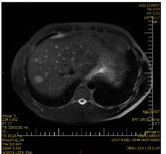
A 35-year-old male patient consulted to us in January 2015 with complaints of abdominal pain, and lassititude. His physical examination revealed subicteric scleras, and on deep abdominal palpation tenderness at the right upper quadrant was detected. Rebound tenderness, and abdominal guarding were not detected. Presence of abdominal pain, and right upper abdominal quadrant tenderness on palpation required abdominopelvic ultrasonographic (US) examination. On US, hypoechoic solid lesions on hepatic segments 2 (8.5 mm), and 7 (12x10 mm) were observed. His hemogram, and biochemical examinations did not reveal any abnormality. For better identification of the hepatic lesion upper abdominal dynamic magnetic resonance (MR) images were obtained. On MR multiple T2 hyperintense, and T1 hypointense nodules in the liver parenchyma, the biggest one measuring 18x16 mm at segment 7 were detected. On dynamic phase series, peripheral diffuse contrast uptake was seen, and washout was not observed in late-phase series. In some of these lesions non-stained areas possibly consistent predominantly with central necrosis were noted (Figure-1). Radiologist interpreted the MR images in favour of metastasis. Because of suspect hepatic metastasis, measurement of hepatitis biomarkers, AFP, CA 19-9, and CEA levels were requested. Biomarkers of hepatitis, and tumor were within normal limits. Chest X-ray, thoracal computed tomography (CT), scrotal US, endoscopic, and colonoscopic examina-tions were requested from the patient in order to search for the primary focus. On scrotal US of the patient whose chest X-Ray, and thoracal CT were unremarkable, slightly varicose veins of 3.5 mm in their largest diameter were detected. Grade-A esophagitis was detected on his endoscopic examination, while his colonoscopy did not reveal any abnormality.
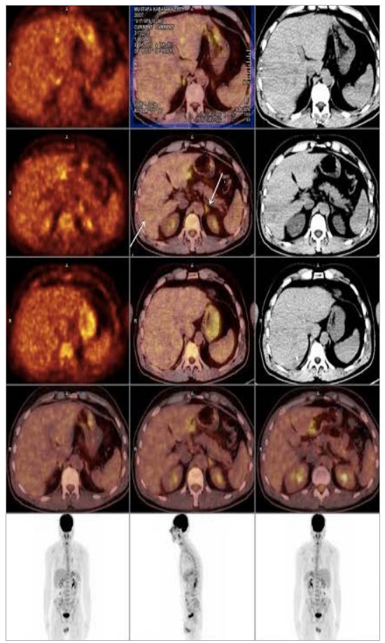
On positron emission tomograms (PET-CT) obtained so as to detect the primary focus, increased FDG uptake suggesting malignancy was detected on the left supraclavicular (SUVmax:4.4), left inferior jugular (SUVmax: 4.6), and in the mediastinum prevasküler (SUVmax: 3.9), right inferior paratracheal (SUVmax: 4.9), left inferior paratracheal (SUVmax:4),right hilar (SUVmax:3.5), left hilar(SUVmax: 5.4) and subcarinal (SUVmax: 5.7) lymph nodes (SUVmax: 5.7, metastasis)(Figure-2).
The liver lesions of the patient were biopsized under the guidance of US. Immunohistochemical examination of the biopsy material disclosed CD34, and vimentin positivity, while pancytokeratine, hepatocyte, CEA, CK7, CK20, TTF-1, CDX-2, S-100, DOG-1, and actin negativity. As a result of immunohistochemical staining the diagnosis of the patient was reported as epithelioid hemangioendothelioma.
The patient with multiple mass lesions who was diagnosed as epithelioid hemangioendothelioma underwent liver transplantation from a live donor in June 2015. Posttrans-plantation follow-up period of the patient is uneventful.
Discussion
Epithelioid hemangioendothelioma is a rarely seen tumor originating from vascular structure of different regions including stomach, spleen, liver, lungs, bone, soft tissue, and skin. It has an intermediate degree of malignancy between benign hemangioma, and malignant hemangiosarcoma (4). Most frequently liver is involved (5). In our patient also hepatic involvement was etected.
The patients most frequently present with symptoms, and signs of abdominal pain, weight loss, ascites, nausea, jundice, hepatic failure, hepatic vein thrombosis, and portal hypertension (6). In our patient, abdominal pain, and jaundice were detected. Imaging modalities is very important in the diagnosis of EHE. On ultrasonographic examination it is frequently (66.3%) seen as a hypoechoic, and occasionally (22.5%) as a heterogenous lesion. On non-contrasted CT, they are seen as hypodense, non-homegenous solid nodules extending into the capsule frequently with a peripheral location. After injection of the contrast material a halo sign formed by a central scar, and a peripheral cell proliferation at the portal venous phase become manifest. On MR, they are seen as non-homogeneous hypointense at T1, and non-homogeneous hyperintense images at T2 sequences (7). On MR of our patient multiple number of nodular lesions in the parenchymal tissue the biggest one with the dimensions of 18x16 mm were observed. These nodules were visualized as hypertense lesions at T2, and hypotense lesions at T1 sequences together with peripheral diffuse contrast uptake in dynamic phase sequences.
For definitive diagnosis histopathological examination is required. On microscopic examination it is very difficult to differentiate these lesions from cholangiocarcinoma, fibrolamellar hepatocellular carcinoma, sarcoma, and metastatic tumors. Definitive diagnosis can be made by confirmation of endothelial cells using immunohistochemical methods (factor VIII, CD34, and CD31 tracers). Presence of increased levels of endothelial cell markers in dendritic and/or epitheloid cells confirms diagnosis (8). Immunohistochemical analysis performed in our patient disclosed CD34, and vimentin positivity.
As a treatment, various methods can be tried based on the location of the tumor in the liver. Among these methods, liver resection, and transplantation are the predominant treatment modalities (9). In EHE cases with diffuse, and unresectable hepatic involvement, even in the presence of distant metastases, liver transplantation is the most suitable treatment modality. (10–12). Also in our case, due to diffuse hepatic involvement, as a treatment modality liver transplantation was preferred.
In the study where 71 patients did not undergo any treatment, while other patients were treated with liver transplantation (n:128), chemotherapy and/or radiotherapy (n:60), and hepatic resection (n:27). In this study, 1-year and 5-year survival rates following liver transplantation were reported as 96, and 54.5%, while in the groups of chemotherapy radiotherapy, and liver resection, corresponding 1-year and 5-year-survival rates were 73.3, and 30%, and 75%, respectively (3).
In conclusion, when multiple liver mass lesions are encountered, EHE should be absolutely considered in the differential diagnosis
Declarations
- Author contributions (CRediT)
- All authors contributed equally to all 14 CRediT roles (Conceptualization, Data curation, Formal analysis, Funding acquisition, Investigation, Methodology, Project administration, Resources, Software, Supervision, Validation, Visualization, Writing – original draft, Writing – review & editing).
- Funding
-
Funding information was not reported in the published version of this article.
- Conflicts of interest
- A conflict of interest statement was not reported in the published version of this article.
- Ethics approval
- Ethics approval details were not reported in the published version of this article.
- Data availability
- A data availability statement was not reported in the published version of this article.
References (12)
8 of 12 references are linked to their DOI.
- Dong K, Wnag X, Feng JL et al. Pathological characteristics of liver biopsies in eight patients with hepatic epithelioid hemangio endothelioma. Int J Clin Exp Pathol 2015;8(9): 11015-23. PubMed Google Scholar
- Pregliasco AG, Wendum D, Goumard C et al. Hepatic epithelioid hemangioendothelioma. Clin Res Hepatol Gastroenterol 2015 Nov PubMed Google Scholar
- pii: S2210-7401(15)00254-5. 3. Mehrabi A, Kashfi A, Fonouni H et al. Primary malignant hepatic epithelioid hemangioendothelioma: A comprehensive rewiew of the literature with emphasis on the surgical therapy. Cancer 2006;107:2108-21. DOI Crossref PubMed Google Scholar
- Weiss SW, Enzinger FM. Epithelioid hemangioendothelioma: A vascular tumor often mistaken for a carcinoma. Cancer 1982;50:970-81. DOI Crossref PubMed Google Scholar
- Sangro B, Inaarrairaegui M, Fernandez-Ros N. Malignant epitheloid hemangioendothelioma of the liver successfully treated with sorafenib. Rare Tumors 2012;4:e34. DOI Crossref PubMed Google Scholar
- Caruso S, Miraglia R, Maruzelli L et al. An unusual presentation of malignant hepatic epithelioid hemangioendothelioma with left pleural and pulmonary localization. Pediatr Radiol 2008;38:1027-30 DOI Crossref PubMed Google Scholar
- Ji ZF, Shen XZ, Zhang DY et al. Hepatic epithelioid hemangio endothelioma: a report of six patients. J Dig Dis 2010;11:254-8. DOI Crossref PubMed Google Scholar
- Tsuneyoshi M, Dorfman HD, Bauer TW. Epithelioid hemangio endothelioms of bone: a clinicopathologic, ultrastructural, and immunohistochemical study. Am J Surg Pathol 1986;10:754-64. PubMed Google Scholar
- Langrehr JM, Petersen I, Pfitzmann R, Lopez-Hanninen E. Malignant epithelioid hemangioendothelioma of the liver. Result of surgical treatment strategies. Chirurg 2005;76:1161-7. PubMed Google Scholar
- Lerut JP, Orlando G, Sempoux C et al. Hepatic hemangio endothelioma in adults: Excellent outcome following liver transplantation. Transpl Int 2004;17:202-07. DOI Crossref PubMed Google Scholar
- Ben-Haim M, Roayaise S, Ye MQ et al. Hepatic epithelioid hemangioendothelioma: Resection or transplantation, which and when? Liver Transpl Surg 1999;5:526-31. DOI Crossref PubMed Google Scholar
- Lerut JP, Orlando G, Adam R. The place of liver transplantation in the treatment of hepatic epitheloid hemangioendothelioma: report of the European liver transplant registry. Ann Surg. 2007 Dec;246(6):949-57. DOI Crossref PubMed Google Scholar
Respond to this article
Comments on a published article are welcome as a Letter to the Editor.
Get an e-mail when new articles in your fields are published
Questions to the authors
Questions are moderated: an editor approves each question, the authors answer, and an editor approves the answer before both appear here.
No answered questions yet.
Ask the authors a question
Discussion
Comments are moderated: they appear after the writer confirms the e-mail address and an editor approves them. Each comment carries a conflict-of-interest statement.
No comments yet.
Write a comment
Tell me when this article is cited
Leave your e-mail address and confirm it. These notices are not being sent yet; your confirmed address is kept for them and used for nothing else.
Read the full text
Open the PDF here
Loading the PDF…
If the PDF does not show here, use the download button above.
How to cite this article
Nayir E. A Rarely Seen Hepatic Tumor: Epithelioid Hemangioendothelioma. Ulutas Med J. 2016;2(1):59-62. doi:10.5455/umj.20160121062843
Nayir, E. (2016). A Rarely Seen Hepatic Tumor: Epithelioid Hemangioendothelioma. The Ulutas Medical Journal, 2(1), 59–62. https://doi.org/10.5455/umj.20160121062843
Nayir, E. (2016) 'A Rarely Seen Hepatic Tumor: Epithelioid Hemangioendothelioma', The Ulutas Medical Journal, 2(1), pp. 59–62. doi: 10.5455/umj.20160121062843.
@article{umj214641,
  title = {A Rarely Seen Hepatic Tumor: Epithelioid Hemangioendothelioma},
  author = {Nayir, Erdinc},
  journal = {The Ulutas Medical Journal},
  year = {2016},
  volume = {2},
  number = {1},
  pages = {59--62},
  doi = {10.5455/umj.20160121062843},
  url = {https://www.ulutasmedicaljournal.com/?mno=214641},
  issn = {2149-388X}
}