The Ulutas Medical Journal · ISSN 2149-0430 (印刷版), 2149-388X (在线版) · https://www.ulutasmedicaljournal.com/?mno=214641
病例报告 · 开放获取
罕见的肝肿瘤:Epithelioid Hemangioendothelioma
标题、摘要和全文为机器翻译。 英文原文
- Department of Medical Oncology, Kahramanmaras Necip Fazil City Hospital, Kahramanmaras, Turkey ROR
- 开放获取
- CC BY-NC 4.0
- ORCID
- 资助
- 伦理批准
- 数据可用性
摘要
导言:Epithelioid Hemangioendothelioma(EHE)是一种罕见的肿瘤,最常发源于肝脏等不同地区的血管结构,其次是胃,脾,肺,骨,软组织,以及皮肤. 案例介绍 : 在本文中,介绍一名肝脏有多重质量损伤的患者,他诊断了接受肝脏移植的EHE. 结论: 它的恶性程度介于良性肝脏瘤和恶性肝脏瘤之间。 它可以表现为非特定症状或肝衰竭. 一般认为这是一种扩散性肝病。 最佳的治疗办法是肝脏重新剖析和移植.
关键词
全文
全文为机器翻译;以英文原文为准。 英文原文
导言
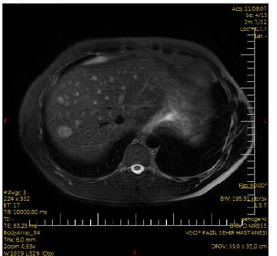
2015年1月,一名35岁的男性患者向我们咨询了腹痛,以及低血压的抱怨. 他的体格检查显示有潜质的scleras,在右上方四角的深腹部微弱处被检测到. 复温柔,腹部卫视未被发现. 腹部疼痛的存在以及右上腹部四肢在发作时的柔软,需要进行腹部多米诺佩维奇超声波检查(美国)。 在美国,观察到肝部分2(8.5毫米)和7(12x10毫米)的低血糖固体损伤。 他的外观和生化检查没有发现任何异常。 为了更好地识别肝损伤上腹部动态磁共振(MR)图像。 在MR的多个 T上2个 高强度和 T页:1 在肝脏间隙中检测到低强度结核,这是第7区最大的18x16毫米结核。 在动态相序上,可以看到外围的散射对比吸收,在后期相序中未观察到冲洗。 在其中一些可能主要与中央坏死相一致的无染病区(图1)。 放射学家诠释MR图像时倾向于元化. 由于疑似肝病,要求测量肝炎生物标记、AFP、CA 19-9和CEA水平。 肝炎和肿瘤的生物标记在正常范围内。 要求病人进行胸X射线、胸腔计算成型摄影、美国眼球、内视和视线检查,以便寻找主要重点。 在胸部X射线和胸腔CT不显眼的病人的血清上,检测到其最大直径为3.5毫米的细血管。 他的眼内膜检查中发现了A级麻黄炎,而他的结肠镜检查没有发现任何异常。
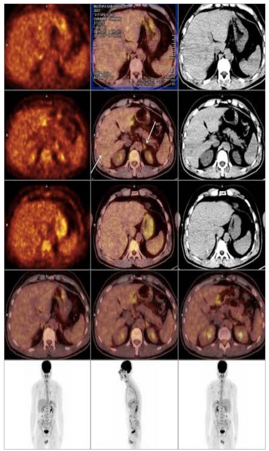
在为探测主要焦点而获得的正电子排放直方图(PET-CT)上,左侧超圆锥体(SUV)检测到的FDG摄入量增加,表明有恶性。最大数:4.4),左下角(SUVmax:4.6),和介于介于介于介于介于介于介于介于介于介于介于介于介于介于介于介于介于介于介于介于介于介于介于介于介于介于介于介于介于介于介于介于介于介于介于介于介于介于介于介于介于介于介于介于介于介于介于介于介于介于介于介于介于介于介于介于介于介于介于介于介于介于介于介于介于介于介于介于介于介于介于介于介于介于介于介于介于介于介于介于介于介于介于介于介于介于介于介于介于介于介于介于介于介于介于介于介于介于介于介于介于介于介于介于介于介于介于介于介于最大数: 3.9),右低级伞兵(SUV)最大数: 4.9),左下低等伞兵(SUVmax:4),右尖(SUV)最大数:3.5),左海拉尔(SUV)最大数: 5.4)和次生(SUV)最大数: 5.7)淋巴结(SUV)最大数: 5.7,元数据(图2).
患者的肝脏损伤在美国的指导下被生物放大. 对活检材料的免疫组织化学检查揭示了CD34和vimentin position,而pancytokeratine、肝细胞、CEA、CK7、CK20、TTF-1、CDX-2、S-100、DOG-1和actin negative。 由于免疫组织化学污染,患者的诊断结果被报告为上位素异构(hemangioendothelioma).
被诊断为上位素血栓瘤(Hemangioendothelioma)的多宗大规模损伤患者于2015年6月接受了一名活体捐献者的肝脏移植. 患者移植后后续期不均匀.
讨论情况
Epithelioid hemangioendothelioma是一种罕见的肿瘤,来源于不同区域的血管结构,包括胃,脾,肝,肺,骨,软组织,以及皮肤. 在良性肝脏瘤和恶性肝脏瘤之间 有中等程度的恶性 (4)最经常涉及肝脏 (5)在我们的病人中 也有肝脏的感染
最常出现症状的病人有腹痛、体重下降、哮喘、恶心、复数、肝衰竭、肝静脉血栓症和门户高血压的迹象。 (6)在我们的病人身上 发现了腹部疼痛和黄油 成像模式在EHE诊断中非常重要. 在超声波检查中,经常(66.3%)被视为低血糖,偶尔(22.5%)被视为异性损伤。 在无接触的CT上,它们被视为低温的、无原生的固体结核,经常以外围位置延伸到胶囊中。 在对对比物进行注射后,中央疤痕形成的光环标志,入口处的旁缘细胞增殖也变得明显. 在MR上,它们被视为T1的非同质下皮素,在T2序列中被视为非同质的超强度图像. (7)在我们的患者MR上,观察到 肠胃组织中多起鼻部损伤 最大的一次是18x16毫米 这些结核被视同T超强度损伤2个以及T的皮下损伤页:1 序列与动态相位序列中外围扩散的对比吸收。
确诊需要组织病理学检查。 在微观检查中,很难将这些损伤与胆固醇癌、纤维性肝细胞瘤、沙科马瘤和元静脉瘤区分开来。 确定性诊断可以通过使用免疫组织化学方法(因子VIII,CD34,和CD31跟踪)确认内皮细胞进行. 登革热和/或上皮细胞内皮细胞标记水平提高证实诊断 (8)在病人身上进行的免疫组织化学分析 揭示了CD34和vimentin正性
作为治疗方法,可以根据肿瘤在肝脏中的位置来尝试各种方法. 在这些方法中,肝脏切除和移植是主要的治疗方式。 (9)在EHE病例中,即使有遥远的元体,肝脏移植也是最合适的治疗方式。 (10–12)同样在我们的情况中,由于肝脏的分散参与,作为治疗方式的肝脏移植更可取。
在研究中,71名患者没有接受任何治疗,而其他患者则通过肝移植(n:128),化疗和/或放射治疗(n:60),以及肝分解(n:27)等方法治疗. 在这项研究中,肝脏移植后的1年和5年存活率分别是96和54.5%,而在化疗放射治疗和肝脏分解中,相应的1年和5年存活率分别为73.3和30%,75%。 (3)。 。 。
总而言之,当遇到多重肝脏质量损伤时,在区别诊断中绝对应考虑EHE
声明
- 作者贡献(CRediT)
- 所有作者对全部 14 项 CRediT 角色贡献均等(Conceptualization, Data curation, Formal analysis, Funding acquisition, Investigation, Methodology, Project administration, Resources, Software, Supervision, Validation, Visualization, Writing – original draft, Writing – review & editing)。
- 资助
-
Funding information was not reported in the published version of this article.
- 利益冲突
- A conflict of interest statement was not reported in the published version of this article.
- 伦理批准
- Ethics approval details were not reported in the published version of this article.
- 数据可用性
- A data availability statement was not reported in the published version of this article.
参考文献 (12)
12 条参考文献中有 7 条已链接 DOI。
- Dong K, Wnag X, Feng JL et al. Pathological characteristics of liver biopsies in eight patients with hepatic epithelioid hemangio endothelioma. Int J Clin Exp Pathol 2015;8(9): 11015-23. PubMed Google Scholar
- Pregliasco AG, Wendum D, Goumard C et al. Hepatic epithelioid hemangioendothelioma. Clin Res Hepatol Gastroenterol 2015 Nov PubMed Google Scholar
- pii: S2210-7401(15)00254-5. 3. Mehrabi A, Kashfi A, Fonouni H et al. Primary malignant hepatic epithelioid hemangioendothelioma: A comprehensive rewiew of the literature with emphasis on the surgical therapy. Cancer 2006;107:2108-21. DOI Crossref PubMed Google Scholar
- Weiss SW, Enzinger FM. Epithelioid hemangioendothelioma: A vascular tumor often mistaken for a carcinoma. Cancer 1982;50:970-81. DOI Crossref PubMed Google Scholar
- Sangro B, Inaarrairaegui M, Fernandez-Ros N. Malignant epitheloid hemangioendothelioma of the liver successfully treated with sorafenib. Rare Tumors 2012;4:e34. DOI Crossref PubMed Google Scholar
- Caruso S, Miraglia R, Maruzelli L et al. An unusual presentation of malignant hepatic epithelioid hemangioendothelioma with left pleural and pulmonary localization. Pediatr Radiol 2008;38:1027-30 DOI Crossref PubMed Google Scholar
- Ji ZF, Shen XZ, Zhang DY et al. Hepatic epithelioid hemangio endothelioma: a report of six patients. J Dig Dis 2010;11:254-8. PubMed Google Scholar
- Tsuneyoshi M, Dorfman HD, Bauer TW. Epithelioid hemangio endothelioms of bone: a clinicopathologic, ultrastructural, and immunohistochemical study. Am J Surg Pathol 1986;10:754-64. PubMed Google Scholar
- Langrehr JM, Petersen I, Pfitzmann R, Lopez-Hanninen E. Malignant epithelioid hemangioendothelioma of the liver. Result of surgical treatment strategies. Chirurg 2005;76:1161-7. PubMed Google Scholar
- Lerut JP, Orlando G, Sempoux C et al. Hepatic hemangio endothelioma in adults: Excellent outcome following liver transplantation. Transpl Int 2004;17:202-07. DOI Crossref PubMed Google Scholar
- Ben-Haim M, Roayaise S, Ye MQ et al. Hepatic epithelioid hemangioendothelioma: Resection or transplantation, which and when? Liver Transpl Surg 1999;5:526-31. DOI Crossref PubMed Google Scholar
- Lerut JP, Orlando G, Adam R. The place of liver transplantation in the treatment of hepatic epitheloid hemangioendothelioma: report of the European liver transplant registry. Ann Surg. 2007 Dec;246(6):949-57. DOI Crossref PubMed Google Scholar
阅读全文
在此打开 PDF
正在加载 PDF…
若 PDF 未在此显示,请使用上方的下载按钮。
如何引用本文
Nayir E. A Rarely Seen Hepatic Tumor: Epithelioid Hemangioendothelioma. Ulutas Med J. 2016;2(1):59-62. doi:10.5455/umj.20160121062843
Nayir, E. (2016). A Rarely Seen Hepatic Tumor: Epithelioid Hemangioendothelioma. The Ulutas Medical Journal, 2(1), 59–62. https://doi.org/10.5455/umj.20160121062843
Nayir, E. (2016) 'A Rarely Seen Hepatic Tumor: Epithelioid Hemangioendothelioma', The Ulutas Medical Journal, 2(1), pp. 59–62. doi: 10.5455/umj.20160121062843.