The Ulutas Medical Journal · ISSN 2149-0430 (print), 2149-388X (online) · https://www.ulutasmedicaljournal.com/?mno=221058
Case Report · Open access
Primary Hydatid Disease within the Quadratus Lumborum Muscle
- Istanbul Training and Research Hospital, Department of General Surgery ROR
- Haseki Training and Research Hospital, Department of General Surgery ROR
Abstract
- Introduction
- Hydatid disease is a parasitic infestation caused by tapeworms of the genus Echinococcus. It may be located anywhere in the human body. The most common organs involved are the liver (52-72%) and the lungs (10-40%). Hydatid disease can rarely develop in muscular structures, accounting for 1-5% of all cases.
- Case Presentation
- In this study, we report the case of a 53-year-old male patient who presented with a swelling on his left lumbar region and was diagnosed with hydatid disease within the quadratus lumborum muscle.
- Conclusion
- If any patient living in endemic countries as Turkey presents with a swelling in soft tissues and if he or she should be undergone investigations such as biopsy or cyst aspiration, then it will be mandatory to make a cautious physical examination and accurate imaging studies by keeping in mind the hydatid disease as a probable diagnosis.
Keywords
Full text
Introduction
Hydatid disease, also called Echinococcosis or hydatid cyst, is a parasitic infestation caused by larvae of the tapeworm Echinococcus. Echinococcus granulosus (E. granulosus) is the most common agent of the disease, whose definitive hosts are carnivores such as dogs, wolves and foxes. Herbivores such as sheep, goats, camels and horses act as intermediate hosts. In humans, following the intake by mouth, the larvae of the tapeworm penetrate the wall of small intestine and then reach the liver via the portal vein. From here, it can pass into the systemic circulation. Hydatid cysts may develop anywhere within the body, with the liver being the mostly involved organ (52-72%) followed by lungs (10-40%) (1). Hydatid cysts can seldom be located within muscles, accounting for 1-5% of all cases (2).
Case Presentation
A 53-year-old male patient presented to our outpatient clinic with a 1-year history of a swelling in his left lower back. His personal medical history and family history were unremarkable. Physical examination revealed an immobile, painless, and well-defined palpable mass in the lumbar region measuring approximately 15x10 cm. The indirect hemagglutination test (IHA) for E. granulosus was positive at 1/512 titration. The complete blood count and biochemical tests were normal. Ultrasonography (US), computed tomography (CT) and Magnetic resonan-ce imaging (MRI) showed a multiloculated, peripheral contrastenhancing cystic lesion of 14x13 mm in size located within the quadratus lumborum muscle suggesting hydatid disease (Figure-1). Based on these findings, the patient was operated on under general anesthesia. The cystic mass was accessed through a transverse incision made on the left lumbar region.
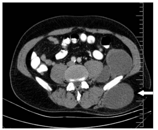
The cyst was aspirated after placement of saline moistened lap pads around it and a purulent fluid was obtained. The cyst wall was unroofed. Daughter vesicles were seen and a diagnosis of infected hydatid cyst was established. A sample of fluid was taken for culture and sensitivity. After content of the cyst was entirely emptied, the space was irrigated with hypertonic saline solution and 10% povidoneiodine. A partial excision of cyst wall was added and the operation was completed. Postoperative course was une-ventful. The culture result was negative and pathological examination was consistent with hydatid cyst.Albendazol treatment was started in early postoperative stage.
Discussion
Although hydatid disease is mostly located in the liver and the lungs, it can also rarely occur in muscular structures especially in the patients from endemic areas (3). Hydatid disease involving the muscles has a prevalance of 3-5% (4, 5) and it usually results from spontaneous or posttraumatic rupture of the hydatid cyst in distant part of the body as well as can occur secondarily to an operation for the hydatid cyst (6).
In diagnosis, US should be the first choice. On the other hand, in case of advanced disease, the relationship of the lesion with the surrounding tissues and adequate resection margins can be determined, and a differential diagnosis can be done by means of MRI (7). When positive the results of serological tests can be taken into account but in half of the cases of primary hydatid cyst of muscle they were reported to be false negative and the sensitivity of IHA was found 67% (8). Our patient presented with a palpable mass which was determined to be a cystic lesion by US and subsequently was found suggestive of hydatid disease by MRI. In addition, the IHA was positive at 1/512 titration by MRI and CT. Diagnostic biopsy or aspiration should be avoided because of risk of rupture and spreading of disease (9). Surgery is currently treatment of choice (10). Our case was also treated using surgery in which the infected cyst was partially excised and completely drained.
In conclusion, in endemic countries like Turkey, it must be remembered that any patient who presents with a swelling in soft tissues should be evaluated cautiously by physical examination and imaging studies in terms of hydatid disease. In addition, no effort should be spent for making biopsy or aspiration unless hydatid disease was ruled out.
Declarations
- Author contributions (CRediT)
- All authors contributed equally to all 14 CRediT roles (Conceptualization, Data curation, Formal analysis, Funding acquisition, Investigation, Methodology, Project administration, Resources, Software, Supervision, Validation, Visualization, Writing – original draft, Writing – review & editing).
- Funding
-
Funding information was not reported in the published version of this article.
- Conflicts of interest
- A conflict of interest statement was not reported in the published version of this article.
- Ethics approval
- Ethics approval details were not reported in the published version of this article.
- Data availability
- A data availability statement was not reported in the published version of this article.
References (10)
7 of 10 references are linked to their DOI.
- Sayek I, Tirnaksiz M. Cystic hydatid disease: Current trends in diagnosis and management. Surg today 2004; 34(8):987-96. DOI Crossref PubMed Google Scholar
- Digesu G, Picone A, La Bianca A, Massaro M, Vetri G. Muscular and subcutanuous hydatidosis. Minerva Med 1987;78(7):835-40. PubMed Google Scholar
- Eckert J, Deplazes P. Biological, epidemiological, and clinical aspects of echinococcosis, a zoonosis of increasing concern. Clin Microbiol Rev 2004;17:107–35. DOI Crossref PubMed Google Scholar
- Gougoulias NE, Varitimidis SE, Bargiotas KA, Dovas TN, Karydakis G, Dailiana ZH. Skeletal muscle hydatid cysts presenting as soft tissue masses. Hippokratia 2010;14:126– 30. PubMed Google Scholar
- García-Alvarez F, Torcal J, Salinas JC, Navarro A, García-Alvarez I, Navarro-Zorraquino M, et al. Musculoskeletal hydatid disease: A report of 13 cases. Acta Orthop Scand 2002;73:227– 31. DOI Crossref PubMed Google Scholar
- Guidelines for treatment of cystic and alveolar echinococcosis in humans. WHO Informal Working Group on Echinococcosis. Bull World Health Organ1996;74:231– 42. PubMed Google Scholar
- Memis A, Arkun R, Bilgen I, Ustun EE. Primary soft tissue hydatid disease: report of two cases with MRI characteristics. Eur Radiol 1999;9:1101– 3. DOI Crossref PubMed Google Scholar
- Durakbasa CU, Tireli GA, Sehiralti V, Sander S, Tosyali AN, Mutus M. An audit on pediatric hydatid disease of uncommon localization: incidence, diagnosis, surgical approach, and outcome. J Pediatr Surg 2006;41:1457–63. DOI Crossref PubMed Google Scholar
- Tatari H, Baran O, Sanlidag T, Gore O, Ak D, Manisalı M, et al. Primary intramuscular hydatidosis of supraspinatus muscle. Arch Orthop Trauma Surg 2001;121:93-4. DOI Crossref PubMed Google Scholar
- Yorganci K, Sayek I. Surgical treatment of hydatid cysts of the liver in the era of percutaneous treatment. Am J Surg 2002;184:63–9. DOI Crossref PubMed Google Scholar
Read the full text
Open the PDF here
Loading the PDF…
If the PDF does not show here, use the download button above.
How to cite this article
Ari A, Buyukasik K, Akce B, Tatar C, Vardar YM. Primary Hydatid Disease within the Quadratus Lumborum Muscle. Ulutas Med J. 2016;2(1):63-65. doi:10.5455/umj.20160317093539
Ari, A., Buyukasik, K., Akce, B., Tatar, C., & Vardar, Y. M. (2016). Primary Hydatid Disease within the Quadratus Lumborum Muscle. The Ulutas Medical Journal, 2(1), 63–65. https://doi.org/10.5455/umj.20160317093539
Ari, A. et al. (2016) 'Primary Hydatid Disease within the Quadratus Lumborum Muscle', The Ulutas Medical Journal, 2(1), pp. 63–65. doi: 10.5455/umj.20160317093539.