The Ulutas Medical Journal · ISSN 2149-0430 (مطبوع), 2149-388X (إلكتروني) · https://www.ulutasmedicaljournal.com/?mno=214641
تقرير حالة · وصول مفتوح
A Rarely Seen Hepatic Tumor: Epithelioid Hemangioendothelioma
ترجمة آلية للعنوان والملخص والنص الكامل. الأصل الإنجليزي
- Department of Medical Oncology, Kahramanmaras Necip Fazil City Hospital, Kahramanmaras, Turkey ROR
الملخص
Introduction: Epithelioid Hemangioendothelioma (EHE) is a rarely seen tumor most frequently originating from vascular structures of different regions including liver, followed by stomach, spleen, lung, bone, soft curriculum, and skin. عرض الحالة: In this paper, a patient with multiple mass lesions in the liver with a diagnosis of EHE who underwent liver transplantation is presented. الاستنتاج: It has an middle degree of malignancy between benign hemangioma, and malignant hemangiosarcoma. يمكن أن تظهر مع الأعراض غير المحددة أو الفشل الكبدي. وعادة ما يُنظر إليه على أنه مرض تنفسي منتشر. وأمثل بدائل العلاج هي إعادة تقطيع الكبد وزرعها.
الكلمات المفتاحية
النص الكامل
ترجمة آلية للنص الكامل؛ النسخة المعتمدة هي الأصل الإنجليزي. الأصل الإنجليزي
مقدمة
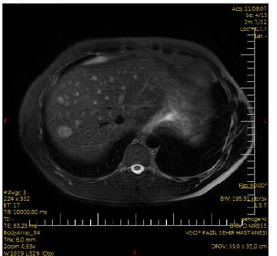
A 35-year-old male patient consulted to us in January 2015 with complaints of abdominal pain, and lassititude. وكشف فحصه البدني عن أنظارات تحتية، وعن عطاءات التشحيم البطني العميقة في الربع الأعلى الأيمن تم اكتشافها. ولم يتم الكشف عن المناقصة العائدة وحراسة البطن. ويستلزم وجود ألم في البطن، والعطاء الكمي في البطن العلوي الأيمن على الشحوم إجراء فحص فوق البدائيين فوق الصوتي (الولايات المتحدة). On US, hypoechoic solid lesions on hepatic segments 2 (8.5 mm), and 7 (12x10 mm) were observed. لم يكشف الأشعة والفحوص الكيميائية الحيوية عن أي شذوذ. For better identification of the hepatic lesion upper abdominal dynamic magnetic resonance (MR) images were obtained. On MR multiple T2 ارتفاع ضغط الدم، وتي1 وقد تم الكشف عن العقيدات الافتراضية في بئر الكبد، وهو أكبر عدد قياسه 18x16 ملم في الجزء 7. وفيما يتعلق بسلسلة المراحل الدينامية، شوهدت زيادة في المنافذ المتباينة للانتشار اللاحق، ولم يلاحظ الغسيل في سلسلة المراحل المتأخرة. ولوحظ في بعض هذه الآفات غير المكتفية التي يمكن أن تكون متسقة بشكل رئيسي مع الزخرفة المركزية )الشكل ١(. علماء الروادي فسروا صور MR لصالح الميستاسيس. ونظراً للاشتباه في وجود التكتل الكبدي، طُلبت قياس العلامات الأحيائية للتهاب الكبد، ومؤشرات التهاب الكبد الوبائي، ورقم المختصر التراكمي، ورقم المختصر التراكمي في المواد الكيميائية 19-9، ومستويات CEA. المؤشرات الحيوية للتهاب الكبد والورم في حدود طبيعية Chest X-ray, thoracal computed tomography (CT), scrotal US, endoscopic, and colonoscopic examina-tions were requested from the patient in order to search for the primary focus. On scrotal US of the patient whose chest X-Ray, and thoracal CT were unremarkable, slightly varicose veins of 3.5 mm in their largest diameter were detected. تم الكشف عن التهاب النسيج من الدرجة الأولى في فحصه الإندوسي، بينما لم يكشف جهازه المستعمر عن أي شذوذ.
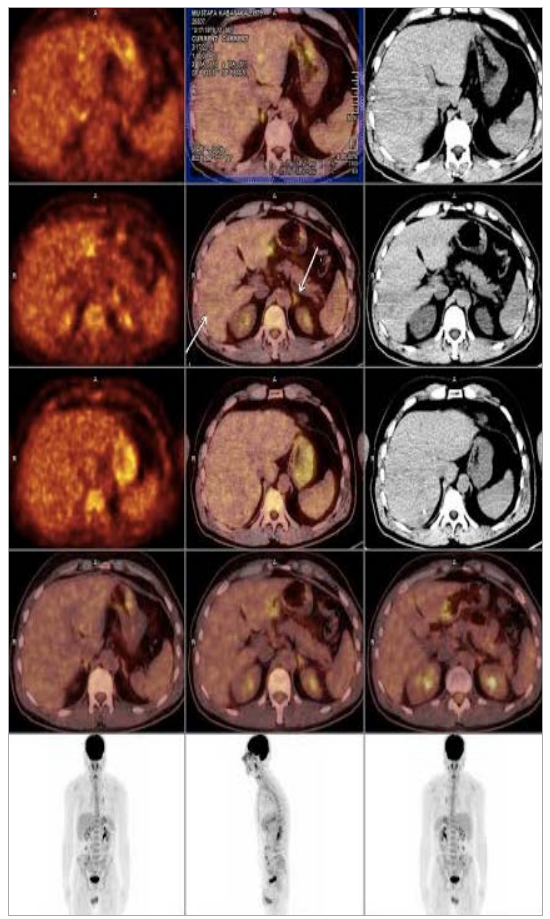
On positron emission tomograms (PET-CT) obtained so as to detect the primary focus, increased FDG uptake suggests malignancy was detected on the left supraclavicular (SUV)الحد الأقصى:4.4, left inferior jugular (SUVmax: 4.6), and in mediastinum prevasküler (SUV)الحد الأقصى: 3.9, right inferior paratracheal (SUV)الحد الأقصى: 4.9, left inferior paratracheal (SUVmax:4),right hilar (SUV)الحد الأقصى:3.5, left hilar(SUV)الحد الأقصى: 5.4) and subcarinal (SUV)الحد الأقصى: 5.7 lymph nodes (SUV)الحد الأقصى: 5.7, metastasis)Figure-2).
The liver lesions of the patient were biopsized under the guidance of US. Immunohistochemical examination of the biopsy material disclosed CD34, and vimentin positivity, while pancytokeratine, hepatocyte, CEA, CK7, CK20, TTF-1, CDX-2, S-100, DOG-1, and actin negativity. As a result of immunohistochemical staining the diagnosis of the patient was reported as epithelioid hemangioendothelioma.
The patient with multiple mass lesions who was diagnosed as epithelioid hemangioendothelioma underwent liver transplantation from a live donor in June 2015. ولا تختلف فترة متابعة المريض بعد عملية التخطيط.
المناقشة
ويندر أن يُنظر إلى الورم الرئوي الرئوي الذي يُنشأ من هيكل الأوعية الدموية لمختلف المناطق، بما في ذلك المعدة والطحال والكبد والرئة والعظم والأنسجة الناعمة والجلد. It has an middle degree of malignancy between benign hemangioma, and malignant hemangiosarcoma (4)في معظم الأحيان يكون الكبد متورطا (5)في مريضنا أيضاً تم إختيار الإرتباط الكبدي
The patients most frequently present with symptoms, and signs of abdominal pain, weight loss, ascites, nausea, jundice, hepatic failure, hepatic vein thrombosis, and gate hypertension (6)في مريضنا، ألم البطن، و(جونديس) تم اكتشافه وتتسم طرائق التصوير بأهمية بالغة في تشخيص الإصابة بفيروس نقص المناعة البشرية. وفي الفحص فوق الصوتي، كثيراً ما يُنظر إليه على أنه افتراضي (66.3 في المائة)، ومن حين لآخر (22.5 في المائة) على أنه آفة غير متجانسة. On non-contrasted CT, they are seen as hypodense, non-homegenous solid nodules extending into the capsule frequently with a peripheral location. وبعد حقن المادة المتناقضة، أصبحت علامة هالو التي تشكلها ندبة مركزية، وأصبح انتشار الخلايا الافتراضية في مرحلة البوّابة منتشرا. On MR, they are seen as non-homogeneous hypointense at T1, and non-homogeneous hyperintense images at T2 sequences (7)- فيما يتعلق بالرنين المغناطيسي لمريضنا، لوحظت عدة إصابات في الأنسجة الرطبة أكبر واحدة ذات أبعاد عيار 18x16 ملم. هذه العقيدات كانت مرئية على أنها مصابة بارتفاع ضغط الدم2"و "الآفات الافتراضية في "تي1 (ج) التسلسلات إلى جانب الانتشار الافتراضي للتفاوتات في مراحل دينامية.
ولا بد من إجراء تشخيص نهائي للمرض. ومن الصعب جداً، فيما يتعلق بفحص الميكروسكوبتر، التمييز بين هذه الآفات من تشولانغيوكارسينوما، وسرطان الخلايا الكبدية الأليفية، وساركوما، وأورام الميثاتية. ويمكن إجراء تشخيص محدد من خلال تأكيد خلايا المسنات باستخدام أساليب غير متجانسة (المفاعل الثامن، CD34، ومتتبعو CD31). وجود مستويات متزايدة من علامات الخلايا الثابتة في الخلايا الدهنية و/أو الخلايا الوبائية تؤكد التشخيص (8)- كشف التحليل الكيميائي الذي أجري في مريضنا عن الوثيقة CD34، وعن استثارة اليقظة.
As a treatment, various methods can be tried based on the location of the tumor in the liver. ومن بين هذه الأساليب إعادة تقطيع الكبد وزرعه طرائق العلاج السائدة (9)In EHE cases with diffuse, and unresectable hepatic involvement, even in the presence of remote metastases, liver transplantation is the most suitable treatment modality. (10–12)وفي حالتنا أيضاً، بسبب انتشار التدخل الكبدي، حيث تم تفضيل عملية زراعة الكبد بطريقة العلاج.
In the study where 71 patients did not undergo any treatment, while other patients were treated with liver transplantation (n:128), chemotherapy and/or radiotherapy (n:60), and hepatic resection (n:27). In this study, 1-year and 5-year survival rates following liver transplantation were reported as 96, and 54.5%, while in the groups of chemotherapy radiotherapy, and liver resection, corresponding oneyear and 5-year-survival rates were 73.3, and 30%, and 75%, respectively (3).
In conclusion, when multiple liver mass lesions are encountered, EHE should be absolutely considered in the differential diagnosis
الإقرارات
- مساهمات المؤلفين (CRediT)
- ساهم جميع المؤلفين بالتساوي في جميع أدوار CRediT الأربعة عشر (Conceptualization, Data curation, Formal analysis, Funding acquisition, Investigation, Methodology, Project administration, Resources, Software, Supervision, Validation, Visualization, Writing – original draft, Writing – review & editing).
- التمويل
-
Funding information was not reported in the published version of this article.
- تضارب المصالح
- A conflict of interest statement was not reported in the published version of this article.
- الموافقة الأخلاقية
- Ethics approval details were not reported in the published version of this article.
- إتاحة البيانات
- A data availability statement was not reported in the published version of this article.
المراجع (12)
7 من أصل 12 مرجعًا مرتبطة بمعرّف DOI.
- Dong K, Wnag X, Feng JL et al. Pathological characteristics of liver biopsies in eight patients with hepatic epithelioid hemangio endothelioma. Int J Clin Exp Pathol 2015;8(9): 11015-23. PubMed Google Scholar
- Pregliasco AG, Wendum D, Goumard C et al. Hepatic epithelioid hemangioendothelioma. Clin Res Hepatol Gastroenterol 2015 Nov PubMed Google Scholar
- pii: S2210-7401(15)00254-5. 3. Mehrabi A, Kashfi A, Fonouni H et al. Primary malignant hepatic epithelioid hemangioendothelioma: A comprehensive rewiew of the literature with emphasis on the surgical therapy. Cancer 2006;107:2108-21. DOI Crossref PubMed Google Scholar
- Weiss SW, Enzinger FM. Epithelioid hemangioendothelioma: A vascular tumor often mistaken for a carcinoma. Cancer 1982;50:970-81. DOI Crossref PubMed Google Scholar
- Sangro B, Inaarrairaegui M, Fernandez-Ros N. Malignant epitheloid hemangioendothelioma of the liver successfully treated with sorafenib. Rare Tumors 2012;4:e34. DOI Crossref PubMed Google Scholar
- Caruso S, Miraglia R, Maruzelli L et al. An unusual presentation of malignant hepatic epithelioid hemangioendothelioma with left pleural and pulmonary localization. Pediatr Radiol 2008;38:1027-30 DOI Crossref PubMed Google Scholar
- Ji ZF, Shen XZ, Zhang DY et al. Hepatic epithelioid hemangio endothelioma: a report of six patients. J Dig Dis 2010;11:254-8. PubMed Google Scholar
- Tsuneyoshi M, Dorfman HD, Bauer TW. Epithelioid hemangio endothelioms of bone: a clinicopathologic, ultrastructural, and immunohistochemical study. Am J Surg Pathol 1986;10:754-64. PubMed Google Scholar
- Langrehr JM, Petersen I, Pfitzmann R, Lopez-Hanninen E. Malignant epithelioid hemangioendothelioma of the liver. Result of surgical treatment strategies. Chirurg 2005;76:1161-7. PubMed Google Scholar
- Lerut JP, Orlando G, Sempoux C et al. Hepatic hemangio endothelioma in adults: Excellent outcome following liver transplantation. Transpl Int 2004;17:202-07. DOI Crossref PubMed Google Scholar
- Ben-Haim M, Roayaise S, Ye MQ et al. Hepatic epithelioid hemangioendothelioma: Resection or transplantation, which and when? Liver Transpl Surg 1999;5:526-31. DOI Crossref PubMed Google Scholar
- Lerut JP, Orlando G, Adam R. The place of liver transplantation in the treatment of hepatic epitheloid hemangioendothelioma: report of the European liver transplant registry. Ann Surg. 2007 Dec;246(6):949-57. DOI Crossref PubMed Google Scholar
اقرأ النص الكامل
افتح ملف PDF هنا
جارٍ تحميل ملف PDF…
إذا لم يظهر ملف PDF هنا، استخدم زر التنزيل أعلاه.
كيف تقتبس هذه المقالة
Nayir E. A Rarely Seen Hepatic Tumor: Epithelioid Hemangioendothelioma. Ulutas Med J. 2016;2(1):59-62. doi:10.5455/umj.20160121062843
Nayir, E. (2016). A Rarely Seen Hepatic Tumor: Epithelioid Hemangioendothelioma. The Ulutas Medical Journal, 2(1), 59–62. https://doi.org/10.5455/umj.20160121062843
Nayir, E. (2016) 'A Rarely Seen Hepatic Tumor: Epithelioid Hemangioendothelioma', The Ulutas Medical Journal, 2(1), pp. 59–62. doi: 10.5455/umj.20160121062843.