The Ulutas Medical Journal · ISSN 2149-0430 (print), 2149-388X (online) · https://www.ulutasmedicaljournal.com/?mno=161134
Original Article · Open access
Estimation of Effective Radiation Dose for Computed Tomography Head in Pediatric Patients at Universal College of Medical Sciences: A Cross-Sectional Study
- Universal College of Medical Sciences and Teaching Hospital, Bhairahawa, Rupandehi, Nepal ROR
Abstract
- Introduction
- In modern medicine, CT has made tremendous advances, but the use of CT has been accompanied by concerns about patient safety since it delivers higher doses of radiation than most other diagnostic imaging procedures. Children are particularly vulnerable because their rapidly dividing cells are more sensitive to radiation, and they will have more years to undergo cancerous changes. This study aimed to estimate the effective radiation dose received by pediatric patients while having CT heads at UCMS-TH.
- Materials and Methods
- This cross-sectional study was conducted in the Department of Radiodiagnosis & Medical Imaging, UCMS-TH, Bhairahawa, Nepal. Seventy-five pediatric patients were included in this study over five months, from November 2020 to March 2021. The radiation doses were estimated using a convenient technique and data were analyzed using SPSS version 20.
- Results
- The average mean age of the participants was 9.85±4.45 years. There were 69.3% males and 30.7% females. The age group 11-15 years had the highest frequency of patients 54.7%, followed by the age group 6-10 years with 24% patients. The mean and standard deviation of CTDIvol, DLP, and ED of NCCT and CECT head were significantly different between males and females throughout the study.
- Conclusion
- In pediatrics, male NCCT head scans had higher ED, whereas female CECT head scans had a maximum ED. The age range of 11-15 years had the maximum ED, followed by the age group of 6-10 years.
Keywords
Full text
Introduction
Computed Tomography (CT) is a significant source of radiation exposure (1). It is estimated that approximately 6% of CT examinations were performed on children under the age of 15 years. Children are more sensitive to ionizing radiation. Therefore, paediatric patients must be protected against it (2–4). The International Commission on Radiological Protection (ICRP) has developed a diagnostic reference level (DRL) for dose optimization (5, 6). Volume CT Dose Index (CTDIvol) quantifies the radiation exposure in CT scans. Dose-length product (DLP) measures energy deposited to the patient during a CT scan in relation to scan length (7). Effective dose (E) is calculated as the sum of the equivalent doses given to each organ multiplied by the tissue weighting factor (8). The aim of this study was to estimate effective radiation dose received by paediatric patients while having CT head at Universal College of Medical Sciences & Teaching Hospital.
Materials and Methods
A cross-sectional study was conducted in the Department of Radiodiagnosis and Medical Imaging, Universal College of Medical Sciences and Teaching Hospital (UCMS-TH), Bhairahawa, Rupandehi, Nepal, using a helical computed tomography scanner (GE 16 slice, Brivo model). Study was approved by UCMS-TH Institutional Review Committee (IRC), with number: UCMS/IRC/028/21. A total of 75 paediatric patients were scanned with CT scans of the head between March 14th, 2021 and June 14th, 2021. A convenient sampling technique was used and the sample size was calculated using formula: n=z(2)p(1-q)/d(2), where n=required sample size, z=1.96 at 95% confidence interval, p=5% estimated prevalence (8) and d=allowable error at 5%. Keeping these values in the above formula, the sample size was calculated to be approximately 73, but we included 75 patients. Tube current (mA), tube voltage (KVp), helical pitch slice thickness, displayed CTDIvol and DLP were the scan parameters employed in the present study. These parameters are required to estimate the radiation dose. However, the tube current was kept at 86 to 150 mA with a fixed tube voltage of 120 KV and a pitch of 0.562:1 mm throughout the study. DLP and CTDIvol was recorded independently. Effective dose was calculated from DLP by applying the conversion factor (f), {using the formula E= DLP.f}. The study protocol for paediatric CT head imaging was routine helical. This protocol was most preferred by the researcher and technologists because of its short scan time and low radiation dose to the patients.
Inclusion Criteria
▪ Patients of age ranging from 1 to 15 years. ▪ Patients of any sex.
▪ Patients whose guardians have given informed consent to the study.
▪ Pediatric patients who have come for head CT examination.
Exclusion Criteria
▪ Patients whose guardians did not give written consent for the study even after explanation.
▪ Patients with any absolute contraindication for CT.
Statistical Analyze
Statistical Package for Social Sciences (SPSS) version 20 was used to analyse the collected data. A volume CT dose index (CTDIvol) & dose product length (DLP) was used to estimate effective doses. We presented the data as mean±standard deviation.
Results
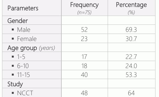
The present study included 75 paediatric patients with an average mean age of 9.85±4.45 (range 1-15) years. There were 52 males (69.3%) and 23 females (30.7%). The age group 11-15 years had the highest frequency of patients 40 (53.3%), followed by 18 (24%) patients in the age group 6-10 years. The age group 1-5 years had the smallest number of patients 17 (22.7%). Out of 75 paediatric patients, 48 (64%) underwent non-contrast computed tomography (NCCT), and 27 (36%) got contrast-enhanced computed tomography (CECT), as shown in Table 1.
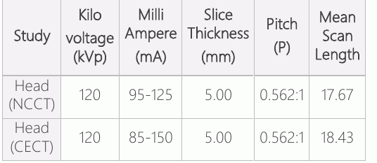
According to the study types (NCCT and CECT), the parameters used in this study were kVp, mA, slice thickness, pitch, and mean scan length (Table 2).
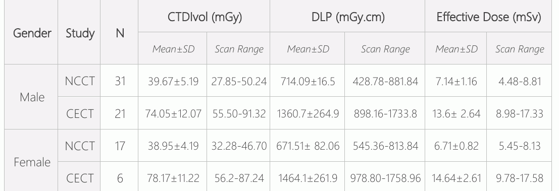
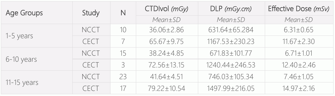
As shown in Table 3, it examines the volumetric CT dose index (CTDIvol), dose length product (DLP), and effective dose (ED) along with NCCT and CECT head examinations of paediatric patients (males and females). There was a significant difference between the mean and standard deviation (SD) of CTDIvol, DLP, and ED of the CECT head between males and females. Table 4 shows the mean values and standard deviation for each age group. The CTDIvol, DLP, and ED were more significant in the 11–15-years age group, followed by 6-10 years age group.
Discussion
This study examined the methods used to estimate radiation doses for paediatric head CT scans. The participants’ average mean age was 9.85±4.45 years, ranging from 1 to 15 years. There were 52 (69.3%) males and 23 (30.7%) females. The majority of the patients were in the age group of 11-15 years, with a frequency of 40 (53.3%), followed by 18 (24%) between the age group of 6-10 years. A similar study was conducted by Inoue Y et al (9).
In comparison to Eddy FK et al.(10), Sulieman A et al.(11), and Kayun Z et al.(8), the findings of our study show that the mean value of the volumetric CT dose index of contrast-enhanced & non-contrast CT scans was 78.17±11.22 mGy & 38.95±4.19 mGy in females, followed by males 74.05±12.07 mGy & 39.67±5.19 mGy; and the mean DLP of contrast-enhanced & non-contrast CT was 1464.16±261.93 mGy.cm & 671.51± 82.06 mGy.cm in females, followed by males 1360.71±264.97 mGy.cm & 714.09± 116.5 mGy.cm; and the mean of an effective dose of contrast-enhanced & non-contrast CT was 14.64±2.61 mSv & 6.71±0.82 mSv in females, followed by males 13.6± 2.64 mSv & 7.14±1.16 mSv, which was seen higher in our study. In our study, the majority of the mean value of contrast-enhanced scans of CTDIvol, DLP, and ED was 79.22±10.54 mGy, 1497.99±216.05 mGy.cm, and 14.97±2.16 mSv in the age groups of 11-15 years, followed by the age groups of 6- 10 years, which was compared to the study by Bouaoun A et al. (12) and Saeed MK et al. (13). ED in the age groups of 10-15, followed by the age groups of 5-10 years. However, our study found no significant variation in radiation dose. A slight difference has been seen due to the difference in the machine and the parameter used.
As limitations, the study was carried out in a single institute, so our sample size was limited. The study was only conducted on patients aged between 1 and 15 years.
Conclusion
The study concluded that male paediatric patients received a higher effective dose for their NCCT head scan, whereas female paediatric patients had a higher effective dose for their CECT head scan. The age range of 11- 15 years had the maximum effective dose, followed by the age group of 6-10 years.
Declarations
- Author contributions (CRediT)
- All authors contributed equally to all 14 CRediT roles (Conceptualization, Data curation, Formal analysis, Funding acquisition, Investigation, Methodology, Project administration, Resources, Software, Supervision, Validation, Visualization, Writing – original draft, Writing – review & editing).
- Funding
-
Funding information was not reported in the published version of this article.
- Conflicts of interest
- A conflict of interest statement was not reported in the published version of this article.
- Ethics approval
- Inclusion Criteria ▪ Patients of age ranging from 1 to 15 years. ▪ Patients of any sex. ▪ Patients whose guardians have given informed consent to the study. ▪ Pediatric patients who have come for head CT examination.
- Data availability
- A data availability statement was not reported in the published version of this article.
References (13)
12 of 13 references are linked to their DOI.
- Gao Y, Quinn B, Mahmood U, Long D, Erdi Y, St. Germain J, Pandit-Taskar N, Xu XG, Bolch WE, Dauer LT. A comparison of pediatric and adult CT organ dose estimation methods. BMC medical imaging. 2017 Dec; 17:1-7. DOI Crossref PubMed Google Scholar
- Alzimami K. Assessment of radiation doses to paediatric patients in computed tomography procedures. Polish journal of radiology. 2014;79:344. DOI Crossref PubMed Google Scholar
- Kadavigere R, Sukumar S. Estimation of radiation dose and establishment of local diagnostic reference levels for computed tomography of head in pediatric population. Journal of X-Ray Science and Technology. 2022 Jan 1(Preprint):1-9. PubMed Google Scholar
- Ogbole GI. Radiation dose in paediatric computed tomography: risks and benefits. Annals of Ibadan postgraduate medicine. 2010;8(2):118-26. DOI Crossref PubMed Google Scholar
- Inoue Y, Itoh H, Shiibashi N, Sasa R, Mitsui K. Sample size and estimation of standard radiation doses for pediatric brain CT. Tomography. 2022 Oct 1;8(5):2486-97. DOI Crossref PubMed Google Scholar
- Khong PL, Ringertz H, Donoghue V, Frush D, Rehani M, Appelgate K, Sanchez R. ICRP publication 121: radiological protection in paediatric diagnostic and interventional radiology. Annals of the ICRP. 2013 Apr;42(2):1-63. DOI Crossref PubMed Google Scholar
- Nievelstein RAJ, Van Dam IM, Van Der Molen AJ. Multidetector CT in children: Current concepts and dose reduction strategies. Pediatr Radiol. 2010;40(8):1324–44. DOI Crossref PubMed Google Scholar
- Kayun Z, Karim MK, Harun HH, Shaari AH, Mahmud R, Hamid HA, Sabarudin A, Chew MT. Radiation doses and size-specific dose estimate from CT brain examinations according to head sizes in a tertiary hospital in Malaysia. Radiation Physics and Chemistry. 2021 Dec 1;189:109694. DOI Crossref PubMed Google Scholar
- Inoue Y, Itoh H, Waga A, Sasa R, Mitsui K. Radiation dose management in pediatric brain CT according to age and weight as continuous variables. Tomography. 2022 Apr 1;8(2):985-98. DOI Crossref PubMed Google Scholar
- Eddy FK, Ngano SO, Jervé FA, Serge A. Radiation dose evaluation of pediatric patients in CT brain examination: multi-center study. Scientific Reports. 2021 Feb 25;11(1):1-6. DOI Crossref PubMed Google Scholar
- Sulieman A, Almogren KS, Tamam N. Radiation exposure estimation in pediatric patients during computed tomography imaging procedures. Radiation Physics and Chemistry. 2023 Mar 1;204:110653. DOI Crossref PubMed Google Scholar
- Bouaoun A, Ben Omrane L, Mogaadi M, Khomsi WD, Zidi A, Hammou A. Pediatric head CT exposure doses in Tunisia: a pilot study towards the establishment of national diagnostic reference levels. Radiation Protection Dosimetry. 2018 Dec 1;182(2):241-51. DOI Crossref PubMed Google Scholar
- Saeed MK, Almalki Y. Assessment of computed tomography radiation doses for paediatric head and chest examinations using paediatric phantoms of three different ages. Radiography. 2021 May 1;27(2):332-9. DOI Crossref PubMed Google Scholar
Read the full text
Open the PDF here
Loading the PDF…
If the PDF does not show here, use the download button above.
How to cite this article
Yadav SC, Dhakal D. Estimation of Effective Radiation Dose for Computed Tomography Head in Pediatric Patients at Universal College of Medical Sciences: A Cross-Sectional Study. Ulutas Med J. 2023;9(4):208-212. doi:10.5455/umj.20230714114808
Yadav, S. C., & Dhakal, D. (2023). Estimation of Effective Radiation Dose for Computed Tomography Head in Pediatric Patients at Universal College of Medical Sciences: A Cross-Sectional Study. The Ulutas Medical Journal, 9(4), 208–212. https://doi.org/10.5455/umj.20230714114808
Yadav, S.C. and Dhakal, D. (2023) 'Estimation of Effective Radiation Dose for Computed Tomography Head in Pediatric Patients at Universal College of Medical Sciences: A Cross-Sectional Study', The Ulutas Medical Journal, 9(4), pp. 208–212. doi: 10.5455/umj.20230714114808.