The Ulutas Medical Journal · ISSN 2149-0430 (print), 2149-388X (online) · https://www.ulutasmedicaljournal.com/?mno=121168
Original Article · Open access
Estimation of Residual Tissue with Thyroid Scintigraphy and Thyroglobulin Level in the Pre-ablative Period of Differentiated Thyroid Carcinoma
Also available in: العربية · Türkçe · 中文
- Hatay Mustafa Kemal University, Faculty of Medicine, Department of Nuclear Medicine, Hatay, Turkey ROR
Abstract
- Introduction
- The aim of the study was to compare the pre-ablative thyroid scintigraphy and serum thyroglobulin (TG) positivity with a therapeutic I-131 scan for the detection of residual tissue.
- Materials and Methods
- Patients included in the study were those with neck residual tissue only and no residual or metastatic tissue in the ablative radioactive iodine (RAI) scan anywhere in the body. Pre-ablative thyroid scintigraphy for the residual tissue in the neck was obtained from all patients. Ablative TSH, TG and anti-TG levels were measured on the same day or one day before the RAI therapy. The evaluation of neck residual tissue detected with thyroid scintigraphy and TG measurement were made in this study accepting I-131 scan as the gold standard.
- Results
- The I-131 scan was positive for residual neck tissue in 67 (93.1%) patients and negative in 5 (6.9%) patients. The sensitivity, specificity, accuracy, positive predictive value, and negative predictive values of thyroid scintigraphy were 70.1%, 80%, 70.8%, 97.9%, and 16.7%, respectively. For the high positive predictive value, the kappa value was 0.182, which showed a slight agreement between Tc-99m thyroid scintigraphy and I-131 scan. The sensitivity, specificity, accuracy, positive predictive value, and negative predictive values of TG measurement were 92.5%, 60%, 90.3%, 96.9%, and 37.5%. There was moderate agreement between the TG measurement and I-131 scan.
- Conclusion
- Negative thyroid scintigraphy should not be noteworthy because of the very low negative predictive value and kappa value. A measurable TG level seems to be more accurate and useful for the detection of residual tissue.
Keywords
Full text
Introduction
Differentiated thyroid cancer, papillary and follicular cancer, are usually curable especially if diagnosed early. The treatment algorithm has changed because there are no prospective randomized trials of treatment because of its prolonged course (1). The aim of treatment is to eradicate the primary tumor, prevent local or distant recurrence, treat the metastases and cure the patients with minimal morbidity (2). In low-risk patients after total thyroidectomy and no evidence of abnormal residual tissue or metastatic lymph nodes, patients are not generally given radioactive iodine (RAI) therapy, but are followed up with neck USG and the measurement of the thyroglobulin (TG) level because of the excellent response (3). I-131 whole body scintigraphy with the combination of serum TG level and neck USG is commonly applied follow-up procedure for differentiated thyroid cancer (4).
In the patients requiring ablative RAI therapy, it is important to define the residual thyroid tissue. After total or near total thyroidectomy, I-131 uptake may persist in the thyroid bed due to residual thyroid tissue. If the residual tissue is macroscopic, the outcome becomes unfavorable (1). Pre-ablative low dose I-131 whole body scan has been used for many years. Despite the beneficial effect of detecting both thyroid remnant and metastasis, there is a rising trend to avoid this, because of the stunning effect of I-131 (5). Avoiding a pre-ablative scan has been shown to result in better radio-ablation, and reduce cost and time (2). The aim of this study was to compare pre-ablative thyroid scintigraphy, with serum thyro globulin positivity with therapeutic I-131 scan for the detection of residual tissue after total thyroidectomy in differentiated thyroid cancer.
Materials and Methods
Patients
The study included 72 patients, comprising 63 females (87.5%) and 9 males (12.5%) with a mean age of 41.56±12 years (min: 19, max:71). Only patients with neck residual tissue and no residual or metastatic tissue anywhere in the body were included in the study. Patients with detectable anti-TG levels (>20 IU/ml) were excluded from the study because of the effect on TG levels. All the patients underwent total thyroidectomy and received ablative I-131 treatment for differentiated thyroid cancer. The therapeutic I-131 scan was applied 5-7 days after therapy. Pre-ablative thyroid scintigraphy for the residual tissue in the neck was applied to all patients. Ablative TSH, TG and anti-TG levels were measured on the same day or one day before the RAI therapy. The evaluation of neck residual tissue detected with thyroid scintigraphy and TG were made in this study accepting the I-131 scan as the gold standard. A TG level below the measurable level (0.2 ng/mL) was accepted as zero. All the Tc-99m pertechnetate thyroid scintigraphy scans were made at least 20 days after the operation. Ablative I-131 therapy was given approximately two weeks after thyroid scintigraphy. This retrospective study was approved by the Ethics Committee of Mustafa Kemal University.
Statistical Analysis
Data obtained in the study were analyzed statistically using SPSS for Windows version 21 software (SPSS, Chicago, IL, USA). Descriptive statistics were stated as mean±standard deviation for continuous variables. Sensitivity, specificity, accuracy, positive predictive value (PPV) and negative predictive value (NPV) of Tc-99m pertechnetate scinti-graphy and TG positivity to detect residual tissue were measured. Tc-99m pertechnetate scintigraphy and serum TG positivity were compared with I-131 scan for the detection of residual tissue with kappa analysis. Interpretation of the kappa test was poor agreement if the value was ≤0.2, fair agreement if 0.21-0.4, moderate agreement if 0.41-0.6, good agreement if 0.61-0.8, and excellent agreement if 0.81-1.
Results
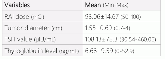
Multifocal tumors were determined in 17 patients. A dose of 50 mCi was given to 5 patients, 75 mCi to 10 patients and 100 mCi to 57 patients with a mean dose of 93.06±1 4.67 mCi. The mean tumor diameter was 1.55± 0.69 cm (min: 0.7-max: 4.0). The mean TSH value was 108.13±72.32µIU/mL (min:30.54-max: 460.06). Stimulated TG level was 6.68±9.59 ng/mL (min: 0 - max: 52.9) (Table 1).
The I-131 scan was positive for residual neck tissue in 67 (93.1%) patients and negative in 5 (6.9%) patients. Thyroid scintigraphy was positive in 48 (66.7%) patients and negative in 24 (33.3%) patients. In 1 patient determined as positive on thyroid scintigraphy, there was no residual tissue on the I-131 scan and this case was evaluated as false (+) thyroid scintigraphy. The sensitivity, specificity, accuracy, PPV and NPV of thyroid scintigraphy were 70.1%, 80%, 70.8%, 97.9% and 16.7%, respectively. The high PPV had a kappa value of 0.182 (p=0.022) indicating slight agreement between Tc-99m scintigraphy and I-131 scan.
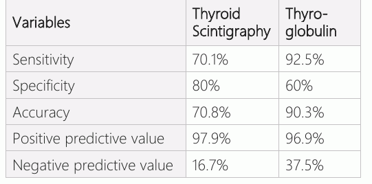
A measurable serum TG (≥0.2) was present in 64 (88.9%) patients and negative in 8 (11.1%) patients. The sensitivity, specificity, accuracy, PPV and NPV of TG measurement were 92.5%, 60%, 90.3%, 96.9% and 37.5%, respectively (Table 2). There was moderate agreement between the TG measurement and I-131 scan (kappa=0.411, p<0.001).
Discussion
Total thyroidectomy, which is the complete macroscopic removal of all grossly apparent thyroid tissue, is applied to the cases of differentiated thyroid carcinoma. However, the postoperative RAI imaging may show incomplete removal of functioning thyroid tissue after total thyroidectomy (6). Following partial thyroidectomy, the risk of malignancy in the residual tissue is high. Tumor multifocality and significant TG elevation are predictive of malignancy in residual tissue (7). Residual thyroid tissue can change significantly based on surgical parameters. Academic centers have reported lower residual thyroid bed uptake on I-131 scans compared to community centers (8). Most patients have some thyroid remnant even after an accurate surgical technique. Salvatori et al. confirmed that 93.1% of patients had thyroid remnant after total thyroidectomy for differentiated thyroid carcinoma with the majority of the remnant tissue observed to be contralateral to the tumor site and outside the thyroid bed. A previous study reported that total thyroidectomy was achieved in 6.9% of patients with negative therapeutic I-131 whole body scan, radioactive iodine uptake <1% and undetectable levothyroxine of TG level (9). Thyroid USG can visualize the thyroid gland and measure the gland accurately, but in the postoperative period before ablative treatment, granulation tissue and edema cannot be discriminated correctly (10). RAI therapy is used after thyroidectomy to ablate residual tissues, prevent recurrences and increase survival (11). Iodides taken orally are almost completely absorbed from the duodenum and excreted through kidneys. In addition to accumulation in thyroid follicular cells, extra-thyroid organs such as salivary glands, the stomach, lactating mammary glands and thymus also accumulate RAI (11–13). Beside the therapeutic effect of RAI, it can also be used for whole body scans in the follow up of thyroid cancer. A whole-body RAI scan is very important in the detection of metastases of well differentiated thyroid cancer during follow up. However, careful evaluation is necessary because of misinterpretation of RAI accumulation lesions. Benign thyroid tissue salivary gland inflammation, or thymic uptake may cause a false positive RAI scan (11, 13, 14). I-131 is also used for the detection of residual thyroid tissue after surgery. RAI planar imaging with I-123 or I-131 is the gold standard imaging method in the follow up after initial surgery (12). Diagnostic I-131 scanning may cause damage to thyroid tissue, thereby impairing the uptake of the therapeutic dose of I-131 which is known as stunning (15). Therefore, because of stunning I- 123 may be used before RAI therapy for the detection of residual tissue. Teoh et al applied pre-ablative whole body RAI scanning, which was positive in 94.9% of patients (2). Routine pre-ablative RAI scan is offered after near total thyroidectomy and if there is remaining thyroid tissue, it should be destroyed with RAI (16). Tc-99m pertechnetate scintigraphy is a low-cost and widely available method for the patients that require postoperative scanning for residual tissue (17). Nadig et al found that Tc-99m pertechnetate SPECT can estimate residual thyroid tissue while performing patient specific dosimetry with the advantage of simple technique (18). Pre-ablative Tc-99m pertechnetate scintigraphy not only provides information about the residual tissue, but can also predict the ablation success, hospitalization time and thyroiditis due to treatment. Pre-ablative Tc-99m pertechnetate scintigraphy is used to predict the outcome of the ablative treatment. A Tc-99m pertechnetate uptake value >1.4% has been reported to predict unsuccessful ablation, longer hospitalization and a higher risk of radioiodine-induced thyroiditis (19). In contrast, Thientunyakit et al stated that Tc-99m pertechnetate scintigraphy and 24 hour I-131 uptake in the thyroid remnant cannot predict the outcome of RAI therapy, although the ablative serum TG level may be a reasonable predictor for success of ablation. In that study, the I-131 whole body scan was negative in 92.9% of patients with negative Tc- 99m pertechnetate scintigraphy (20).
The synthesis and release of TG from normal thyroid tissue is controlled by TSH, and thyroid tissue size and serum TSH levels are correlated to serum TG levels. The serum TG level after total thyroidectomy should be undetectable or within the normal range in patients without residual or metastatic thyroid tissue (21). Charles et al reported that serum TG levels are more sensitive than RAI scan during follow up and TG analyses may replace or become an important adjunct to RAI scan (22).
Residual thyroid tissue volume should be determined correctly when assessing for the completion thyroidectomy. Erbil et al., recommended that USG, radioactive iodine uptake and serum TSH level should be assessed to estimate the postoperative thyroid remnant volume for completion thyroidectomy in the patients with incidental thyroid carcinoma detected after the surgery (23). Zhao et al recommended serial stimulated serum TG measurement in a short period, such as 8 days. Serial measurements may be more informative after thyroidectomy before ablative treatment than a single stimulated TG measurement (24). In cases with negative serum TG and anti-TG levels with positive I-131 scan, this may be related to limitations in the immunoassays. Both stimulated serum TG and anti-TG levels with I- 131 scan are recommended in the follow up of patients with high-risk thyroid cancer (25). In the current study, patients with measurable anti-TG levels were excluded because the anti- TG level may have interfered with the TG levels and TG levels may not have been measurable despite the high level of TG.
Filesi et al. recommended whole body I-131 scan and TG measurements for all patients after total thyroidectomy of differentiated thyroid carcinoma. In the presence of residual tissue, analysis of both scan and TG levels can provide reliable and early detection of metastases (26). Khammash et al compared Tc-99m and I-131 scans for the detection of residual and metastatic thyroid tissue in the follow up of patients, and Tc-99m pertechnetate was reported to have sensitivity, specificity and accuracy of 87%, 97% and 92.5%, respectively. Negative Tc-99m images cannot exclude residual or metastatic functioning thyroid tissue because of the false negative rate of 15.6% (27). In the current study, the sensitivity, specificity and accuracy values were not as high as in the study by Khammash et al, but the results were similar to those of the Ozdemir et al study, in which the residual tissue was evaluated with Tc-99m pertechnetate scintigraphy and therapeutic I-131 whole body scan. The post-operative Tc-99m pertechnetate scan was positive in 69.6% and negative in 30.4% of patients whereas therapeutic I-131 scan was positive in 93.9% and negative in 6.1% of patients. The sensitivity, specificity, accuracy, PPV and NPV of Tc-99m pertechnetate scan were 72.2%, 70.5%, 72%, 97.4% and 14.2%, respectively when the I-131 scan was accepted as the standard test (10). In the current study, despite the relatively high sensitivity, specificity, and accuracy and the excellent PPV, the NPV was very low, so the kappa value was 0.182 (p=0.022), indicating slight agreement between Tc-99m thyroid scintigraphy and I-131 scan. In the current study, measurable TG levels were more concordant with the I-131 scan with better sensitivity, accuracy and NPV than thyroid scintigraphy. The kappa was 0.411, showing moderate agreement between measurable TG levels and the I-131 scan. Kueh et al compared Tc-99m pertechnetate and I-131 scan for the detectionof remnant tissue after thyroidectomy. Tc-99m pertechnetate was seen to have moderately high sensitivity (90%) and a very high PPV (100%). A positive scan is reliable for the presence of residual tissue, but in case of a negative scan additional scanning might be needed (17). In a study by Chantadisai et al, they compared Tc-99m pertechnetate whole body scan, neck and chest SPECT/CT with post ablative I-131 whole body scan and SPECT/CT for the presence of remnant or metastatic tissue.The percentage of concordance between the two scans was 96.4% per patient analysis with a good correlation of the unweighted kappa value of 0.7358. Better diagnostic parameters were determined with SPECT/CT. In the current study, only static images were acquired (5).
Conclusion
In conclusion, the sensitivity, specificity and accuracy of thyroid scintigraphy is relatively high and positive predictive value is excellent. However, because of the very low negative predictive value and kappa value, negative thyroid scintigraphy should not be given too much attention. A measurable TG level seems to be more accurate and useful for detection of residual tissue.
Declarations
- Author contributions (CRediT)
- All authors contributed equally to all 14 CRediT roles (Conceptualization, Data curation, Formal analysis, Funding acquisition, Investigation, Methodology, Project administration, Resources, Software, Supervision, Validation, Visualization, Writing – original draft, Writing – review & editing).
- Funding
-
Funding information was not reported in the published version of this article.
- Conflicts of interest
- The authors declared no conflict of interest.
- Ethics approval
- As a nature of being a retrospective study, there is no need for consent of patient for the present study.
- Data availability
- A data availability statement was not reported in the published version of this article.
References (27)
23 of 27 references are linked to their DOI.
- Mazzaferri EL, Kloos RT. Clinical review: Current approaches to primary therapy for papillary and follicular thyroid cancer. J Clin Endocrinol Metab. 2001;86:1447 –63 DOI Crossref PubMed Google Scholar
- Teoh CM, Rohaizak M, Chan KY, Jasmi AY, Fuad I. Preablative diagnostic whole-body scan following total thyroidectomy for well-differentiated thyroid cancer: is it necessary? Asian J Surg. 2005;28:90– 6 DOI Crossref PubMed Google Scholar
- Janovsky CC, Maciel RM, Camacho CP, Padovani RP, Nakabashi CC, Yang JH, et al. A Prospective Study Showing an Excellent Response of Patients with Low-Risk Differentiated Thyroid Cancer Who Did Not Undergo Radioiodine Remnant Ablation after Total Thyroidectomy. Eur Thyroid J. 2016;5:44-9 DOI Crossref PubMed Google Scholar
- Triggiani V, Giagulli VA, Iovino M, De Pergola G, Licchelli B, Varraso A, et al. False positive diagnosis on (131)iodine whole-body scintigraphy of differentiated thyroid cancers. Endocrine. 2016;53:626-35 DOI Crossref PubMed Google Scholar
- Chantadisai M, Kingpetch K. Usefulness of (99m)Tc-pertechnetate whole body scan with neck and chest SPECT/CT for detection of post-surgical thyroid remnant and metastasis in differentiated thyroid cancer patients. Ann Nucl Med. 2014;28:674–82 DOI Crossref PubMed Google Scholar
- Holsinger FC, Ramaswamy U, Cabanillas ME, Lang J, Lin HY, Busaidy NL, et al. Measuring the extent of total thyroidectomy for differentiated thyroid carcinoma using radioactive iodine imaging: relationship with serum thyroglobulin and clinical outcomes. JAMA Otolaryngol Head Neck Surg. 2014;140:410-5 DOI Crossref PubMed Google Scholar
- Alzahrani AS, Al Mandil M, Chaudhary MA, Ahmed M, Mohammed GE. Frequency and predictive factors of malignancy in residual thyroid tissue and cervical lymph nodes after partial thyroidectomy for different thyroid cancer. Surgery. 2002;131:443– 9 DOI Crossref PubMed Google Scholar
- Song JSA, Moolman N, Burrell S, Rajaraman M, Bullock MJ, Trites J, et al. Use of radioiodine-131 scan to measure influence of surgical discipline, practice, and volume on residual thyroid tissue after total thyroidectomy for differentiated thyroid carcinoma. Head Neck. 2018;40(:2129-2136 DOI Crossref PubMed Google Scholar
- Salvatori M, Raffaelli M, Castaldi P, Treglia G, Rufini V, Perotti G, et al. Evaluation of the surgical completeness after total thyroidectomy for differentiated thyroid carcinoma. Eur J Surg Oncol. 2007;33:648-54 DOI Crossref PubMed Google Scholar
- Ozdemir D, Cuhaci FN, Ozdemir E, Aydin C, Ersoy R, Turkolmez S, Cakir B. The role of postoperative Tc-99m pertechnetate scintigraphy in estimation of remnant mass and prediction of successful ablation in patients with differentiated thyroid cancer. Nucl Med Commun. 2016;37:640-5 DOI Crossref PubMed Google Scholar
- Wilson LM, Barrington SF, Morrison ID, Kettle AG, O’Doherty MJ, Coakley AJ. Therapeutic implications of thymic uptake of radioiodine in thyroid carcinoma. Eur J Nucl Med.1998;25:622-8. DOI Crossref PubMed Google Scholar
- Ahmed N, Niyaz K, Borakati A, Marafi F, Birk R, Usmani S. Hybrid SPECT/CT Imaging in the Management of Differentiated Thyroid Carcinoma. Asian Pac J Cancer Prev. 2018;19:303– 8 PubMed Google Scholar
- Kim S, Park CH, Yoon KN, Hwang K. A false positive I-131 whole body scan in chronic parotitis: a case report. Clin Nucl Med 2001;26:536– 7 DOI Crossref PubMed Google Scholar
- Coover LR. False positive result of a total body scan caused by benign thyroidal tissue after I- 131 ablation. Clin Nucl Med 1999;24:182– 3 DOI Crossref PubMed Google Scholar
- Iwano S, Kato K, Nihashi T, Ito S, Tachi Y, Naganawa S. Comparisons of I- 123 diagnostic and I-131 post-treatment scans for detecting residual thyroid tissue and metastases of differentiated thyroid cancer. Ann Nucl Med. 2009;23:777–82 DOI Crossref PubMed Google Scholar
- Utiger RD. Follow-up of patients with thyroid carcinoma. N Engl J Med 1997;337:928–30 DOI Crossref PubMed Google Scholar
- Kueh SS, Roach PJ, Schembri GP. Role of Tc-99m for remnant scintigraphy post-thyroidectomy. Clin Nucl Med. 2010;35:671–74 DOI Crossref PubMed Google Scholar
- Nadig MR, Pant GS, Bal C. Usefulness of 99mTc-pertechnetate single-photon emission computed tomography in remnant mass estimation of postsurgical patients of differentiated thyroid cancer during internal dosimetry. Nucl Med Commun. 2008;29:809– 14 DOI Crossref PubMed Google Scholar
- Giovanella L, Suriano S, Ricci R, Ceriani L, Anton Verburg F. Postsurgical thyroid remnant estimation by Tc99-pertechnetate scintigraphy predicts radioiodine ablation effectiveness in patients with differentiated thyroid carcinoma. Head Neck. 2011;33:552– 6 PubMed Google Scholar
- Thientunyakit T, Pusuwan P, Tuchinda P, Khiewvan B. The correlation of post-operative radioiodine uptake and Tc-99m pertechnetate thyroid scintigraphy and the result of thyroid remnant ablation [published correction appears in J Med Assoc Thai. 2014 Jan;97(1):138. Kiewvan, Benjapa [corrected to Khiewvan, Benjapa]]. J Med Assoc Thai. 2013;96:1199–207 PubMed Google Scholar
- Girelli ME, De Vido D. Serum thyroglobulin measurements in differentiated thyroid cancer. Biomed Pharmacother. 2000;54:330– 3 DOI Crossref PubMed Google Scholar
- Charles MA, Dodson LE Jr, Waldeck N, Hofeldt F, Ghaed N, Telepak R, et al. Serum thyroglobulin levels predict total body iodine scan findings in patients with treated well-differentiated thyroid carcinoma. Am J Med. 1980;69:401-7 DOI Crossref PubMed Google Scholar
- Erbil Y, Barbaros U, Salmaslioglu A, Issever H, Tukenmez M, Adalet I, et al. Determination of remnant thyroid volume: comparison of ultrasonography, radioactive iodine uptake and Residual Tissue in Differentiated Thyroid Carcinoma Atilgan et al. serum thyroid-stimulating hormone level. J Laryngol Otol. 2008;122:615-22 DOI Crossref PubMed Google Scholar
- Zhao T, Liang J, Li T, Guo Z, Vinjamuri S, Lin Y. Value of serial preablative thyroglobulin measurements: can we address the impact of thyroid remnants? Nucl Med Commun 2016;37:632-9 DOI Crossref PubMed Google Scholar
- Cherk MH, Francis P, Topliss DJ, Bailey M, Kalff V. Incidence and implications of negative serum thyroglobulin but positive I-131 whole-body scans in patients with well-differentiated thyroid cancer prepared with rhTSH or thyroid hormone withdrawal. Clin Endocrinol (Oxf). 2012;76:734–40 DOI Crossref PubMed Google Scholar
- Filesi M, Signore A, Ventroni G, Melacrinis FF, Ronga G. Role of initial iodine-131 whole-body scan and serum thyroglobulin in differentiated thyroid carcinoma metastases. J Nucl Med. 1998;39:1542– 6 PubMed Google Scholar
- Khammash NF, Halkar RK, Abdel-Dayem HM. The use of technetium-99m pertechnetate in postoperative thyroid carcinoma. A comparative study with iodine-131. Clin Nucl Med. 1988;13:17–22 DOI Crossref PubMed Google Scholar
Respond to this article
Comments on a published article are welcome as a Letter to the Editor.
Get an e-mail when new articles in your fields are published
Questions to the authors
Questions are moderated: an editor approves each question, the authors answer, and an editor approves the answer before both appear here.
No answered questions yet.
Ask the authors a question
Discussion
Comments are moderated: they appear after the writer confirms the e-mail address and an editor approves them. Each comment carries a conflict-of-interest statement.
No comments yet.
Write a comment
Tell me when this article is cited
Leave your e-mail address and confirm it. These notices are not being sent yet; your confirmed address is kept for them and used for nothing else.
Read the full text
Open the PDF here
Loading the PDF…
If the PDF does not show here, use the download button above.
How to cite this article
Atilgan HI, Yalcin H. Estimation of Residual Tissue with Thyroid Scintigraphy and Thyroglobulin Level in the Pre-ablative Period of Differentiated Thyroid Carcinoma. Ulutas Med J. 2020;6(3):169-175. doi:10.5455/umj.20200805104357
Atilgan, H. I., & Yalcin, H. (2020). Estimation of Residual Tissue with Thyroid Scintigraphy and Thyroglobulin Level in the Pre-ablative Period of Differentiated Thyroid Carcinoma. The Ulutas Medical Journal, 6(3), 169–175. https://doi.org/10.5455/umj.20200805104357
Atilgan, H.I. and Yalcin, H. (2020) 'Estimation of Residual Tissue with Thyroid Scintigraphy and Thyroglobulin Level in the Pre-ablative Period of Differentiated Thyroid Carcinoma', The Ulutas Medical Journal, 6(3), pp. 169–175. doi: 10.5455/umj.20200805104357.
@article{umj121168,
  title = {Estimation of Residual Tissue with Thyroid Scintigraphy and Thyroglobulin Level in the Pre-ablative Period of Differentiated Thyroid Carcinoma},
  author = {Atilgan, Hasan Ikbal and Yalcin, Hulya},
  journal = {The Ulutas Medical Journal},
  year = {2020},
  volume = {6},
  number = {3},
  pages = {169--175},
  doi = {10.5455/umj.20200805104357},
  url = {https://www.ulutasmedicaljournal.com/?mno=121168},
  issn = {2149-388X}
}