The Ulutas Medical Journal · ISSN 2149-0430 (印刷版), 2149-388X (在线版) · https://www.ulutasmedicaljournal.com/?mno=161134
原创论著 · 开放获取
估计世界医学院儿科病人有效放射剂量:跨部门研究
标题、摘要和全文为机器翻译。 英文原文
- Universal College of Medical Sciences and Teaching Hospital, Bhairahawa, Rupandehi, Nepal ROR
- 开放获取
- CC BY-NC 4.0
- ORCID
- 资助
- 伦理批准
- 数据可用性
摘要
导言: 在现代医学中,CT取得了巨大进步,但CT的使用伴随着对患者安全的担忧,因为它提供的辐射剂量高于大多数其他诊断成像程序. 儿童特别脆弱,因为他们的迅速分裂的细胞对辐射更加敏感,而且他们将有更多的时间接受癌症的改变。 这项研究旨在估计儿科病人在UCMS-TH中头部为CT时得到的有效辐射剂量。 材料和方法: 这项跨部门研究是在尼泊尔Bhairahawa的无线电诊断和医疗图像部、UCMS-TH进行的。 2020年11月至2021年3月,在5个月的时间里,75名儿科患者被纳入本研究. 使用一种方便的技术估计了辐射剂量,使用SPSS版本20分析了数据。 结果:参加者的平均平均年龄为9.85±4.45岁。 男性占69.3%,女性占30.7%。 11-15岁年龄组的病人频率最高,占54.7%,其次是6-10岁年龄组,占24%。 在整个研究过程中,CTDIvol、DLP和NCCT和CECT头的ED的平均值和标准偏差在男性和女性之间有很大不同。 结论: 在儿科中,男性NCCT头部扫描有较高的ED,而女性ECT头部扫描有最高ED. 11-15岁年龄组的ED最高,其次是6-10岁年龄组.
关键词
全文
全文为机器翻译;以英文原文为准。 英文原文
导言
CT)是辐射照射的重要来源 (1)据估计,大约6%的CT考试是针对15岁以下儿童进行的。 儿童对电离辐射更为敏感。 因此,必须防止儿科病人感染 (2–4)国际辐射防护委员会(辐射防护委员会)为优化剂量制定了诊断参考水平(DRL) (5, 6). 卷CT剂量指数(CTDIvol)在CT扫描中量化辐射照射. 剂量长产品(DLP)测量在CT扫描期间与扫描长度相关的沉积给病人的能量 (7)有效剂量(E)按给每个器官的等效剂量乘以组织权重系数计算 (8)这项研究的目的是估计儿科病人在通用医学院和教学医院接受有效辐射剂量。
材料和方法
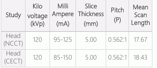
在尼泊尔Rupandehi的Bhairahawa世界医学科学与教学医院(UCMS-TH)无线电诊断和医学成像系进行了跨部门研究,使用直升机计算成像扫描仪(GE 16片,Brivo模型)。 研究得到了天主教卫生联合会机构审查委员会的批准,编号:UCMS/IRC/028/21。 2021年3月14日至2021年6月14日期间,共对75名儿科患者进行了头部CT扫描. 使用一种方便的取样技术,并使用公式n=z计算样本大小。(2)p(1-q)/d (中文(简体) ).(2),其中n=需要样本大小,z=1.96在95%的置信间隔,p=5%的估计流行率 (8) d=5%的错误。 将这些数值保留在上述公式中,样本大小被计算为约73个,但我们包括75个病人. 显示的管电流(mA)、管电压(KVp)、螺旋管片厚度(CTDIvol)和DLP是本研究采用的扫描参数。 需要这些参数来估计辐射剂量。 然而,管电流一直保持在86至150mA,固定管电压为120KV,整个研究的波段为0.562:1毫米. DLP和CTDIvol是独立录制的. 有效剂量通过采用换算系数(f)计算,{使用公式E=DLP.f}。 儿科CT头部成像的研究协议是常规的直升机. 这一协议由于扫描时间短,对患者的辐射剂量低,因此最受研究人员和技术人员青睐.
包含标准
* 1至15岁的病人。 * 任何性别的病人。
* 监护人知情同意研究的病人。
* 来接受CT头部检查的儿科病人。
排除标准
* 监护人即使在解释后也没有书面同意研究的病人。
· 任何绝对禁止治疗的病人。
统计分析
社会科学统计包第20版被用来分析收集的数据。 使用了体积CT剂量指数和剂量产品长度(DLP)来估计有效剂量。 我们提出数据为平均标准偏差。
结果
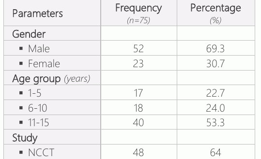
本研究包括75名儿科病人,平均平均年龄为9.85±4.45岁(范围为1-15岁)。 男性52人(69.3%),女性23人(30.7%)。 11-15岁年龄组的患者频率最高,为40人(53.3%),其次是6-10岁年龄组的18人(24%)。 1-5岁年龄组的患者人数最少,为17人(22.7%)。 在75名儿科病人中,48人(64%)接受了非连续计算成像法(NCCT),27人(36%)接受了对比计算成像法(CCT),如表1所示。
根据研究类型(NCCT和CCT),本研究使用的参数为kVp,mA,切片厚度,投球,以及平均扫描长度(表2).
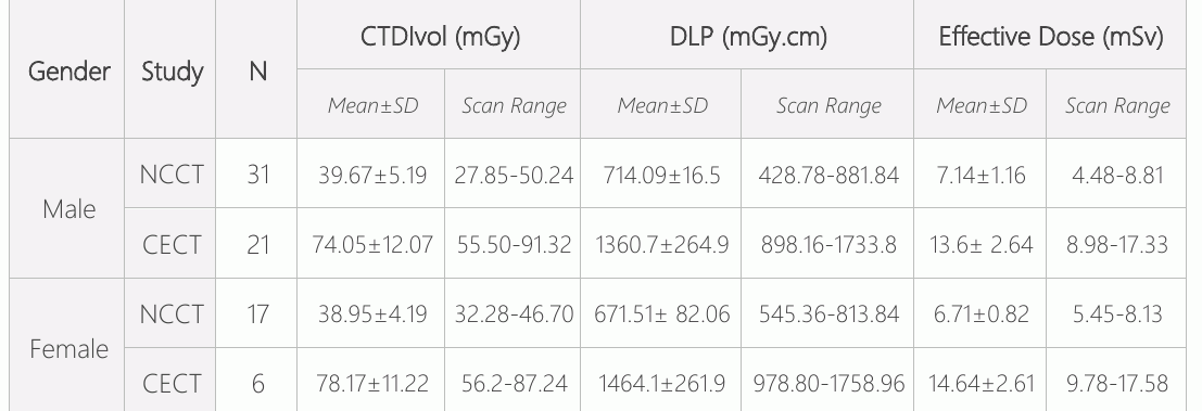
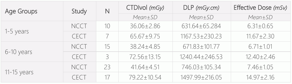
如表3所示,它审查了体积CT剂量指数(CTDIvol)、剂量长度产物(DLP)和有效剂量(ED),以及对儿科病人(男性和女性)的NCCT和ECT头部检查。 CTDIvol、DLP和ECT头部的平均值和标准偏差(SD)在男性和女性之间有很大差别。 表4显示每个年龄组的平均值和标准差。 CTDIvol、DLP和ED在11-15岁年龄组中更为重要,其次是6-10岁年龄组。
讨论情况
这项研究审查了用于估计儿科头部CT扫描辐射剂量的方法。 参加者的平均平均年龄为9.85±4.45岁,从1岁到15岁不等。 男性52人(69.3%),女性23人(30.7%)。 患者大多在11-15岁年龄组,频率为40人(53.3%),其次是6-10岁年龄组的18人(24%)。 Inoue Y等人进行了类似的研究。 (9)。 。 。
与Eddy FK等人比较.(10), Sulieman A等人著.(11),还有Kayun Z等人.(8),我们的研究结果显示,雌性对比增生和无连续CT扫描的体积CT剂量指数平均值为78.17±11.22 mGy & 38.95±4.19 mGy,其次是雄性74.05±12.07 mGy & 39.67±5.19 mGy;以及雌性对比增生和无连续CT的平均DLP为1464.16±261.93 mGy.cm & 671.51±82.06 mGycm,其次是雄性1360.71±264.97 mGycm & 714.09±116.5 mGy.cm;以及雌性对比增生和无连续CT的有效剂量平均值为14.64±2.61 mSv & 6.71±08.2 mSv,其次是雄性13.6±2.64 mSv & 7.14±1 mv. 在我们的研究中,CTDIvol,DLP,和ED的对比增强扫描的多数平均值是79.22±10.54 mGy,1497.99±216.05 mGy.cm,11-15岁年龄组的14.97±2.16 mSv,其次是6-10岁年龄组,这与Bouaoun A等人的研究比较. (12) 和赛义德·MK等人。 (13)10-15岁年龄组的ED,其次是5-10岁年龄组。 然而,我们的研究发现,辐射剂量没有显著变化。 由于所用机器和参数的不同,出现了微小差异.
作为限制,研究是在一个研究所进行的,所以我们的样本规模有限。 这项研究只针对1至15岁的病人。
结论
研究结论认为,男性儿科患者接受NCCT头部扫描的有效剂量较高,而女性儿科患者接受CECT头部扫描的有效剂量较高. 11-15岁年龄段的剂量最大,其次是6-10岁年龄组。
声明
- 作者贡献(CRediT)
- 所有作者对全部 14 项 CRediT 角色贡献均等(Conceptualization, Data curation, Formal analysis, Funding acquisition, Investigation, Methodology, Project administration, Resources, Software, Supervision, Validation, Visualization, Writing – original draft, Writing – review & editing)。
- 资助
-
Funding information was not reported in the published version of this article.
- 利益冲突
- A conflict of interest statement was not reported in the published version of this article.
- 伦理批准
- Inclusion Criteria ▪ Patients of age ranging from 1 to 15 years. ▪ Patients of any sex. ▪ Patients whose guardians have given informed consent to the study. ▪ Pediatric patients who have come for head CT examination.
- 数据可用性
- A data availability statement was not reported in the published version of this article.
参考文献 (13)
13 条参考文献中有 12 条已链接 DOI。
- Gao Y, Quinn B, Mahmood U, Long D, Erdi Y, St. Germain J, Pandit-Taskar N, Xu XG, Bolch WE, Dauer LT. A comparison of pediatric and adult CT organ dose estimation methods. BMC medical imaging. 2017 Dec; 17:1-7. DOI Crossref PubMed Google Scholar
- Alzimami K. Assessment of radiation doses to paediatric patients in computed tomography procedures. Polish journal of radiology. 2014;79:344. DOI Crossref PubMed Google Scholar
- Kadavigere R, Sukumar S. Estimation of radiation dose and establishment of local diagnostic reference levels for computed tomography of head in pediatric population. Journal of X-Ray Science and Technology. 2022 Jan 1(Preprint):1-9. PubMed Google Scholar
- Ogbole GI. Radiation dose in paediatric computed tomography: risks and benefits. Annals of Ibadan postgraduate medicine. 2010;8(2):118-26. DOI Crossref PubMed Google Scholar
- Inoue Y, Itoh H, Shiibashi N, Sasa R, Mitsui K. Sample size and estimation of standard radiation doses for pediatric brain CT. Tomography. 2022 Oct 1;8(5):2486-97. DOI Crossref PubMed Google Scholar
- Khong PL, Ringertz H, Donoghue V, Frush D, Rehani M, Appelgate K, Sanchez R. ICRP publication 121: radiological protection in paediatric diagnostic and interventional radiology. Annals of the ICRP. 2013 Apr;42(2):1-63. DOI Crossref PubMed Google Scholar
- Nievelstein RAJ, Van Dam IM, Van Der Molen AJ. Multidetector CT in children: Current concepts and dose reduction strategies. Pediatr Radiol. 2010;40(8):1324–44. DOI Crossref PubMed Google Scholar
- Kayun Z, Karim MK, Harun HH, Shaari AH, Mahmud R, Hamid HA, Sabarudin A, Chew MT. Radiation doses and size-specific dose estimate from CT brain examinations according to head sizes in a tertiary hospital in Malaysia. Radiation Physics and Chemistry. 2021 Dec 1;189:109694. DOI Crossref PubMed Google Scholar
- Inoue Y, Itoh H, Waga A, Sasa R, Mitsui K. Radiation dose management in pediatric brain CT according to age and weight as continuous variables. Tomography. 2022 Apr 1;8(2):985-98. DOI Crossref PubMed Google Scholar
- Eddy FK, Ngano SO, Jervé FA, Serge A. Radiation dose evaluation of pediatric patients in CT brain examination: multi-center study. Scientific Reports. 2021 Feb 25;11(1):1-6. DOI Crossref PubMed Google Scholar
- Sulieman A, Almogren KS, Tamam N. Radiation exposure estimation in pediatric patients during computed tomography imaging procedures. Radiation Physics and Chemistry. 2023 Mar 1;204:110653. DOI Crossref PubMed Google Scholar
- Bouaoun A, Ben Omrane L, Mogaadi M, Khomsi WD, Zidi A, Hammou A. Pediatric head CT exposure doses in Tunisia: a pilot study towards the establishment of national diagnostic reference levels. Radiation Protection Dosimetry. 2018 Dec 1;182(2):241-51. DOI Crossref PubMed Google Scholar
- Saeed MK, Almalki Y. Assessment of computed tomography radiation doses for paediatric head and chest examinations using paediatric phantoms of three different ages. Radiography. 2021 May 1;27(2):332-9. DOI Crossref PubMed Google Scholar
阅读全文
在此打开 PDF
正在加载 PDF…
若 PDF 未在此显示,请使用上方的下载按钮。
如何引用本文
Yadav SC, Dhakal D. Estimation of Effective Radiation Dose for Computed Tomography Head in Pediatric Patients at Universal College of Medical Sciences: A Cross-Sectional Study. Ulutas Med J. 2023;9(4):208-212. doi:10.5455/umj.20230714114808
Yadav, S. C., & Dhakal, D. (2023). Estimation of Effective Radiation Dose for Computed Tomography Head in Pediatric Patients at Universal College of Medical Sciences: A Cross-Sectional Study. The Ulutas Medical Journal, 9(4), 208–212. https://doi.org/10.5455/umj.20230714114808
Yadav, S.C. and Dhakal, D. (2023) 'Estimation of Effective Radiation Dose for Computed Tomography Head in Pediatric Patients at Universal College of Medical Sciences: A Cross-Sectional Study', The Ulutas Medical Journal, 9(4), pp. 208–212. doi: 10.5455/umj.20230714114808.