The Ulutas Medical Journal · ISSN 2149-0430 (print), 2149-388X (online) · https://www.ulutasmedicaljournal.com/?mno=171894
Original Article · Open access
Pisidium Guajava Ameliorates Testicular Distortions Triggered by Aluminium Chloride in Adult Male Wistar Rats
- Anatomy Department, Facultry of Basic Medical Sciences, College of Health Sciences, University of Ilorin, Ilorin Kwara State-Nigeria ROR
- Anatomy Department, Faculty of Basic Medical Sciences, College of Health Sciences, Benue State University Makurdi, Nigeria ROR
Abstract
- Introduction
- Aluminium (Al) is a trivalent cation that does not undergo redox changes. Extensive experimental shreds of evidence demonstrate in-vitro and in-vivo, that high Al concentrations cause oxidative stress; an imbalance between free radical generation and the antioxidant defense system. Oxidative stress induced by Aluminium is one of the major contributing factors to male reproductive disorders. Psidium guajava, the guava tree is widely distributed as a native plant in Latin America and Sub-Saharan Africa, with its leaves being deployed in folk medicine to manage several diseased conditions such as; diarrhoea, stomach ache, and hepatic problems. The study was conducted to evaluate the effects of an aqueous extract of Pisidium guajava on Aluminium (AlCl3) induced testicular damage.
- Materials and Methods
- Thirty adult male Wistar rats were divided randomly into six (6) groups of five (5) animals each and used for this study. Group A was orally administered with 0.5 ml of normal saline from the 1st to the 14th day. Group B got 0.5 ml of 100mg/kg body weight of aluminium chloride (AlCl3) only, from the 1st to 14th day. Group C got 0.5 mls of 250mg/kg body weight of Psidium gaujava leaf extract from the 1st to 14th day, Group D got 0.5 mls of 500 mg/kg body weight of Psidium guajava from the 1st to 14th day. Group E got 0.5 mls of 100mg/kg body weight of aluminum chloride from the 1st to 14th day, and 0.5 mls of 250 mg/kg body weight of Psidium guajava leaf extract from the 15th to 28th day. Group F got 0.5mls of 100 mg/kg body weight of aluminum chloride from the 1st to the 14th day, and 0.5mls of 500 mg/kg body weight of Psidium guajava leaf extract from the 15th to the 28th day. Administration was via the oral gavage.
- Results
- Oral administration of aluminum chloride adversely compromised the testicular histomorphology of Wistar rats, causing testicular atrophy associated with degeneration of germ cells within the seminiferous tubules, and enlarged lumen with fewer spermatids compared with the non-treated animals.
- Conclusion
- Guava leaf extract reversed the damage caused by aluminum chloride, restoring the distorted seminiferous tubules and presenting the lumen with densely packed primary spermatocytes.
Keywords
Full text
Introduction
Man is exposed to many environmental contaminants at different stages of life and exposure to aluminium (Al) due to industrial and environmental pollution poses severe ecological hazards (1). It has been adjudged the most widely distributed metal in the environment. It is believed to be the most prevalent metal and the third most crucial abundant element in the earth's crust; only oxygen (49.5%) and silicon (26%) occur more commonly than Al(8%)(2). In biological systems, Al is present only in trace amounts, shown to have no definite biological function and is poorly absorbed but efficiently eliminated when absorption does occur. Al is described mainly in bone, liver, testes, kidney, and brain, while its accumulation in tissues and organs results in dysfunction and toxicity (3).
The wide distribution of this element ensures the potential to exert harmful effects on human health upon exposure (4). Al compounds are widely used in medicines as antacids, vaccines, anti-diarrhea, phosphate binders, and allergen injections (5); food additives and toothpaste (6); water purification agents (7). It is still a metal of choice in making various household cookware and storage utensils.
Al contributes to various cognitive impairments in mice, rabbits and rats (8, 9). Epidemiological studies have indicated a link between Al in drinking water and Alzheimer’s disease, and a variety of human and animal studies have implicated learning and memory deficits post- Al exposure (10, 11). AlCl3 can generate reactive oxygen species (ROS) (12). In another instance, Al has been reported to have induced toxicity in the epididymis, vas deferens, seminal vesicle, and ventral prostrate of mice. AlCl3-induced free radicals and inhibited antioxidant enzymes in blood and seminal plasma, liver, testes, kidney, lung, and brain of rabbits (12, 13).
Antioxidants are substances that protect cells against the adverse effects of xenobiotics, carcinogens, drugs, and toxic agents (14). Since spermatogenesis and Leydig steroidogenesis are vulnerable to oxidative stress, the low oxygen tension that characterizes this issue may be an essential component of the mechanisms by which the testis protects itself from free radical-mediated damage (15).
The use of plants as medicines predates written human history. Nigeria has a vast repository of medicinal plants. It has thus paved the way for synchronizing traditional medicine into the healthcare delivery system, and the guava plant is one such example (16). The use of herbs to treat disease is almost universal among non-industrialized societies and is often more affordable than purchasing expensive modern pharmaceuticals (17).
Psidium guajava, the common guava tree, is widely distributed as a native plant in Africa and America from Mexico to Brazil. Whose leaves have been used in folk medicine for many years in different parts of the globe in managing various disease conditions and overcome severaldisorders, such as;rheumatism, diarrhea, diabetes mellitus, cough, and ulcers (18–21). The plant has also been reported to exhibit antimicrobial, analgesic and anti-inflammatory properties (22, 23).
Materials and Methods
Ethical approval for the study was given by the ethical committee of the College of Health Sciences, University of Ilorin, in line with the globally approved guidelines for the use of animals for scientific studies.
Thirty male adult Wistar rats, weighing between 160 to 250g, were obtained from a private animal farm in Ogbomoso, Oyo State, Nigeria, and maintained in the animal house of the College of Health Sciences, University of Ilorin.They were housed in cages with adequate space to encourage free movement under natural and dark cycles (12hr light and 12hr dark) at room temperature. They were bred on conventional commercial standard rat pellets. They were also supplied with water ad libitum and allowed to acclimatize to experimental conditions for 14 days before commencement of the experiments. Interval weighing of animals was done throughout. Strict hygiene was maintained by clearing their wastes daily.
Fresh leaves of P. guajava were obtained from a private farm at Tanke, a suburb of Ilorin, Kwara State-Nigeria. Aqueous extraction of guava leaf was achieved according to an existing procedure (24). In this procedure, fresh guava leaves were harvested from the botanical garden of the University of Ilorin, washed thoroughly, air-dried and pulverized using an electric blender. Two hundred grams (200g) of the powder obtained was macerated with 600mls of distilled water for 72 hours at 30±4oC. The mixture was sieved and filtered with Whatman No. 1 filter paper and the filtrate was concentrated in an electrically regulated oven at 45°C. Finally, concentrated crude aqueous extract was dried to a solid mass. The testing samples were prepared by dissolving the solid mass with normal saline to obtain a final concentration of 250 mg/kg body weight (bw) and 500 mg/kg body weight (bw) used for the experiment. Laboratory grade AlCl3 was locally sourced from Dennis and Dennis Laboratory, Taiwo Road, Ilorin-Nigeria. Other reagents & chemicals were of analytical grade. The thirty (30) adult male Wistar rats were divided randomly into six (6) groups of five (5) animals per group. Group A was orally administered with 0.5 ml of normal saline from the 1st to 14th day and served as the control group; Group B was orally administered with 0.5 ml of 100 mg/kg body weight of AlCl3 only from the 1st to 14th day. Group C was orally administered with 0.5 mls of 250mg/kg body weight of Psidium gaujava leaf extract from the 1st to 14 day; Group D was orally administered with 0.5mls of 500 mg/kg body weight of Psidium guajava leaf extract from 1st to 14th day, Group E was orally administered with 0.5 mls of 100 mg/kg body weight of AlCl 3 from 1st to 14 day, followed by the oral administration of 0.5 mls of 250 mg/kg body weight of Psidium guajava leaf extract from 15th to 28 day, Group F was orally administered with 0.5mls of 100 mg/kg body weight of AlCl3from 1st to 14th day, followed by the oral administration of 0.5mls of 500 mg/kg body weight of Psidium guajava leaf extract from 15th to 28th day. All administration was achieved via oral gavage.
While all the rats from Groups A to D were sacrificed on the 15th day of the experiment, Groups E and F were sacrificed on the 29th day of the experiment. At the end of the treatment protocols, the animals were euthanized with intramuscular injection of ketamine hydro-chloride (20 mg/kg). The testes were identified, removed, cleared of fat, and weighed using a sensitive analytical weighing balance and were placed in a solution of Bouin’s fluid and processed for histological light microscopic examination using routine hematoxylin and eosin, and immunohistological examination using Ki67 marker.
The right testes were placed in 0.25 mM of sucrose solution in the ratio of 1:4. Each of these tissues was separately transferred to a glass homogenizer containing the cold sucrose solution and were homogenized using an electrical homogenizer (Remi 8000 RPM). The unbroken cells and cell debris were removed by centrifuging at 3000 RPM for 15 minutes. The obtained supernatant(s) were used for the enzymatic estimations (antioxidant enzyme levels). Determination of follicle stimulating hormone (FSH), luteinizing hormones (LH), and testosterone were quantitatively measured by adopting an enzyme-linked immunosorbent assay (ELISA) technique using the Accu-Bind Microwell kit. Lake Forest, CA 92630, USA.
Results
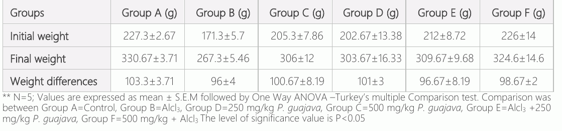
The effect of the oral administration of aqueous extract of P. guajava leaves on body weight of Alcl3-induced testicular toxicity is presented in Table 1. The weight of the experimental animals was measured once a week, and the analysis of the data obtained throughout the experiment indicates an increase in body weight in the control group and the P. guajava groups (250mg/kg and 500 mg/kg bw). The extract showed no significant changes (p<0.05) in body weight at all doses administered compared to the control group. However, the animals orally administered Alcl3 for 14 days recorded the lowest body weight, which was reversed when treated with the aqueous extract of guava leaves (250 mg/kg and 500mg/kg bw). However, the reversal effect was dose-dependent.
The effect of the oral administration of aqueous extract of P. guajava leaves on testicular weight of Alcl3-induced testicular toxicity is presented in Table 2. There was an increase in testicular weight in the control and P. guajava groups (250 and 500 mg/kg). The extract showed no significant changes (p<0.05) in testicular weight at all doses administered compared to control. However, the animals given oral administration of AlCl3 for 14 days recorded the lowest testicular weight, which was reversed when treated with an aqueous extract of guava leaves (250 and 500 mg/kg); however, the reversal effect was dose-dependent.
Biochemical evaluation
Quantification of reproductive hormones, follicle-stimulating hormone (FSH), luteinizing hormone (LH), and testosterone (T) was achieved via competitive enzyme immunoassay technique utilizing a polyclonal anti-LH, anti- FSH, and anti-T. These were done with ELISA at 450nm using the microplate readers, according to manufacturer’s specifications.
FSH
Follicle-stimulating hormones act directly on the seminiferous tubules, critical in stimulating both mitotic and meiotic DNA synthesis in spermatogonia (25). The effect of the oral administration of an aqueous extract of P. guajava leaves on FSH of the AlCl3-induced testicular toxicity is presented in Chart 1. An increment in FSH level was observed in the control group and the P. guajava groups (250 mg/kg and 500 mg/kg). The extract showed no significant changes (p<0.05) in FSH at all doses administered compared to control. However, the animals given oral administration of AlCl3 for 14 days recorded the lowest level of follicle-stimulating hormone, which was reversed when treated with an aqueous extract of guava leaves (250 mg/kg and 500 mg/kg). However, reversal effect was dose-dependent.
LH
Luteinizing hormone is a gonadotropic hormone primarily secreted by the anterior pituitary gland. It is a glycoprotein that regulates testosterone production from the Leydig cells' interstitial cells. The effect of the oral administration of an aqueous extract of P. guajava leaves on the luteinizing hormone of Alcl3-induced testicular toxicity is presented in chart 2. There was an increase in the level of luteinizing hormone in the control group and the P. guajava groups (250 mg/kg and 500 mg/kg). The extract showed no changes in luteinizing hormone at all doses administered compared with control. However, the animals given oral administration of AlCl3 for 14 days recorded the lowest luteinizing hormone, which was reversed when treated with an aqueous extract of guava leaves (250 and 500 mg/kg); however, reversal effect was dose-dependent.

TESTOSTERONE
Testosterone (T) is a C19 steroid and the most potent naturally secreted androgen. In the male, it is synthesized in the interstitial Leydig cells and regulated by luteinizing hormone. It is responsible for the development of secondary sexual characteristics. The effect of the oral administration of aqueous extract of P. guajava leaves on testosterone level of AlCl3-induced testicular toxicity is presented in chart 3. There was an increase in the level of testosterone in the control group and the P. guajava groups (250 and 500 mg/kg). The extract showed no changes in T levels at all doses administered compared to control. However, the animals given oral administration of AlCl3 for 14 days recorded the lowest testosterone, which was reversed when treated with an aqueous extract of guava leaves (250 and 500 mg/kg); however, the reversal effect was dose-dependent.
SOD
Superoxide Dismutase (SOD) presents the first line of defense against superoxide.It dismutases (O2-) anion to form O2 and H2O2. SOD protects spermatozoa against spontaneous O2 toxicity and Lipid peroxidation (26). Several reports suggest that AlCl3 may inhibit the activity of SOD and CAT (27, 28). The effect of the oral administration of an aqueous extract of P. guajava leaves on SOD activity of AlCl3-induced testicular toxicity is presented in chart 4. There was an increase in the level of SOD in the control group and the P. guajava groups (250 mg/kg and 500 mg/kg). The extract showed no changes in SOD at all doses administered when compared with control. However, the animals given oral administration of AlCl3 for 14 days recorded the lowest SOD, which was reversed when treated with an aqueous extract of guava leaves (250 mg/kg and 500 mg/kg); however, the reversal effect was again, dose-dependent.
MDA
Malondialdehyde (MDA) is a well-known lipid peroxidation indicator that increases in the testis and epididymis after Al exposure (29, 30). Increased lipid peroxidation might be caused by interference with mineral metabolism distribution. The capacity of Al to displace other biological cations such as calcium, iron, zinc, copper, and magnesium from their binding sites is a potential target for the adverse effects of Al (31). The effect of the oral administration of aqueous extract of P. guajava leaves on MDA activity of AlCl3-induced testicular toxicity is presented in chart 5. There was a decrease in the level of MDA in the control group and the P. guajava groups (250 mg/kg and 500 mg/kg). The extract showed no significant changes (p<0.05) in MDA level at all doses administered compared to control. However, the animals given oral administration of AlCl3 for 14 days recorded the highest level of MDA activity, which was reversed when treated with an aqueous extract of guava leaves (250 mg/kg and 500 mg/kg). However, the reversal effect was equally dose-dependent.
Discussion
Body and testicular weight observations
During close observation, the group of rats given 0.5ml of normal saline, 250mg/kg, and 500mg/kg B.W of Psidium guajava leaves extract orally for 14 days appeared normal and active all through the experiment, while the group treated with aluminum chloride only for 14 days were generally weak for about 5 to 10 minutes before picking up again, this contributed to the reduction of feed intake and in essence decrease in weight gain compared to the control, 250mg/kg and 500mg/kg BW of Psidium guajava. However, Psidium guajava-
Serum FSH Level
3
0 A
GROUPS
F
Serum LH Level
8
) 6 I/mI 4 (mU LH 2
0 A
GROUPS
F
Serum Testosterone Level
I) /mg 6 n e ( no 4 er st o 2 test
GROUPS
F
SOD ACTIVITY
0.25
I) 0.20
I/mo 0.15 M
(uD 0.10
O
S 0.05
GROUPS
F
MDA ACTIVITY
0.0025
I) 0.0020
I/m 0.0015
Mou
(A 0.0010
MD 0.0005
0.0000 A
GROUPS
F
treated rats were normal all through the experiment, depicting that aqueous extract of guava leaves ameliorated the inactiveness caused by aluminum chloride. The inactiveness observed in the aluminum-treated rats might be due to the induction of oxidative stress. Oxidative stress is recognized as the major mode of action of aluminum exposure as this has been observed in vitro in several types of cells and also in vivo in soft tissues such as the liver, kidney, brain, lung, and testes of experimental animals (7). Guava leaves, with its antioxidant properties, were observed to have ameliorated the effect induced by AlCl3, as all the groups that took 250 mg/kg and 500 mg/kg bw aqueous extract of the leaves appeared active and healthy. Also, the ultimate effect of testicular damage on body weight indicates its toxicity to health (32). However, there have been conflicting reports of correlations between testicular damage and weight changes in the body. In this current study, the weight of the experimental animals was measured once a week. Analysis of the data obtained from animal body weights monitored throughout the experiment indicates that animals in all the experimental groups gained weight. The group that gained weight most was the control group, which had a weight gain of 103.33±3.71. An increase was observed in the group given 250 mg/kg and 500 mg/kg BW of Psidium guajava leaves extract only, which had a rise of 100.67 ±8.19 and 101±3, respectively. A similar increase was observed in the treated groups given 250 mg/kg and 500 mg/kg BW of Psidium guajava leaves extract after a 100 mg/kg dosage of Alcl3 for 14 days. The last increase in weight was that of the group given 100 mg/kg of Alcl3 only for 14 days. However, the animals given oral administration of Alcl3 only for 14 days recorded the lowest body weight gain; this result is in tandem with previous findings that reported that during three months of observation of rats receiving AlCl3, a decrease in water and food intake and transient diarrhea occurred, which led to lowering of final body mass of animals compared to controls (33).The low body weight was reversed when treated with an aqueous extract of guava leaves (250 mg/kg and 500 mg/kg bw); however, the reversal effect was dose-dependent, and the weight gained showed no changes. This could be attributed to Psidium guajava leaf extract exerting its antioxidant effect; hence, it lessened the effect of AlCl3 on weight. This result corroborates the findings of another study, which also reported an increase in the weight of rats when administered 250 mg/kg, 500 and 750 mg/kg, aqueous extract of guava leaves for 14 days (34). Oral administration of AlCl3 significantly decreased testicular weight with 100 mg/kg bw. At 14 days compared to the control, the groups were administered 250 and 500 mg/kg bw. Aqueous extract of guava leaves. These findings aligned with another study where a decrease in absolute and relative testes and seminal vesicle weights after AlCl3 ingestion was reported (35). The decrease in the reproductive organs' weights could be due to the decrease in testosterone that may be attributed to oxidative damage induced in the rat testes. The decrease in testicular weight was noted to have been reversed when treated with an aqueous extract of guava leaves (250 and 500 mg/kg); however, reversal effect was dose-dependent. These findings are in tandem with another independent study where oral administration of 250 mg/kg and 500 mg/kg of aqueous extract of guava leaves increased body weight (36).
Hormonal and biochemical observations
The complex functioning of reproductive hormones is essential and closely linked with the temperance of spermatogenesis and sperm development. Co-secreted by gonadotrophin cells of the anterior pituitary, LH and FSH trigger T release by the Leydig cells of the testes in the male reproductive system, which affords a handy pointer to hormonal events in scenarios of significant distortions of sperm quality.
The results of the present study indicate that the treated rats with AlCl3 in a dose of 100 mg/kg daily for 14-day periods have shown a significant decrease in the serum FSH, LH, and testosterone, which are represented in bar charts 1-3, compared with the control group and the groups orally administered with 250 mg/kg and 500 mg/kg of guava leaves only. It also showed a significant decrease compared to the group treated with varying doses of aqueous guava leaves previously administered AlCl3 orally. AlCl3 can significantly adversely affect reproductive performance in the animal model (38). This effect can be exerted centrally, peripherally, or both. Aluminium is a voltage-sensitive calcium channel blocker and can affect neurotransmitter release (39). The results aligned with the earlier report that AlCl3 could generate reactive oxygen species in rabbit testes (12). Another finding equally showed that; exposure to aluminum lowered plasma and testicular testosterone in mice (9). A decrease in the LH release will lead to a reduction in the serum concentration of testosterone, as it is shown in this study. A decrease in testosterone level will eventually be reflected in peripheral sex organs functions, including epididymis and testis (40). The decrease in LH, FSH, and testosterone levels was reversed when treated with an aqueous extract of guava leaves (250 and 500 mg/kg bw). However, the reversal effect was dose-dependent. This finding is in line with a previous investigation, which showed that guava leaf extract improves semen quality in Adriamycin-induced testicular toxicity (41). Malondialdehyde (MDA) is a well-known lipid peroxidation indicator that increased in testis and epididymis after Al exposure (29, 30). Increased lipid peroxidation might be caused by the interference with mineral metabolism distribution. The capacity of Al to displace other biological cations such as calcium, iron, zinc, copper and magnesium from their binding sites is a potential target for the adverse effects of Al (31). Initial findings on intraperitoneal injection of Al chloride in male mice for two weeks significantly increased the testicular Al burden, broke the balance of essential minerals Zn, Cu and Fe which lead to increased lipid peroxidation, as well as decreased testis ACE (testis-specific angiotensin-converting enzyme) activity. Al ions might gradually displace Zn from induced MT or MT-like proteins and release Zn capable of synthesizing more thionein proteins with further binding of Al and other metals, including copper. Reports point to aluminum’s interference with iron homeostasis and severely impeding cellular metabolism. Aluminum also favors the generation of an oxidative environment because it creates a labile iron pool and interacts with membrane lipids (9). In this study, it was observed that testicular SOD activity of orally administered 100 mg/kg bw of AlCl3 was lower. At the same time, the MDA level was higher compared to the control, 250 mg/kg bw and 500 mg/kg bw of aqueous extract of guava leaves only. The group treated with 250 mg/kg and 500 mg/kg bw after oral administration of Alcl3 also showed an increased level of SOD and a decreased level of MDA compared to the AlCl3- treated group; however, the increase was dose-dependent. These findings aligned with those of a previous study where Al was shown to reduce antioxidants and increase lipid peroxidation (42). Another previous study had also reported Al-induced oxidative damage and the ability of Al to cross the blood-testis barrier after inducing oxidative stress and lipid peroxidation that damaged the biological membrane of the testes (43).
Histoarchitectural observations
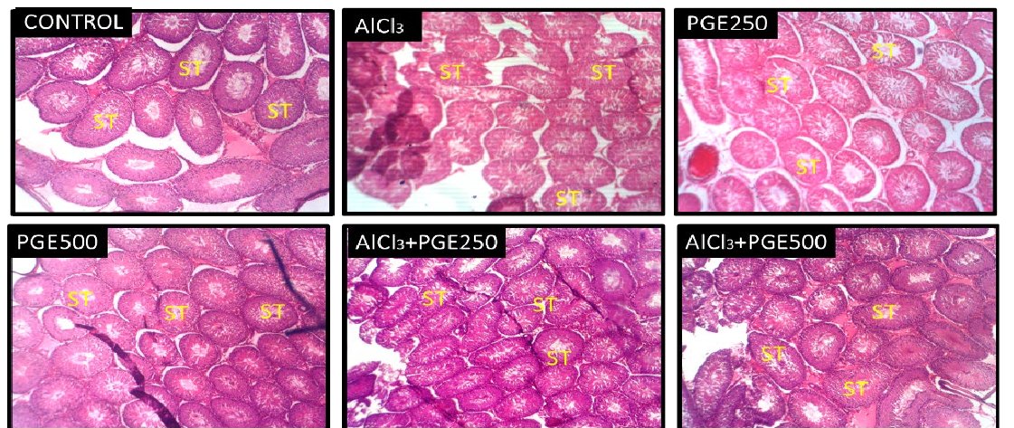
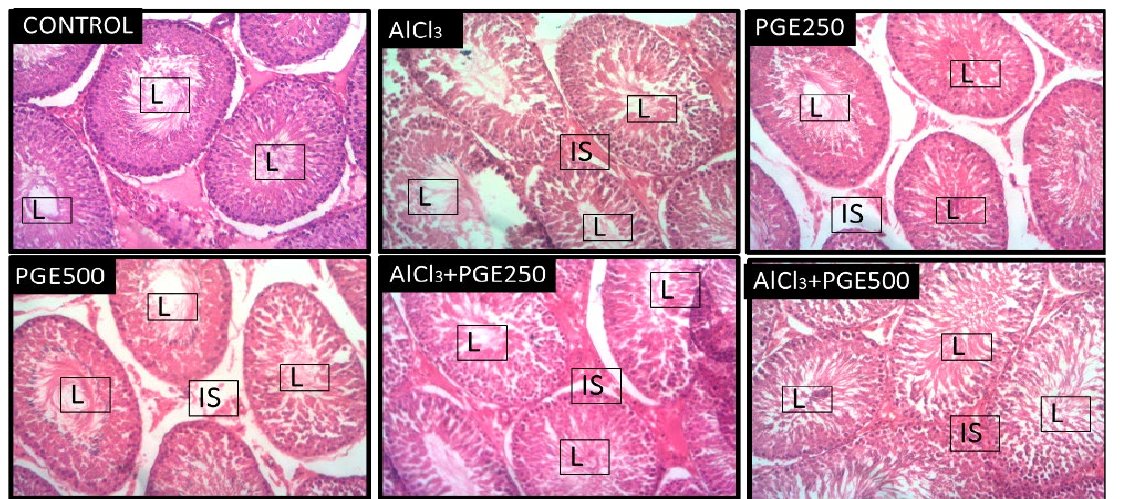
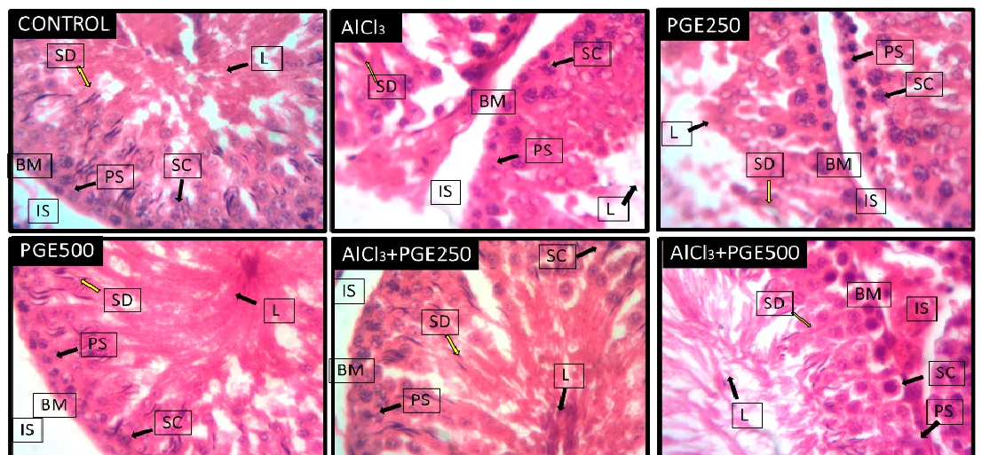
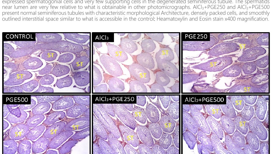
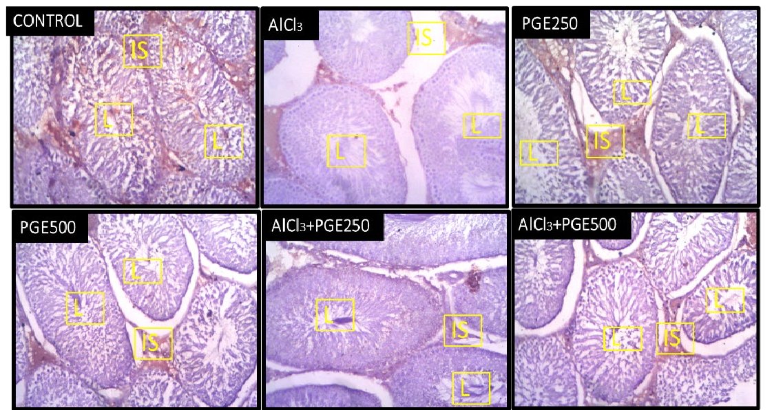
Testicular toxicity of AlCl3 was further confirmed by immunohistological observation in testicular tissues. These immunohistological observations revealed induced testicular atrophy associated with degeneration of germ cells within the seminiferous tubules, enlarged lumen and distortions in normalspermatogenic maturation when compared to the standard testicular architecture with a conspicuous orderly arrangement of germinal cells and Sertoli cells as observed in the control group and the groups administered 250 mg/kg and 500 mg/kg of aqueous extract of guava leaves. Testes treated with 100 mg/kg bw present intense necrosis, spermatogenesis disrupts, spermatozoa were absent in the lumen, and the seminiferous tubules' germinal epithelium was degraded. These findings agree with the data obtained from a previous study where in AlCl3- treated groups, histopathological examinations revealed apparent alterations in the testes, which induced marked lesions in seminiferous tubules (30). In addition, other documented findings reported that administering sodium fluoride and AlCl3 for 30d caused disorganized epithelium cell debris in the lumen and reduced sperm density (44). The immune-histological staining for Ki67 also revealed that the AlCl3 treated group was poorly stained, thereby revealing lesser cell proliferation than the control and treated groups.
Treatment with aqueous extract of Psidium guajava (250 and 500 mg/kg bw) showed both the drastic reduction in the histopathological features (testicular atrophy and vascular intimal erosion) and also expressed a large population of immunopositive cells as was obtained in the control groups.
Conclusion
Oral administration of AlCl3 distorted the testicular histomorphology of Wistar rats, causing testicular atrophy associated with degeneration of germ cells within seminiferous tubules, enlarged lumen with fewer spermatids compared with the non-treated animals. Aqueous extract of guava leaves was able to reverse the damage caused by AlCl3, reversing the distorted seminiferous tubules and presenting lumen with densely packed primary spermatocytes. A reversal in weight reduction caused by AlCl3 was also established.
Declarations
- Author contributions (CRediT)
- All authors contributed equally to all 14 CRediT roles (Conceptualization, Data curation, Formal analysis, Funding acquisition, Investigation, Methodology, Project administration, Resources, Software, Supervision, Validation, Visualization, Writing – original draft, Writing – review & editing).
- Funding
-
Funding information was not reported in the published version of this article.
- Conflicts of interest
- There are no conflicts of interest.
- Ethics approval
- Ethical approval for the study was given by the ethical committee of the College of Health Sciences, University of Ilorin, in line with the globally approved guidelines for the use of animals for scientific studies.
- Data availability
- A data availability statement was not reported in the published version of this article.
References (44)
32 of 44 references are linked to their DOI.
- McLachlan RI, O’Donnell L, Meachem SJ, Stanton PG, De-Kretser DM, Pratis K, Robertson DM. Identification of specific sites of hormonal regulation in spermatogenesis in rats, monkeys and man. Recent Prog Horm Res. 2002;57:149-179. DOI Crossref PubMed Google Scholar
- Kumar V, Gill KD. Aluminium neurotoxicity: neurobehavioural and oxidative aspects. Arch Toxicol. 2009;83:965-978. DOI Crossref PubMed Google Scholar
- Rahimzadeh MR, Rahimzadeh RM, Kazemi S, Amiri RJ, Pirzadeh M, Moghadamnia AA. Aluminium Poisoning with Emphasis on Its Mechanism and Treatment of Intoxication. Emerg Med Int. 2022:1480553. DOI Crossref PubMed Google Scholar
- Zhang K, Zhou Q. Toxic effects of Al-based coagulants on Brassica chinensis and Raphanus sativus growing in acid and neutral conditions. Environ Toxicol. 2005;20(2):179-87. PubMed Google Scholar
- Lione A. Aluminium toxicity and the aluminium containing medication. J Pharmacol Ther. 1985;29:255-285. PubMed Google Scholar
- Abbasali KM, Zhila T, Farshad N. Developmental Toxicity of aluminium from High Doses of AlCl3 in Mice. J Appl Res. 2005;5:575-579. PubMed Google Scholar
- Newairy AS, Salama AF, Hussien HM, Yousef MI. Propolis alleviate aluminium-induced lipid peroxidation and biochemical parameters in male rats. Food Chem Toxicol. 2009;47(6):1093-8. DOI Crossref PubMed Google Scholar
- Bilkei- Gorzo A. Neurotoxic effect of enteral Aluminium. Food Chem Toxicol. 1993;31:357-361. DOI Crossref PubMed Google Scholar
- Guo C, Huang C, Chen S, Wang HG. Serum and testicular testosterone and nitric oxide products in aluminum-treated mice. Environ Toxicol Pharmacol. 2001;10:53-60. DOI Crossref PubMed Google Scholar
- Exley C. The aluminium-amyloid cascade hypothesis and Alzheimer’s disease aluminium and β-amyloid. Alzheimer’s Dis. 2005;38:225-234. DOI: 10.1007/0-387-23265-11. DOI Crossref PubMed Google Scholar
- Buraimoh AA, Ojo SA, Hambolu JO, Adebisi SS. Histological study of the effects of aluminium chloride exposure on the testis of Wistar rats. Am Int J Contemp Res. 2012;2:114-122. PubMed Google Scholar
- Yousef MI. Aluminium- induced changes in hemato-biochemical parameters, lipid peroxidation and enzyme activities of male rabbits: protective role of ascorbic acid. Toxicology. 2004;199:47-57. DOI Crossref PubMed Google Scholar
- Yousef MI, El-Morsy AMA, Hassan MS. Aluminium-induced deterioration in reproductive performance and seminal plasma biochemistry of male rabbits: protective role of ascorbic acid. Toxicology. 2005;215:97-107. DOI Crossref PubMed Google Scholar
- Kurutas EB. The importance of antioxidants which play the role in cellular response against oxidative/nitrosative stress: current state. Nutr J. 2016;15:71. DOI Crossref PubMed Google Scholar
- Aitken RJ, Roman SD. Antioxidant systems and oxidative stress in the testes. Oxid Med Cell Longev. 2008;1(1):15-24. DOI Crossref PubMed Google Scholar
- Ogbadoyi EO, Abdulganiy AO, Adama TZ, Okogun JI. In vivo trypanocidal activity of Annona senegalensis Pers. leaf extract against Trypanosoma brucei brucei. J Ethnopharmacol. 2007;112(1):85-89. DOI Crossref PubMed Google Scholar
- Lifongo LL, Simoben CV, Ntie-Kang F, Babiaka SB, Judson NP. A Bioactivity Versus Ethnobotanical Survey of Medicinal Plants from Nigeria, West Africa. Nat Prod Bioprospect. 2014;4:1-19. DOI Crossref PubMed Google Scholar
- Gutiérrez RMP, Mitchell S, Solis RV. Psidium guajava: A review of its traditional uses, phytochemistry and pharmacology. J Ethnopharmacol. 2008;117:1-27. 226 77 Pisidium Guajava Ameliorates Testicular Distortions Kiekwe et al. DOI Crossref PubMed Google Scholar
- Sanda KA, Grema HA, Geidam YA, Bukar -Kolo YM. Pharmacological aspects of P. guajava: An update. Int J Pharmacol. 2011;7:316-324. PubMed Google Scholar
- Shruthi SD, Roshan A, Sharma S, Sunita S. A review on the medicinal plant Psidium guajava Linn. (Myrtaceae). J Drug Deliv Ther. 2013;3:162-168. DOI Crossref PubMed Google Scholar
- Morais- Braga MFB, Carneiro JNP, Machado AJT, Dos Santos ATL, Sales DL, Lima LF, Figueredo FG, Coutinho HDM. Psidium guajava L., from ethnobiology to scientific evaluation: Elucidating bioactivity against pathogenic microorganisms. J Ethnopharmacol. 2016;194:1140–1152. DOI Crossref PubMed Google Scholar
- Aarti R, Vikas M, Nitin S. Phytochemical screening, antimicrobial, antioxidant and cytotoxic potential of different extracts of Psidium guajava leaves. Int J Plant Res Biotechnol. 2020. PubMed Google Scholar
- Olajide OA, Awe SO, Makinde JM. Pharmacological studies on the leaf of Psidium guajava. Fitoterapia. 1999;70:25-31. DOI Crossref PubMed Google Scholar
- Ofodile LN, Nwakanma CNM, Mordi M, Ademolu O, Ezimoke I, Owoso J. Genotoxic and antimicrobial studies of the leaves of Psidium guajava. Eurasia J Biosci. 2013;7:60-68. DOI Crossref PubMed Google Scholar
- Oduwole OO, Peltoketo H, Huhtaniemi IT. Role of Follicle-Stimulating Hormone in Spermatogenesis. Front Endocrinol. 2018;9:763. DOI Crossref PubMed Google Scholar
- Alvarez JG, Touchstone JC, Blasco L, Storey BT. Spontaneous lipid peroxidation and production of hydrogen peroxide and superoxide in human spermatozoa: superoxide dismutase as a major enzyme protectant against oxygen toxicity. J Androl. 1987;8:336-348. DOI Crossref PubMed Google Scholar
- Al-Hashem FH. Camel’s Milk Alleviates Oxidative Stress and Lipid Peroxidation Induced by Chronic Aluminium Chloride Exposure in Rat’s Testes. Am J Appl Sci. 2009;6:1868-1875. DOI Crossref PubMed Google Scholar
- Anand P, Nehru B. Alterations in glutathione system in adult and pup rat brains following chronic aluminium exposure. Indian J Occup Environ Med. 2006;10:128-132. DOI Crossref PubMed Google Scholar
- Yousef MI, Kamel KI, El-Gvendi MI, El-Demerdash FM. An in vitro study on reproductive toxicity of aluminium chloride on rabbit sperm: the protective role of some antioxidants. Toxicology. 2007;239(3):213-223. DOI Crossref PubMed Google Scholar
- Hala AH, Khattab I, Abdallaha ZA, Kamel GM. Grape seed extract alleviate reproductive toxicity caused by aluminium chloride in male rats. J Am Sci. 2010;6:1200-1209. PubMed Google Scholar
- Yokel RA. Brain uptake, retention, and efflux of aluminum and manganese. Environ Health Perspect. 2002;110:699-704. DOI Crossref PubMed Google Scholar
- Ebong PE, Eyong EU, Eteng MU, Ukwe CN. Influence of chronic administration of chloroquine on Leydig cell integrity and testosterone profile of albino Wistar rats. Afr J Rep Heath. 1999;3(2):97-107. DOI Crossref PubMed Google Scholar
- Kowalczyk E, Kopff A, Kędziora J, Błaszczyk J, Kopff M, Niedworok J, Fijałkowski P. Effect of Long-Term Aluminium Chloride Intoxication on Selected Biochemical Parameters and Oxidative-Antioxidative Balance in Experimental Animals. Pol J Environ Stud. 2004;13(1):41-43. PubMed Google Scholar
- Udemezue OO, Ukoha U, Ezejindu DN, Okafor JI, Obilor AD. The histological effects of guava leaf aqueous extract on kidneys of adult wistar rats. Int J Res Med Health Sci. 2014;4(3):ISSN 2307-2083. PubMed Google Scholar
- Bataineh H, Al-Hamood MH, Elbetieha AM. Assessment of aggression, sexual behavior and fertility in adult male rat following long-term ingestion of four industrial metals salts. Hum Exp Toxicol. 1998;17:570-576. DOI Crossref PubMed Google Scholar
- Choudhury S, Sinha MP. Effects of Psidium guajava Aqueous Extract on Testosterone and Serum Lipid Profile of Albino Rats. Middle-East J Sci Res. 2014;21(10):1893-1897. DOI: 10.5829/idosi.mejsr.2014.21.10.84298. DOI Crossref PubMed Google Scholar
- Vigeh M, Smith DR, Hsu PC. How does lead induce male infertility? Iran J Reprod Med. 2011;9:1-8. PubMed Google Scholar
- Yousef MI, El-Morsy AMA, Hassan MS. Aluminium-induced deterioration in reproductive performance and seminal plasma biochemistry of male rabbits: Protective role of ascorbic acid. Toxicology. 2005. DOI Crossref PubMed Google Scholar
- Yousef MI, Salama AF. Propolis protection from reproductive toxicity caused by aluminium chloride in male rats. Food Chem Toxicol. 2009;47:1168–1175. DOI Crossref PubMed Google Scholar
- Shahraki MR, Palan MJ. Effect of Aluminium on Testosterone Hormones in Male Rat. J Med Sci. 2006;6:296-299. DOI Crossref PubMed Google Scholar
- Ola-Davies OE, Oloye AA, Oyeyemi MO. Ameliorating effects of guava (psidium guajava) extract on adriamycin induced reproductive toxicities. Folia Veterinaria. 2015;59(3):153-158. PubMed Google Scholar
- Anane R, Creppy EE. Lipid peroxidation as pathway of aluminium cytotoxicity in human skin fibroblast cultures: Prevention by superoxide dismutase plus catalase and vitamins E and C. Hum Exp Toxicol. 2001;20:477–481. DOI Crossref PubMed Google Scholar
- Kutlubay R, Oguz EO, Can B, Guven MC, Sinik Z, Tuncay OL. Vitamin E protection from testicular damage caused by intraperitoneal aluminium. Int J Toxicol. 2007;26:297-306. DOI Crossref PubMed Google Scholar
- Chinoy NJ, Momin R, Jhala DD. Fluoride and aluminium induced toxicity in mice epididymis and its mitigation by vitamin C. Fluoride. 2005;38:115–121. PubMed Google Scholar
Read the full text
Open the PDF here
Loading the PDF…
If the PDF does not show here, use the download button above.
How to cite this article
Oyeyemi AO, Olawepo A, Kiekwe V. Pisidium Guajava Ameliorates Testicular Distortions Triggered by Aluminium Chloride in Adult Male Wistar Rats. Ulutas Med J. 2023;9(4):213-226. doi:10.5455/umj.20231003030105
Oyeyemi, A. O., Olawepo, A., & Kiekwe, V. (2023). Pisidium Guajava Ameliorates Testicular Distortions Triggered by Aluminium Chloride in Adult Male Wistar Rats. The Ulutas Medical Journal, 9(4), 213–226. https://doi.org/10.5455/umj.20231003030105
Oyeyemi, A.O., Olawepo, A. and Kiekwe, V. (2023) 'Pisidium Guajava Ameliorates Testicular Distortions Triggered by Aluminium Chloride in Adult Male Wistar Rats', The Ulutas Medical Journal, 9(4), pp. 213–226. doi: 10.5455/umj.20231003030105.