The Ulutas Medical Journal · ISSN 2149-0430 (مطبوع), 2149-388X (إلكتروني) · https://www.ulutasmedicaljournal.com/?mno=161134
مقالة أصلية · وصول مفتوح
Estimation of Effective Radiation Dose for Computed Tomography Head in Pediatric Patients at Universal College of Medical Sciences: A Cross-Sectional Study
ترجمة آلية للعنوان والملخص والنص الكامل. الأصل الإنجليزي
- Universal College of Medical Sciences and Teaching Hospital, Bhairahawa, Rupandehi, Nepal ROR
الملخص
مقدمة: In modern medicine, CT has made enormous advances, but the use of CT has been accompanied by concerns about patient safety since it delivers higher doses of radiation than most other diagnostic imaging procedures. فالأطفال معرضون للخطر بوجه خاص لأن خلاياهم المتفرقة بسرعة أكثر حساسية للإشعاع، وسيكون أمامهم سنوات أكثر لإجراء تغييرات سرطانية. This study aimed to estimate the effective radiation dose received by pediatric patients while having CT heads at UCMS-TH. المواد والأساليب: وقد أُجريت هذه الدراسة الشاملة لعدة قطاعات في إدارة التصوير الطبي بالراديو، وشركة UCMS-TH، وبراهاوا، ونيبال. وقد أُدرج في هذه الدراسة خمسة وسبعون مريضا من مرضى الأطفال على مدى خمسة أشهر، من تشرين الثاني/نوفمبر 2020 إلى آذار/مارس 2021. وقُدرت الجرعات الإشعاعية باستخدام تقنية ملائمة وجرى تحليل البيانات باستخدام النسخة 20 من نظام SPSS. النتائج: بلغ متوسط عمر المشاركين 9.854.45 سنة. وكان هناك 69.3 في المائة من الذكور و 30.7 في المائة من الإناث. The age group 11-15 years had the highest frequency of patients 54.7%, followed by the age group 6-10 years with 24% patients. The mean and standard deviation of CTDIvol, DLP, and ED of NCCT and CECT head were significantly different between males and females throughout the study. الاستنتاج: In pediatrics, male NCCT head scans had higher ED, whereas female CECT head scans had a maximum ED. The age range of 11-15 years had the maximum ED, followed by the age group of 6-10 years.
الكلمات المفتاحية
النص الكامل
ترجمة آلية للنص الكامل؛ النسخة المنشورة الرسمية هي الأصل الإنجليزي. الأصل الإنجليزي
مقدمة
الترجيح الحاسوبي هو مصدر هام للتعرض للإشعاع (1)وتشير التقديرات إلى أن نحو 6 في المائة من الفحوصات التي أجريت على الأطفال دون سن الخامسة عشرة. الأطفال أكثر حساسية للإشعاع المؤين لذلك يجب حماية مرضى الأطفال منه (2–4)- وضعت اللجنة الدولية للحماية الإشعاعية مستوى مرجعي تشخيصي لتعظيم الجرعة (5, 6)- يصف مؤشر الجرعة المقطعية (CTDIvol) التعرض للإشعاع في المسح الأشعة المقطعية. مقياس استخدام الجرعات (DLP) للطاقة المودعة للمريض خلال المسح الأشعة المقطعية فيما يتعلق بمسح طوله (7)تُحسب الجرعة الفعالة (هاء) على أنها مجموع الجرعات المكافئة الممنوحة لكل عضو مضروبة بمعامل وزن الأنسجة (8)وكان الهدف من هذه الدراسة هو تقدير الجرعة الإشعاعية الفعالة التي يتلقاها مرضى الأطفال أثناء توليهم رئاسة CT في مستشفى الكلية العالمية للعلوم الطبية.
المواد والأساليب
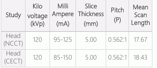
وأجريت دراسة شاملة لعدة قطاعات في إدارة التشخيص الإشعاعي والتصوير الطبي، والكلية العالمية للعلوم الطبية، ومستشفى التعليم، والبراهوة، وروبانديهي، ونيبال، باستخدام جهاز مسح للتصوير الضوئي الحاسبي المشوي (النموذج 16 من نوع " بريفو " ). وقد وافقت لجنة الاستعراض المؤسسي المشتركة بين الولايات المتحدة والولايات المتحدة الأمريكية (IRC) على الدراسة، مع الرقم: UCMS/IRC/028/21. وتم مسح ما مجموعه 75 مريضا من مرضى الأطفال بأشعة مقطعية من الرأس بين 14 آذار/مارس و 2021 و 14 حزيران/يونيه 2021. استُخدمت تقنية لأخذ العينات المناسبة، وحُسب حجم العينة باستخدام الصيغة:(2))ف(/د(2)حيث يُشترط حجم العينة، z=1.96 في 95% من فترة الثقة، p=5 في المائة (8) وخطأ متدني بنسبة 5 في المائة وحافظنا على هذه القيم في الصيغة المذكورة أعلاه، حسب حجم العينة بأنه نحو 73 مريضاً، ولكننا ضمنا 75 مريضاً. وكان تيار توبي (MA)، وفولط الأنابيب، وسماكة الشرائح الشائكة، وظهرت CTDIvol و DLP البارامترات المسحية المستخدمة في هذه الدراسة. وهذه البارامترات مطلوبة لتقدير جرعة الإشعاع. غير أن تيار الأنابيب كان يُحتفظ به في 86 إلى 150 ميغاواط مع تركيبة أنبوبية ثابتة قوامها 120 كيلو فولت أمبير وبطاقة تبلغ 0.562:1 ملم طوال الدراسة. وسُجِّل كل من حزب تحرير شعب الهادي وشركة CTDIvol بصورة مستقلة. (و) حُسبت الجرعة الفعالة من الـ DLP بتطبيق معامل التحويل (و)، {استعمال الصيغة E= DLP.f}. The study protocol for paediatric CT head imaging was routine helical. وكان الباحثون وعلماء التكنولوجيا يفضلون هذا البروتوكول أكثر من غيرهم نظراً لقصر فترة المسح وللجرعة الإشعاعية المنخفضة للمرضى.
معايير الإدماج
:: المرضى الذين تتراوح أعمارهم بين سنة واحدة و 15 سنة. :: مرضى أي جنس.
:: المرضى الذين وافق أوصياؤهم على الدراسة.
:: مرضى الأطفال الذين أتوا للفحص بالأشعة المقطعية.
معايير الاستبعاد
:: المرضى الذين لم يوافق أولياؤهم على الدراسة حتى بعد شرحها.
:: المرضى الذين يعانون من أي إهانة مطلقة بالاختصار بالنسبة للقطع العنقودية.
التحليل الإحصائي
واستُخدمت النسخة 20 من مجموعة الإحصاءات المتعلقة بالعلوم الاجتماعية لتحليل البيانات المجمعة. واستُخدم رقم قياسي للجرعات من طراز CT لتقدير الجرعات الفعلية. عرضنا البيانات على أنها انحراف عن المعايير
النتائج
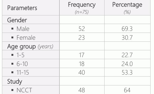
وشملت هذه الدراسة 75 مريضاً من الأطفال يبلغ متوسط أعمارهم 9.854.45 في المتوسط (من 1 إلى 15 سنة). وكان هناك 52 ذكرا (69.3 في المائة) و 23 امرأة (30.7 في المائة). The age group 11-15 years had the highest frequency of patients 40 (53.3%), followed by 18 (24%) patients in the age group 6-10 years. The age group 1-5 years had the smallest number of patients 17 (22.7%). ومن بين 75 مريضاً من الأطفال، أصيب 48 مريضاً (64 في المائة) بتصوير محاسبي غير مطابق، وحصل 27 (36 في المائة) على رسم قياسي مقارن معزز، على النحو المبين في الجدول 1.
According to the study types (NCCT and CECT), the parameters used in this study were kVp, mA, slice fishness,play, and mean scan length (Table 2).
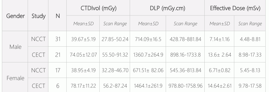
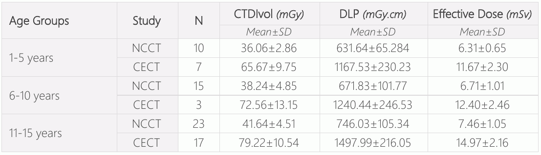
وكما هو مبين في الجدول 3، يفحص الرقم القياسي لجرعات الأشعة المقطعية (CTDIvol)، ومنتج طول الجرعة (DLP)، والجرعة الفعالة (ED) إلى جانب الفحوصات التي تجريها اللجنة الوطنية للوقاية من الأمراض المنقولة بالاتصال الحاسوبي المباشر والفحص الرأسي للإصابة بأمراض الأطفال (الذكور والإناث). وكان هناك فرق كبير بين الانحراف الحاد والتقليدي لـ " CTDIvol " و DLP و ED of the CECT head between males and females. ويبين الجدول 4 القيم والانحراف المعياري لكل فئة عمرية. The CTDIvol, DLP, and ED were more significant in the 11-15-years age group, followed by 6-10 years age group.
المناقشة
وفحصت هذه الدراسة الأساليب المستخدمة لتقدير الجرعات الإشعاعية للأشعة المقطعية لرأس الأطفال. وكان متوسط عمر المشاركين هو 9.854.45 سنة، تتراوح بين سنة واحدة و 15 سنة. وكان هناك 52 ذكرا (69.3 في المائة) و 23 امرأة (30.7 في المائة). The majority of the patients were in the age group of 11-15 years, with a frequency of 40 (53.3%), followed by 18 (24%) between the age group of 6-10 years. A similar study was conducted by Inoue Y et al (9).
بالمقارنة مع إددي FK et al.(10)(سليمان أ وآخرون)(11)و(كايون زي وآخرون)(8)الفقرتان الفرعيتان: وفي دراستنا، كانت غالبية القيمة الدنيوية للمسح المقارن للأشعة السيكولوجية (CTDIvol) وDLP 79.2210.54 mGy, 1497.99216.05 mGy.cm, and 14.972.16 mSv in the age groups of 11-15 years, followed by the age groups of 6- 10 years, which was compared to the study by Bouaoun A et al. (12) and Saeed MK وآخرون (13)ED in the age groups of 10-15, followed by the age groups of 5-10 years. غير أن دراستنا لم تجد أي تغير كبير في الجرعة الإشعاعية. وقد لوحظ اختلاف طفيف بسبب الفرق في الآلة والبارامترات المستخدمة.
As limitations, the study was carried out in a single institute, so our sample size was limited. The study was only conducted on patients aged between 1 and 15 years.
خاتمة
The study concluded that male paediatric patients received a higher effective dose for their NCCT head scan, whereas female paediatric patients had a higher effective dose for their CECT head scan. The age range of 11- 15 years had the maximum effective dose, followed by the age group of 6-10 years.
الإقرارات
- مساهمات المؤلفين (CRediT)
- ساهم جميع المؤلفين بالتساوي في جميع أدوار CRediT الأربعة عشر (Conceptualization, Data curation, Formal analysis, Funding acquisition, Investigation, Methodology, Project administration, Resources, Software, Supervision, Validation, Visualization, Writing – original draft, Writing – review & editing).
- التمويل
-
Funding information was not reported in the published version of this article.
- تضارب المصالح
- A conflict of interest statement was not reported in the published version of this article.
- الموافقة الأخلاقية
- Inclusion Criteria ▪ Patients of age ranging from 1 to 15 years. ▪ Patients of any sex. ▪ Patients whose guardians have given informed consent to the study. ▪ Pediatric patients who have come for head CT examination.
- إتاحة البيانات
- A data availability statement was not reported in the published version of this article.
المراجع (13)
12 من أصل 13 مرجعًا مرتبطة بمعرّف DOI.
- Gao Y, Quinn B, Mahmood U, Long D, Erdi Y, St. Germain J, Pandit-Taskar N, Xu XG, Bolch WE, Dauer LT. A comparison of pediatric and adult CT organ dose estimation methods. BMC medical imaging. 2017 Dec; 17:1-7. DOI Crossref PubMed Google Scholar
- Alzimami K. Assessment of radiation doses to paediatric patients in computed tomography procedures. Polish journal of radiology. 2014;79:344. DOI Crossref PubMed Google Scholar
- Kadavigere R, Sukumar S. Estimation of radiation dose and establishment of local diagnostic reference levels for computed tomography of head in pediatric population. Journal of X-Ray Science and Technology. 2022 Jan 1(Preprint):1-9. PubMed Google Scholar
- Ogbole GI. Radiation dose in paediatric computed tomography: risks and benefits. Annals of Ibadan postgraduate medicine. 2010;8(2):118-26. DOI Crossref PubMed Google Scholar
- Inoue Y, Itoh H, Shiibashi N, Sasa R, Mitsui K. Sample size and estimation of standard radiation doses for pediatric brain CT. Tomography. 2022 Oct 1;8(5):2486-97. DOI Crossref PubMed Google Scholar
- Khong PL, Ringertz H, Donoghue V, Frush D, Rehani M, Appelgate K, Sanchez R. ICRP publication 121: radiological protection in paediatric diagnostic and interventional radiology. Annals of the ICRP. 2013 Apr;42(2):1-63. DOI Crossref PubMed Google Scholar
- Nievelstein RAJ, Van Dam IM, Van Der Molen AJ. Multidetector CT in children: Current concepts and dose reduction strategies. Pediatr Radiol. 2010;40(8):1324–44. DOI Crossref PubMed Google Scholar
- Kayun Z, Karim MK, Harun HH, Shaari AH, Mahmud R, Hamid HA, Sabarudin A, Chew MT. Radiation doses and size-specific dose estimate from CT brain examinations according to head sizes in a tertiary hospital in Malaysia. Radiation Physics and Chemistry. 2021 Dec 1;189:109694. DOI Crossref PubMed Google Scholar
- Inoue Y, Itoh H, Waga A, Sasa R, Mitsui K. Radiation dose management in pediatric brain CT according to age and weight as continuous variables. Tomography. 2022 Apr 1;8(2):985-98. DOI Crossref PubMed Google Scholar
- Eddy FK, Ngano SO, Jervé FA, Serge A. Radiation dose evaluation of pediatric patients in CT brain examination: multi-center study. Scientific Reports. 2021 Feb 25;11(1):1-6. DOI Crossref PubMed Google Scholar
- Sulieman A, Almogren KS, Tamam N. Radiation exposure estimation in pediatric patients during computed tomography imaging procedures. Radiation Physics and Chemistry. 2023 Mar 1;204:110653. DOI Crossref PubMed Google Scholar
- Bouaoun A, Ben Omrane L, Mogaadi M, Khomsi WD, Zidi A, Hammou A. Pediatric head CT exposure doses in Tunisia: a pilot study towards the establishment of national diagnostic reference levels. Radiation Protection Dosimetry. 2018 Dec 1;182(2):241-51. DOI Crossref PubMed Google Scholar
- Saeed MK, Almalki Y. Assessment of computed tomography radiation doses for paediatric head and chest examinations using paediatric phantoms of three different ages. Radiography. 2021 May 1;27(2):332-9. DOI Crossref PubMed Google Scholar
اقرأ النص الكامل
افتح ملف PDF هنا
جارٍ تحميل ملف PDF…
إذا لم يظهر ملف PDF هنا، استخدم زر التنزيل أعلاه.
كيف تقتبس هذه المقالة
Yadav SC, Dhakal D. Estimation of Effective Radiation Dose for Computed Tomography Head in Pediatric Patients at Universal College of Medical Sciences: A Cross-Sectional Study. Ulutas Med J. 2023;9(4):208-212. doi:10.5455/umj.20230714114808
Yadav, S. C., & Dhakal, D. (2023). Estimation of Effective Radiation Dose for Computed Tomography Head in Pediatric Patients at Universal College of Medical Sciences: A Cross-Sectional Study. The Ulutas Medical Journal, 9(4), 208–212. https://doi.org/10.5455/umj.20230714114808
Yadav, S.C. and Dhakal, D. (2023) 'Estimation of Effective Radiation Dose for Computed Tomography Head in Pediatric Patients at Universal College of Medical Sciences: A Cross-Sectional Study', The Ulutas Medical Journal, 9(4), pp. 208–212. doi: 10.5455/umj.20230714114808.