The Ulutas Medical Journal · ISSN 2149-0430 (print), 2149-388X (online) · https://www.ulutasmedicaljournal.com/?mno=25847
Case Report · Open access
Ossifying Subperiosteal Hematoma of the Iliac Bone: Imaging Findings
- Cumhuriyet University, School of Medicine, Department of Radiology, Sivas, Turkey ROR
- Nazilli State Hospital, Department of Radiology, Nazilli, Aydın, Turkey ROR
Abstract
- Background
- Subperiosteal hematoma (SPH) of the iliac bone is a rarely reported posttraumatic lesion that predominantly affects young men. SPH often involves the skull, tibia, and iliac bones.
- Case report
- Herein, we aimed to report the plain radiography, computerized tomography (CT) and magnetic resonance imaging (MRI) findings of an ossified SPH of the left iliac bone in a 53-year-old man with a history of anticoagulant medication.
- Conclusions
- SPH of the iliac bone is typically an asymptomatic condition. Observing an ossified lesion in the shape of a lens on the inner aspect of the iliac wing in radiological examinations is diagnostic of this condition.
Keywords
Full text
Introduction
Subperiosteal hematoma (SPH) of iliac bone is a rarely reported posttraumatic lesion that predominantly affects young men. SPH often involves the skull, tibia, and iliac bones. The majority of these lesions have been reported as case reports, and the number of radiological reported cases is extremely small. Detected as biconvex lesions on the medial aspect of the iliac bone, these lesions are often detected incidentally by a computerized tomography examination (1–3). Herein, we aimed to report plain radiography, computerized tomography (CT) and magnetic resonance imaging (MRI) findings of an ossified SPH of the left iliac bone in a 53-year-old man with a history of anticoagulant medication.
Case Presentation
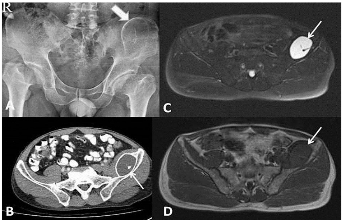
A 53-year-old man was referred by an outside center for CT examination for lower abdominal pain. He had no history of trauma but had used an anticoagulant agent for some time 12 months earlier. Laboratory findings were within normal limits. On physical examination, deep palpation showed rigidity in the left pelvic area. A plain pelvic radiogram showed a lesion of 8x6 cm in size that was superimposed on the left iliac bone and had a sclerosed periphery and a radiolucent center (Figure-1A). A CT examination revealed an ovoid-shaped lesion with an ossified periphery and a hypodense center at the level of the left iliac fossa, posterior to the iliac muscle, which made a wide angle with the inner tabula of the iliac bone. The lesion contained osseous septae in its central portion (Figure 1B). A pelvic MRI showed a smooth-bordered hyperintense area in the left iliac region that contained iso-/hypointense reas on T1-weighted series and patchy hyperintense areas on T2-weighted series and which compressed the iliacus muscle in the postero-anterior direction (Figure-1C and 1D). The adjacent bone marrow showed normal signal properties. There was no edema in neighboring soft tissue and bone. On diffusion-weighted images, there was no pathological limitation of diffusion. He was radiological diagnosed with a chronic ossified SPH.
Discussion
Subperiosteal hematoma of the iliac bone has been rarely reported, and usually occurs as a rare posttraumatic lesion affecting young males predominantly. It typically affects the skull, tibia, and iliac bones. SPH is seen in children and young adults and mainly results from any trauma. Affected patients are asymptomatic. The underlying pathophysiological mechanism is a loose attachment of periosteum to bone, causing vascular detachment during traumatic movements performed by young person’s (2–4). Acute hematoma may be completely healed but may also progress to ossification of variable degree. Peripheral curvilinear calcifications procedures. Observing an ossified lesion in the shape of a lens on the inner aspect of the iliac wing in radiological examinations is diagnostic of this condition.
develop 10-14 days after hematoma formation. Conflict of Interests As hematoma gradually becomes organized and is a resorbed, peripheral calcification increase (5, 6). It may be seen as a crescentric peripheral calcification surrounding uncalcified, irregular, and radiolucent center on plain radiograms. As the subperiostal area is a limited dense potential area, the hematoma assumes a typical double concave lens configuration, much like other subperiostal hematomas developing elsewhere. “Ghost native cortex'' sign has been recently defined and may aid in confirming its diagnosis. The latter is defined as the presence of a dense line between a lesion and residual bone marrow and has not been previously reported for other bone lesions. The presence of combination of all features on CT and MRI is almost pathognomonic (1, 7, 8). In addition to be radiation-free, MRI offers several advantages over CT in the diagnosis of subperiostal hematoma of the iliac bone. MRI may be superior to CT in acute and subacute phases for confirming nature of a hematoma based on its signal properties. Bone contusion and occult fractures can be easily detected by MRI. In chronic phase, subperiostal hematoma of the iliac bone can be reliably diagnosed by both CT and MRI (1, 4). In differential diagnosis of SPH, aneurysmal bone cyst, simple bone cyst and monostatic form of bone dysplasia should be considered (1, 2, 5).
In conclusion, SPH of the iliac bone is typically an asymptomatic condition, the typical form of which should be recognized using radiological studies to avoid possible unnecessary invasive The authors declare that they have no conflict of interest in the current study.
Declarations
- Author contributions (CRediT)
- All authors contributed equally to all 14 CRediT roles (Conceptualization, Data curation, Formal analysis, Funding acquisition, Investigation, Methodology, Project administration, Resources, Software, Supervision, Validation, Visualization, Writing – original draft, Writing – review & editing).
- Funding
-
Funding information was not reported in the published version of this article.
- Conflicts of interest
- The authors declare that they have no conflict of interest in the current study.
- Ethics approval
- Ethics approval details were not reported in the published version of this article.
- Data availability
- A data availability statement was not reported in the published version of this article.
References (8)
8 of 8 references are linked to their DOI.
- Guillin R, Moser T, Koob M, Khoury V, Chapuis M, Ropars M, et al. Subperiosteal hematoma of the iliac bone: imaging features of acute and chronic stages with emphasis on pathophysiology. Skelet Radiol. 2012; 41: 667– 75 DOI Crossref PubMed Google Scholar
- Ben Zakoun J, Dallaudière B, Palazzo E, Lefere M, Monteil J, Dieudé P. Chronic ossified subperiosteal hematoma of the iliac bone. Diagn Interv Imaging. 2014; 95: 889-91 DOI Crossref PubMed Google Scholar
- Pirouzmand F, Midha R. Subacute femoral compressive neuropathy from iliacus compartment hematoma. Can J Neurol Sci. 2001; 28: 155– 8 DOI Crossref PubMed Google Scholar
- 4.Patel A, Calfee R, Thakur N, Eberson C. Non-operative management of femoral neuropathy secondary to a traumatic iliacus haematoma in an adolescent. J Bone Joint Surg Br. 2008; 90: 1380– 1 DOI Crossref PubMed Google Scholar
- Gitto S, Pescatori LC, Aliprandi A, Sconfienza LM. Subperiosteal hematoma of the ilium: an unusual complication of acetabular fracture. doi: 10.1594/EURORAD/ CASE.14182. DOI Crossref PubMed Google Scholar
- Naude RJ, Thomson SR. Femoral nerve palsy following a subperiosteal haematoma. Injury. 1993; 24: 62 –3 DOI Crossref PubMed Google Scholar
- Wada Y, Yanagihara C, Nishimura Y. Bilateral iliopsoas hematomas complicating anticoagulant therapy. Intern Med. 2005; 44: 641– 3 DOI Crossref PubMed Google Scholar
- Higashi R, Hayashi Y, Watanabe T, Hirota Y, Park CH, Hamada J. Huge recurrent subperiosteal hematoma in a 9-year-old boy: case report with special reference to pathologic and anatomic studies. Childs Nerv Syst. 2008; 24: 403– 5 DOI Crossref PubMed Google Scholar
Read the full text
Open the PDF here
Loading the PDF…
If the PDF does not show here, use the download button above.
How to cite this article
Atalar MH, Baspinar N. Ossifying Subperiosteal Hematoma of the Iliac Bone: Imaging Findings. Ulutas Med J. 2019;5(1):107-109. doi:10.5455/umj.20190113113349
Atalar, M. H., & Baspinar, N. (2019). Ossifying Subperiosteal Hematoma of the Iliac Bone: Imaging Findings. The Ulutas Medical Journal, 5(1), 107–109. https://doi.org/10.5455/umj.20190113113349
Atalar, M.H. and Baspinar, N. (2019) 'Ossifying Subperiosteal Hematoma of the Iliac Bone: Imaging Findings', The Ulutas Medical Journal, 5(1), pp. 107–109. doi: 10.5455/umj.20190113113349.