The Ulutas Medical Journal · ISSN 2149-0430 (print), 2149-388X (online) · https://www.ulutasmedicaljournal.com/?mno=256234
Original Article · Open access
The Other Breast Matters: Unexpected Pathologies in Oncoplastic Symmetry Procedures
Also available in: العربية · Türkçe · 中文
- Department of General Surgery, University of Health Sciences, İzmir Training and Research Hospital, İzmir, Turkey ROR
- Department of General Surgery, Bartın State Hospital, Bartın, Turkey ROR
- Department of General Surgery, Çiğli Kent Hospital, İzmir, Turkey ROR
Abstract
- Background
- Oncoplastic breast surgery allows high-volume excision without compromising aesthetics. We aimed to analyze the probability of identifying high-risk malignant lesions in the contralateral breast.
- Materials and Methods
- The study analyzed patients who underwent oncoplastic surgery between January 2000 and December 2023. All the patients underwent a preoperative assessment of the contralateral breast using mammography, ultrasonography, and breast MRI. Pathology reports were reviewed to identify malignant and high-risk lesions.
- Results
- 38 patients had left-sided breast cancer, and 44 had right-sided breast cancer. The distribution of the pathology results was invasive ductal carcinoma in 68 patients, invasive lobular carcinoma in 11 patients, malignant phylloid tumor in one patient, borderline phylloid tumor, and metaplastic carcinoma in one patient. In contralateral breast pathology, 12 patients had low-risk lesions and 12 patients had high-risk lesions. Two patients had invasive tumors in the contralateral breast.
- Conclusion
- Although detailed preoperative imaging is performed in patients undergoing oncoplastic breast surgery, a significant number of high-risk or malignant lesions may be present in the contralateral breast.
Keywords
Full text
Introduction
Oncoplastic breast surgery has evolved from breast-conserving surgery by expanding the indications for breast cancer surgery to achieve wide resection margins without compromising cosmetic and oncological principles (1). Contralateral breast symmetry should be considered as an important component of oncoplastic surgery. Major procedures include volume replacement techniques depending on breast volume, tumor size, and location. Moreover, these procedures are more complex and timeconsuming than breast-conserving surgery. Women with breast cancer on the opposite side of the breast have a high risk of developing a new tumor. A significant portion of the opposite breast may also be diagnosed with breast cancer. It can be removed surgically and a fairly large sample can be taken for pathological examination (2–4). Complex fibroadenoma, simple ductal hyperplasia, sclerosing adenosis, papillamatosis, and radial scar were breast lesions in the low-risk group, but these lesions increased the risk of breast cancer by 1,5-2 times (5). Fibrocystic changes, mild hyperplasia, and fibroadenomas are considered benign lesions. Breast (6). Atypical ductal hyperplasia (ADH), Ductal carcinoma in situ (DCIS) and atypical lobular hyperplasia (ALH) are high-risk breast lesions, and invasive carcinoma is considered malignant (7, 8). The aim of the study was to identify malignant or high-risk lesions in the contralateral breast tissue of patients with breast cancer using reduction mammoplasty.
Materials and Methods
This retrospective analysis of oncoplastic reduction mammoplasty performed between January 2000 to December 2023 included 82 patients with breast cancer and macromastia. The pathology reports of the patients from the hospital automation system and cases diagnosed with malignant and high-risk lesions were examined.
Patients with benign lesions and those in the low-risk group were excluded from the study. These patients were excluded to ensure the study focused on clinically significant, high-risk, or malignant lesions that could impact treatment planning, prognosis, and surgical decision-making. Including low-risk or benign findings would have diluted the statistical power and clinical relevance of the analysis. Patients diagnosed with breast cancer underwent physical examinations, laboratory tests, and radiological imaging. Breast ultrasound, mammography, abdominal ultrasound, Breast MRI and direct lung radiography were performed in all patients. In the last ten years, this examination has been performed by a breast-specific radiologist.
All patients were evaluated at the oncology council before surgery. Surgery was performed in T1-T2 patients, and T3 and T4 patients were referred for neoadjuvant therapy. Suitable patients were included in the study after neoadjuvant treatment. Oncoplastic surgery was performed in all the patients without compromising oncological principles.
Patients’ selection Inclusion criteria: 1. Breast cancer patients with macromastia underwent oncoplastic surgery; 2. Mammography and breast ultra-sonography, Breast MRI were performed for preoperative contralateral breast evaluation; 3. Jan 2000 to Dec 2023 surgery women with macro-mastia ≥18 years old who had breast cancer surgery; 4. Histologically confirmed primary unilateral breast cancer.
Exclusion criteria: 1. Patients with clinically or radiologically suspicious lesions in the opposite breast were excluded from the study; 2. Stage IV breast cancer; 3. Patients with BRCA mutation; 4. Patients who have undergone mastectomy; 5. Patients with benign lesions or in the low-risk group were excluded from the study; 6. Patients who do not have sufficient data and are lost to follow-up.
Statistical Methodology
Data were collected using IBM SPSS Statistics Standard Concurrent User v26 (IBM Corp., Armonk, New York, USA) and evaluated using the statistical package program. Descriptive statistics are given as number of units (n), percentage, mean±standard deviation, median (interquartile range), and minimum and maximum values. The normality of the data was evaluated using the Shapiro-Wilk test. The breast size and number of lesions removed in the low- and high-risk groups with no normal side pathology were compared using the Kruskal-Wallis test. Fisher’s exact test was used to compare the groups with categorical variables. If the Fisher’s exact test result was found to be significant, subgroup analyses were performed usingtwo Bonferroni-corrected ratio z tests. Differences were considered statistically significant at p<0.05.
Results
Eighty-two cases were retrospectively analyzed in the study. The mean age of the patients was 51.1±9.7 years. The number of menopausal patients is 31 (37.8%) were menopausal. Thirty-eight patients (46.3%) had left breast cancer, and 44 (53.7%) had right side. Mortality occurred in 11 patients (13.4%) during follow-up period. Demographic data, radiological and pathological stages, localization, multicentricity, tumor size, axillary status, surgical information, neoadjuvant treatment, receptor, removed breast volume, pathological diagnoses of the tumor side, pathological findings of the nontumor side, complication data, and mortality are shown in Table 1. In 56 of 82 patients, no pathology was detected in the contralateral breast pathology.
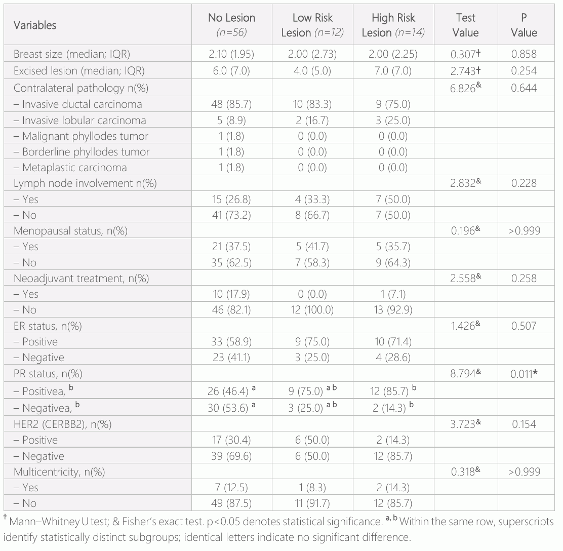
In the pathological examination of the contralateral breast of 26 patients who were reported as normal on imaging, low-risk lesions were detected in 12 patients, high-risk lesions in 12 patients and invasive tumors in 2 patients. The findings related to the contralateral pathologies of the patients are given in Table 2. There was no statistical difference in breast size and the presence of incidental lesions (p=0.858). When evaluated according to histopathologic results, there was no statistically significant difference in the likelihood of malignant or high-risk lesions on the contralateral side (p=0.644). There was no significant difference in the distribution of lymph node involvement between the groups (p=0.228). Again, the distribution of menopausal and non-menopausal patients did not differ significantly (p=0.999). No patient in the low-risk group received neoadjuvant treatment. There is no significant difference between the neoadjuvant treatment distributions of the groups. There is no difference between ER positivity (p=0.507). The number of patients with PR positivity in the high-risk group was statistically higher than the group without pathology (p=0.011). The CERBB2 distributions of the groups are not different (p=0.154). The multicentricity distributions of the groups are not different (p=0,999). Multicentricity was observed in 12.2% of the overall cohort not limited to patients with contralateral lesions. This rate reflects the presence of multiple tumor foci within the same breast rather than across both breasts. Among the 11 patients who received neoadjuvant treatment, a partial response was observed in 7 patients (63.6%), stable disease in 2 (18.2%), and complete radiologic response in 2 patients (18.2%). Despite the tumor size reduction in some cases, incidental lesions in the contralateral breast were still detectable during surgery and histopathological analysis.
A comparison of the breast size and lesion no difference between the rates of lesions in the opposite breast in terms of menopausal status (pre/postmenopausal patients). However, no significant differences were found in terms of breastsize, contralateral pathology, lymph node involvement, menopausal status, neoadjuvant treatment distribution, ER positivity, and multicentricity.
Given the small number of patients with high-risk or malignant lesions in the contralateral breast (n=3), the statistical power for subgroup analyses was limited. Therefore, findings related to these subgroups should be interpreted with caution.
Discussion
Oncological principles require the safe removal of breast cancer in such a way as not to leave cancerous tissue behind. Sometimes, there are deformities in the breast that are left behind, which severely impair cosmetic appearance. Therefore, new operations are required to correct these deformities. The inability of the correction surgeries performed to provide results at the expected level have also caused oncoplastic surgery to come to the fore (9). In the case of removal of 20% or more of the breast volume, oncoplastic techniques should be used to prevent the formation of deformities and achieve cosmetically successful results. The method combines the principles of oncological and plastic surgery. Thus, breast reconstruction surgery is planned simultaneously for women who will undergo surgery for breast cancer, and it is ensured that women who have previously lost their breasts due to cancer regain their breasts (10, 11). In a study of patients with breast cancer and macromastia, the mean age was 59.6 years (12). In our study, the mean age was 51.1±9.7 year. The youngest patient was 25 years of age. Ten patients were aged <40 years; therefore, not only patients aged ≥40 years but also all patients who are scheduled for surgery should be examined in more detail. The study's sample size of 82 patients may compromise the statistical power, particularly when conducting subgroup analyses. For instance, malignant or high-risk lesions detected in the contralateral breast was minimal (n=3). Although similar sample sizes are documented in the literature, the small number of patients with high-risk or malignant lesions in the contralateral breast limits the statistical power of subgroups. It reduces its ability for detecting meaningful associations and may explain the lack of differences in some variables.
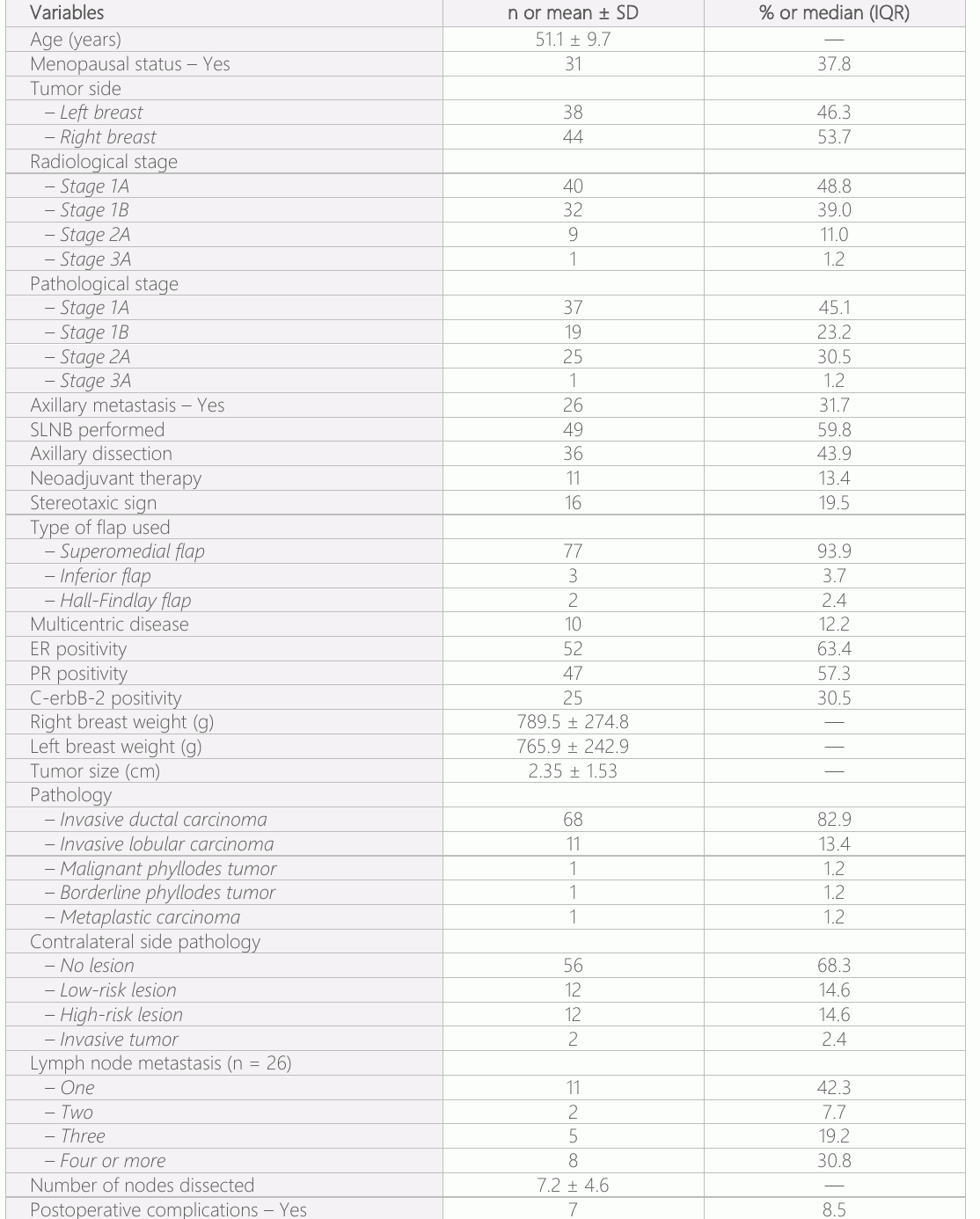
Regarding the relationship between breast size and tumor presence, Kusano et al. reported that the incidence of breast cancer was higher in women with bra cup D and larger than in those with bra cup A and smaller (13). All breast volumes of the patients in our study consisted of bra D and above patients, and the extracted breast volume was found to be 789.5±274.8 g for the right breast and 765.9±242.9 g for the left breast. A study by Boughey JC et al (14). on bilateral breast cancer reported that the tumour characteristics of patients with bilateral breast cancer differed from those of patients with unilateral breast cancer. It was highlighted that there were differences, particularly in terms of tumour subtypes and biological behavior. Approximately 15% of all breast cancers are lobular carcinomas, and approximately 6-28% of these are bilateral. Later, contralateral carcinoma development occurs in 9-14% of cases, and 50% of these were invasive (15, 16) our study, although not statistically significant, high-risk disease was detected in 27.3% with lobular cancer (17).
The bilateral incidence of lobular carcinomas varies between 6-28% (18). No pathology was detected in the contralateral breast in 8 of the 11 patients with invasive lobular carcinoma in our study. Ductal carcinoma was detected in one of the three patients, atypical ductal hyperplasia in the other, and atypical lobular hyperplasia in the third. In our study, lymph node involvement (50%) was found in breast cancers with high-risk lesions on the contralateral side (19).
Neoadjuvant therapy is an important practice that has been discussed and applied worldwide since the 1960s and has started to enter into routine after 2000 years (20). Neoadjuvant therapy were applied in our clinic in accordance with the guidelines since 2010, in which 11 patients underwent neoadjuvant chemotherapy and all patients had a complete or partial response to chemotherapy. The outcomes of neoadjuvant treatment may influence the detectability and evaluation of contralateral lesions. While tumor shrinkage can improve surgical margins and visibility, it does not eliminate the risk of incidental lesions in the contralateral breast. Therefore, routine preoperative imaging and intraoperative assessment remain essential, regardless of neoadjuvant response.
There are studies in the literature indicating that the prognosis of ER-positive patients is better, but there are no studies indicating a relationship between hormone receptor status or C-erbB-2 positivity and bilaterality (21). In our study, PR positivity was significantly associated with a higher incidence of high-riskor malignant lesions in the contralateral breast (p=0.011). This finding is noteworthy, as progesterone receptor expression is considered a favorable prognostic marker in breast cancer due to its association with hormone receptor-positive, indolent tumors that respond well to endocrine therapy. However, our results suggest that PR positivity may also indicate an increased likelihood of harboring synchronous high-risk or malignant lesions in the contralateral breast.
This association has important implications. Patients with the PR-positive tumors may benefit from more comprehensive preoperative evaluation of contralateral breast, potentially including advanced imaging modalities such as breast MRI, which offers high sensitivity for detecting occult lesions. Identifying such lesions preoperatively could influence surgical decisions, such as considering bilateral surgery over a unilateral approach. More rigorous surveillance strategies may be warranted in PR-positive patients during follow-up.
The observed association may reflect an underlying hormonal responsiveness that predisposes both breasts to proliferative changes. It is possible that PR-positive tumors arise in a hormonal microenvironment that promotes multifocality or bilateral disease. Further studies are needed to elucidate the biological mechanisms involved and to determine whether PR status could serve as a marker for increased contralateral breast risk, ultimately guiding screening, surgical planning, and individualized patient care.
Although the hormone receptor status of ER and C-erbB-2 was not significant in either group, the incidence of lesions in the breast increased significantly compared with PR positivity. The prevailing consensus in the extant literature supports the notion that positive public relations are a known prognostic factor. However, the association between positive PR and bilateral high-risk lesions remains to be elucidated. They are predictive and prognostic factors of breast cancer. Progesterone-positive patients are considered to have a better prognosis. It is important to note that this finding is independent of contralateral breast lesions and represents intramammary tumor multiplicity, which may have implications for surgical planning and risk stratification. In our study, the incidence of multicentric lesions was 12.2%, which is consistent with the literature (22, 23), extending beyond those with contralateral breast lesions. This percentage indicates the occurrence of multiple tumor foci within a single breast, rather than involvement of both breasts. In oncoplastic breast surgery, surgical treatment of cancer without compromising oncological principles is the primary goal. Improving the patient's quality of life and preserving body integrity during cancer surgery requires leaving a breast with an acceptable aesthetic appearance. The aim was to reconstruct the opposite breast in order to ensure symmetry (24, 25).
A study by Jackson et al (26). examined the rate of malignancy in the opposite breast in symmetry procedures performed during oncoplastic breast-conserving surgery, finding it to be approximately 5%. The study analyzed 289 patients, detecting malignant lesions in 5 patients (36%) in the contralateral breast tissues obtained from 100 patients. This finding underscores the significance of incidental detection of malignancy in the contralateral breast, emphasizing the need for vigilance in such cases. A retrospective study by De Rambukwella et al (27). analyzed 84 patients who underwent large-volume oncoplastic surgery, revealing that 16.7% of malignant or high-risk lesions were detected in the contralateral breast. These lesions included invasive ductal carcinoma (IDC), ductal carcinoma in situ (DCIS), lobular carcinoma in situ (LCIS) and atypical hyperplasia. The study emphasizes that the possibility of incidental malignancy should be considered when tissue sampling is performed during symmetry procedures in the opposite breast. In our study’s sample size of 82 patients may compromise the statistical power, particularly when conducting subgroup analyses. For instance, the number of patients with malignant or high-risk lesions detected in the contralateral breast was minimal. However, some similar studies with the same number of patients are documented in the literature (27).
Our analysis found no relationship between premenopausal and postmenopausal patients in terms of incidental lesion occurrence in the contralateral breast. This finding suggests that menopausal status may not be a distinguishing factor in predicting the presence of occult high-risk or malignant lesions in the opposite breast. Therefore, premenopausal and postmenopausal patients may warrant equally thorough preoperative evaluation and pathological assessment during symmetry procedures.
These studies support the hypothesis that the likelihood of finding incidental lesions increases with an increase in the amount of tissue removed during surgery. Consequently, it is recommended that a meticulous examination and histopathological analysis of excised tissues during symmetry procedures be considered. This study makes an important contribution to the detection of malignant or high-risk lesions in the opposite breast in breast cancer patients with macromastia. However, the retrospective design of the study, the limited number of patients, and the lack of detailed examination of some clinical parameters are some of the shortcomings of this study. Elimination of these deficiencies in future studies will make the results of the study more reliable and generalizable. Furthermore, a more detailed examination of the effect of genetic factors and neoadjuvant treatment on lesion development in the opposite breast may make important contributions to clinical practice.
Conclusion
Incidental lesions detected during contralateral breast assessment underscore the importance of thorough preoperative imaging and histopathologic evaluation in patients undergoing oncoplastic breast surgery. These findings may support future considerations for more standardized protocols or guidelines to ensure early detection and appropriate management, potentially improving patient outcomes
Declarations
- Author contributions (CRediT)
- All authors contributed equally to all 14 CRediT roles (Conceptualization, Data curation, Formal analysis, Funding acquisition, Investigation, Methodology, Project administration, Resources, Software, Supervision, Validation, Visualization, Writing – original draft, Writing – review & editing).
- Funding
-
This research received no specific grant from any funding agency in the public, commercial, or not-for-profit sectors.
- Conflicts of interest
- The authors reported no conflicts of interest.
- Ethics approval
- All procedures followed the ethical rules and the principles of the Declaration of Helsinki. The study was initiated with the approval of the İzmir Training and research Hospital, Clinical Research Ethics Committee (Ethical no:2020- 11- 50). Patient’s demographic and characteristics were evaluated. Since the study was designed retrospectively, the need for written informed consent from the patients was waived.
- Data availability
- A data availability statement was not reported in the published version of this article.
References (27)
19 of 27 references are linked to their DOI.
- Fitzal F, Bolliger M, Dunkler D, Geroldinger A, Gambone L, et al. Retrospective, multicenter analysis comparing conventional with oncoplastic breast conserving surgery: oncological and surgical outcomes in women with high-risk breast cancer from the OPBC-01/iTOP2 study. Ann Surg Oncol. 2022;29(3):1061-70. DOI Crossref PubMed Google Scholar
- Macmillan RD, McCulley SJ. Oncoplastic breast surgery: what, when and for whom? Curr Breast Cancer Rep. 2016;8(2):112-7. DOI Crossref PubMed Google Scholar
- Munhoz AM, Montag E, Gemperli R. Oncoplastic breast surgery: indications, techniques and perspectives. Gland Surg. 2013;2(3):143-57. PubMed Google Scholar
- Berry MG, Fitoussi AD, Curnier A, Couturaud B, Salmon RJ. Oncoplastic breast surgery: a review and systematic approach. J Plast Reconstr Aesthet Surg. 2010;63(8): 1233-43. DOI Crossref PubMed Google Scholar
- Lokuhetty D, White VA, Watanabe R, Cree IA. WHO classification of tumours of the breast. 5th ed. Lyon (France): IARC Press; 2019. PubMed Google Scholar
- Kuehner G, Darbinian J, Habel L, Axelsson K, Butler S, et al. Benign papillary breast mass lesions: favorable outcomes with surgical excision or imaging surveillance. Ann Surg Oncol. 2019;26(6):1695-703. DOI Crossref PubMed Google Scholar
- Falomo E, Adejumo C, Carson KA, Harvey S, Mullen L, et al. Variability in the management recommendations given for high-risk breast lesions detected on image - guided core needle biopsy at U.S. academic institutions. Curr Probl Diagn Radiol. 2019;48(5):462-6. DOI Crossref PubMed Google Scholar
- Ramakrishnan R, Bhandare D, Fine N, Khan S, Lal A, et al. Pathologic findings in contralateral reduction mammaplasty specimens in patients with breast cancer. Breast J. 2005;11(5):372-3. Pathologies in Oncoplastic Symmetry Procedures Semra Salimoğlu et al. DOI Crossref PubMed Google Scholar
- Iwuchukwu OC, Harvey JR, Dordea M, Critchley AC, Drew PJ. The role of oncoplastic therapeutic mammoplasty in breast cancer surgery: a review. Surg Oncol. 2012;21(2):133-41. DOI Crossref PubMed Google Scholar
- Nahabedian MY. History of oncoplastic surgery of the breast. In: Nahabedian MY, editor. Oncoplastic surgery of the breast. Philadelphia (PA): Saunders; 2009. p1-8. DOI Crossref PubMed Google Scholar
- Piper M, Peled AW, Sbitany H. Oncoplastic breast surgery: current strategies. Gland Surg. 2015;4(2):154-63. PubMed Google Scholar
- Crown A, Handy N, Rocha FG, Grumley JW. Oncoplastic reduction mammaplasty, an effective and safe method of breast conservation. Am J Surg. 2018;215(5):910-5. DOI Crossref PubMed Google Scholar
- Kusano AS, Trichopoulos D, Terry KL, Chen WY, Willett WC, et al. A prospective study of breast size and premenopausal breast cancer incidence. Int J Cancer. 2006;118(8):2031-4. DOI Crossref PubMed Google Scholar
- Schulze AK, Hoskin TL, Moldoveanu D, Sturz JL, Boughey JC. Tumor characteristics of bilateral breast cancer compared with unilateral breast cancer. Ann Surg Oncol. 2024;31(2):947-56. DOI Crossref PubMed Google Scholar
- Eusebi V, Pich A, Macchiorlatti E, Bussolati G. Morpho-functional differentiation in lobular carcinoma of the breast. Histopathology. 1977;1(4):301-14. DOI Crossref PubMed Google Scholar
- Baelen KV, Geukens T, Maetens M, Tjan-Heijnen V, Lord CJ, et al. Current and future diagnostic and treatment strategies for patients with invasive lobular breast cancer. Ann Oncol. 2022;33(8):769-85. PubMed Google Scholar
- Wilson N, Ironside A, Diana A, Oikonomidou O. Lobular breast cancer: a review. Front Oncol. 2020;10: 591399. DOI Crossref PubMed Google Scholar
- Visscher DW, Frost MH, Hartmann LC, Frank RD, Vierkant RA, et al. Clinicopathologic features of breast cancers that develop in women with previous benign breast disease. Cancer. 2016;122(3):378-85. DOI Crossref PubMed Google Scholar
- Kim H, Park W, Kim SS, Ahn SJ, Kim YB, et al. Prognosis of patients with axillary lymph node metastases from occult breast cancer: analysis of multicenter data. Radiat Oncol J. 2021;39(2):107-12. DOI Crossref PubMed Google Scholar
- Charfare H, Limongelli S, Purushotham AD. Neo adjuvant chemotherapy in breast cancer. Br J Surg. 2005;92(1):14-23. DOI Crossref PubMed Google Scholar
- Liedtke C, Broglio K, Moulder S, Hsu L, Kau SW, et al. Prognostic impact of discordance between triple-receptor measurements in primary and recurrent breast cancer. Ann Oncol. 2009;20(12):1953-8. DOI Crossref PubMed Google Scholar
- Yetkin Dİ, Akpınar MG, Durhan G, Demirkazik FB. Comparison of clinical and magnetic resonance imaging findings of triple-negative breast cancer with non-triple-negative tumours. Pol J Radiol. 2021; 86: 269-76. PubMed Google Scholar
- Masanna YA, Agrawal A, Maraqa L, Fuller M, Down SK, et al. Multifocal and multicentric breast cancer, is it time to think again? Ann R Coll Surg Engl. 2020;102(1):62-6. PubMed Google Scholar
- Ettinger RE, Agarwal S, Izenberg PH, Beil RJ, Sherick DG. Bilateral reduction mammaplasty as an oncoplastic technique for the management of early-stage breast cancer in women with macromastia. Eplasty.2016;16:e 5. PubMed Google Scholar
- Liang Y, Muse-Fisher C, Rambukwella M, Naber SP, Chatterjee A. Malignant and high-risk lesions in the contralateral breast symmetry mastopexy and reduction specimens when performing large-volume displacement oncoplastic surgery. Ann Plast Surg. 2019;82(4 Suppl 3):S185-91. DOI Crossref PubMed Google Scholar
- Jackson A, Smith B, Taylor C, et al. Incidental malignancy in contralateral breast following onco-plastic breast-conserving surgery: a retrospective review. Ann Surg Oncol. 2023;30(4):1452-61. PubMed Google Scholar
- Muse-Fisher C, Rambukwella M, Liang Y, Naber S, Chatterjee A. Rates of high risk and malignant lesions incidentally detected in the contralateral breast after large volume displacement oncoplastic breast surgery [abstract]. In: Proceedings of the 2018 San Antonio Breast Cancer Symposium; 2018 Dec 4–8; San Antonio, TX. Cancer Res. 2019;79(4 Suppl):p 22-02. DOI Crossref PubMed Google Scholar
Respond to this article
Comments on a published article are welcome as a Letter to the Editor.
Get an e-mail when new articles in your fields are published
Questions to the authors
Questions are moderated: an editor approves each question, the authors answer, and an editor approves the answer before both appear here.
No answered questions yet.
Ask the authors a question
Discussion
Comments are moderated: they appear after the writer confirms the e-mail address and an editor approves them. Each comment carries a conflict-of-interest statement.
No comments yet.
Write a comment
Tell me when this article is cited
Leave your e-mail address and confirm it. These notices are not being sent yet; your confirmed address is kept for them and used for nothing else.
Read the full text
Open the PDF here
Loading the PDF…
If the PDF does not show here, use the download button above.
How to cite this article
Salimoglu S, Dogan Y, Demirli S. The Other Breast Matters: Unexpected Pathologies in Oncoplastic Symmetry Procedures. Ulutas Med J. 2025;11(2):25-33. doi:10.5455/umj.20250505094058
Salimoglu, S., Dogan, Y., & Demirli, S. (2025). The Other Breast Matters: Unexpected Pathologies in Oncoplastic Symmetry Procedures. The Ulutas Medical Journal, 11(2), 25–33. https://doi.org/10.5455/umj.20250505094058
Salimoglu, S., Dogan, Y. and Demirli, S. (2025) 'The Other Breast Matters: Unexpected Pathologies in Oncoplastic Symmetry Procedures', The Ulutas Medical Journal, 11(2), pp. 25–33. doi: 10.5455/umj.20250505094058.
@article{umj256234,
  title = {The Other Breast Matters: Unexpected Pathologies in Oncoplastic Symmetry Procedures},
  author = {Salimoglu, Semra and Dogan, Yuksel and Demirli, Semra},
  journal = {The Ulutas Medical Journal},
  year = {2025},
  volume = {11},
  number = {2},
  pages = {25--33},
  doi = {10.5455/umj.20250505094058},
  url = {https://www.ulutasmedicaljournal.com/?mno=256234},
  issn = {2149-388X}
}