The Ulutas Medical Journal · ISSN 2149-0430 (print), 2149-388X (online) · https://www.ulutasmedicaljournal.com/?mno=206985
Review Article · Open access
Review: Descending Necrotizing Mediastinitis
Also available in: العربية · Türkçe · 中文
- Thoracic Surgery, Hitit University Corum Education and Research Hospital, Corum Turkey ROR
- Pulmanory Medicine, Viransehir State Hospital, Sanliurfa Turkey ROR
Abstract
Mediastinitis is an infection of the mediastinal connective tissue and surrounding structures, the course of which can be serious and lethal. Descending necrotizing mediastinitis (DNM) is one of the most lethal forms of mediastinitis. It occurs as a result of a complication of cervical necrotizing fasciitis resulting from infections in the floor of the mouth or the cervical region and spreads to the mediastinum through the fascial planes without primary skin or muscle involvement. Necrosis and tissue damage develops from the infection spreading from this region through the fascial and deep cervical planes. It can lead mortality, if not prevented at the early stage. When early diagnosis is delayed, surgical drainage and debridement may be required.
Keywords
Full text
Introduction
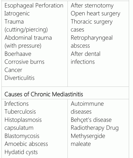
Mediastinitis is a lethal infection of the mediastinal connective tissue. It can be infectious or non-infectious and acute or chronic depending on the etiology. If it is not diagnosed and treated during the early stages, this infection is life threatening. A majority of acute mediastinitis cases develop secondary to esophageal perforation or previous mediastinal and cardiac operations. In its chronic form, this infection develops secondary to tuberculosis and bacterial and fungal infections like histoplasmosis or sarcoidosis (Table-1).
MTHFR has an important Descending necrotizing mediastinitis (DNM) occurs as a result of a complication of cervical necrotizing fasciitis resulting from infections in the floor of the mouth or in the cervical region spreading to the mediastinum through the fascial planes without primary skin or muscle involvement. Clinically, this infection is quite different from mediastinitis that develops after esophageal perforation or cardiac surgery. It is a rare infection, but the mortality rate is very high (15–50%). It is a clinical condition, the course of which still has a high mortality rate despite intensive treatments.
DNM, which is caused by necrotizing soft tissue infections (NSTI) in general, was first identified by Estrea et al. in 1983 (1). The most common causes of DNM are dental, retropharyngeal, peritonsillar, and odontogenic abscesses (2–5). In a wide series comprising DNM cases, it has been reported that the most common cause of DNM was of odontogenic origin (58%) (6).
Anatomy
Primarily, the main fascial pathways in neck region should be studied to understand the spread of necrotizing soft tissue infections to mediastinal compartments. The infection spreads to the mediastinal compartment via three main fascial pathways (7). These are pretracheal (superficial) ending in the anterior mediastinum, latero pharyngeal (perivascular) ending in middle mediastinum, and retropharyngeal (prevertebral space) ending in the posterior mediastinum (8).
The retropharyngeal space is the main pathway for oropharyngeal infections to reach the mediastinum.
Retropharyngeal Pathway
The infection spreads to the posterior mediastinum via this pathway. This region is a limited space above the hyoid bone and extends up to the T1 vertebra. The cervical prevertebral space has two compartments. The retropharyngeal region in the anterior compartment is the most common dissemination pathway of DNM (7–10).
The posterior compartment is known as the danger space. Following rupture of the alar fascia due to an abscess in retropharyngeal region, the infection spreads to the posterior mediastinal region after reaching the danger space. Since this region extends from the skull base to coccyx, transdiaphragmatic dissemination of the infection has been shown (8).
The pretracheal pathway is the most superficial of the cervical fascial pathways. It is limited to the anterior trachea and thyroid cartilage (6) and is the main pathway for spread of thyroid-related infections to the mediastinum. Parietal pleura constitute the inferior part of this region in the pericardium and carina.
The lateropharyngeal pathway is the main pathway followed for draining necrotizing soft tissue infections from the head and neck region to the middle mediastinum. This region consists of the internal jugular veins, carotid arteries, and vagus nerves. Arterial hemorrhage may develop because of perivascular dissemination.
Epidemiology and Risk Factors
DNM is commonly observed during odontogenic infections of the second and third molar teeth (Ludwig’s angina) and spreads to mediastinum due to negative thoracic pressure along the cervical fascia in the lateropharyngeal region. On the other hand, DNM may further develop due to infections originating from the teeth, parotid glands, and tonsils and otitis, mastoiditis, or epiglottitis rarely occur. Diabetes, advanced age, obesity, peripheral vascular disease, cancer, immunodeficiency, hypoalbuminemia, chronic renal failure, corticosteroid use, and malnutrition are risk factors for DNM (8–11).
Microbiology
Generally, aerobic and anaerobic micro-organisms co-exist (Table 2) and are the most commonly observed causative organisms in one of the three cases. The most commonly detected aerobes are Streptococ pyogenes, Staphylococcus. aureus, Escherichia coli, and Pseudomonas aeruginosa as well as anaerob such as Bacteroides fragilis and Clostridium species (12).
Diagnosis
Early diagnosis of mediastinitis is difficult due to uncertain symptoms. History and physical examinations are important for diagnosis. The most important step in the diagnosis of the disease is to consider NSTI. Deep cervical infections with the abovementioned predisposing factors in the patient’s history should indicate necrotizing fasciitis. Estrera et al. reported the diagnostic criteria, which includes severe oropharyngeal infection residuals, characteristic radiologic findings, necrotizing mediastinitis infection during surgery, and a relationship between DNM and the oropharyngeal process (1–10).
Beta-hemolytic oral streptococcus
Prevotella
Peptostreptococcus
Fusobacterium
Veillonella
Actinomyces
Bacteroides
Staphylococcus
Alpha-hemolytic streptococcus
Klebsiella
Porphyromonas
Viridans streptococci
Serratia
Enterobacter
Neisseria
Pseudomonas aeruginosa
Escherichia coli

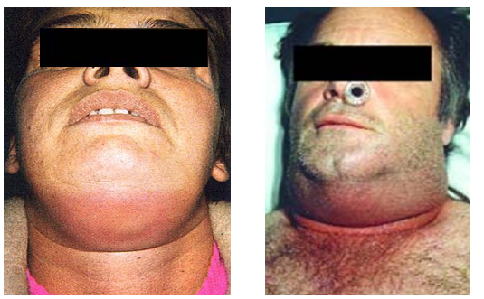
the first 48 h. Mediastinal involvement is typically observed between 12 h and 2 weeks. Retrosternal pain, swelling, redness, rigidity in the neck and anterior thoracic wall (Figure 1 and 2), sepsis symptoms such as shivering and fever, cough, and dyspnea are observed. Dysphagia may develop. Pleural fluid and tamponade may develop because of pleural and/or pericardial involvement. In leukocytosis and sepsis, elevation in the platelet count is observed during the early stage, and if disseminated, intravascular coagulation occurs during later, with a decrease in the platelet count (Figures 1 and 2).
Radiology
Radiologically, the main finding is Estrea et al. mediastinal extension.
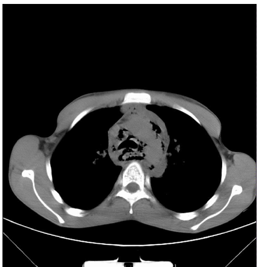
summarized the radiological findings in mediastinitis, which include widening with or without air-fluid level in the retropharyngeal region (Picture 3), mediastinal emphysema (Picture 4), widening of the cervical lordosis, deviation of the tracheal air colon towards the front, pneumothorax due to pleural involvement, and hydropneumothorax.
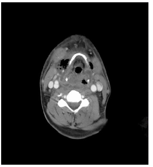
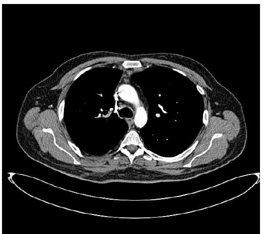
Computerized tomography (CT) findings include mediastinal air sacs, localized mediastinal fluid collection or abscess collection, decrease in the normal adipose tissue and soft tissue infiltration, no lymphadenopathy, internal jugular vein thrombosis, and carotid pseudoaneurysm (Figure 3 and 4) (13, 14).
Treatment
The main treatment principle in DNM is suitable antibiotic use and surgical drainage. Antibiotic treatment is not effective without surgical drainage and debridement. The necrotic tissues should be debridged and the mediastinal and pleural regions should be irrigated.
The level and method of surgical drainage depends on the infection site. Here, the fourth thoracic vertebra was an important location. If it is above this level, anterior cervical mediastinotomy and placing a soft drain are beneficial; however, in infections below this level, the visceral compartment should be drained as well. Drainage to the pleural space is performed by opening the mediastinal pleura. This can be performed by thoracotomy or video-assisted thoracic surgery (VATS) (Picture 5). If the infection includes bilateral thoracic cavities, bilateral VATS or unilateral posterior thoracotomy and exploration with contralateral VATS or “Clamshell” procedure can be performed for bilateral exploration. The subxiphoid trans thoracic approach may be performed concomitantly with the transcervical approach in anterior mediastinal collections (3).
Median sternotomy for drainage is not recommended because of the development of DNM posteriorly and difficulty in accessing the site and also the risk of osteomyelitis. The “Clamshell” incision (bilateral thoracotomy and median sternotomy) allows good exploration; however, it is a highly invasive method. With the Clamshell, the bilateral thorax can be reached in one session; however, it is a risky procedure for critical patients (Figure 5).
Hyperbaric Oxygen Therapy
Hyperbaric oxygen therapy can be initiated after the patient is stabilized and is performed in repeated sessions. The main purpose of this therapy is to prevent infections from spreading by increasing the oxygen perfusion to tissues and also to accelerate healing (15).
Prognosis
The mortality rate was approximately 50% before antibiotic treatment (16). The most common cause of mortality is late diagnosis and insufficient drainage (17). Death may occur due to fulminant sepsis, bleeding, aspiration, metastatic cranial infection, emphysema and purulent pericarditis, and tamponade.
Conclusion
Descending necrotizing mediastinitis (DNM) is one of the most lethal forms of mediastinitis. It occurs as a result of a complication of cervical necrotizing fasciitis resulting from infections in the floor of the mouth or the cervical region and spreads to the mediastinum through the fascial planes without primary skin or muscle involvement. Necrosis and tissue damage develops from the infection spreading from this region through the fascial and deep cervical planes. It can lead mortality, if not prevented at the early stage. When early diagnosis is delayed, surgical drainage and debridement may be required.
Declarations
- Author contributions (CRediT)
- All authors contributed equally to all 14 CRediT roles (Conceptualization, Data curation, Formal analysis, Funding acquisition, Investigation, Methodology, Project administration, Resources, Software, Supervision, Validation, Visualization, Writing – original draft, Writing – review & editing).
- Funding
-
Funding information was not reported in the published version of this article.
- Conflicts of interest
- A conflict of interest statement was not reported in the published version of this article.
- Ethics approval
- Ethics committee approval was not required for this article, as it is based on previously published literature and does not involve human participants, patient data or animals.
- Data availability
- Data sharing is not applicable to this article as no new data were created or analysed in this study.
References (17)
10 of 17 references are linked to their DOI.
- Estrera AS et al. Descending necrotizing mediastinitis. Surg Gynecol Obstet 157;545:1983. PubMed Google Scholar
- Mihos P, Potaris K, Gakidis I, Papadakis D, Rallis G. Management of descending necrotizing mediastinitis. J Oral Maxillofac Surg 2004;62:966–72. DOI Crossref PubMed Google Scholar
- Mitjans MS, Sanchıs JB, Padro XB, Garcıa I, Garolera JM, Garay MR. Descending necrotizing mediastinitis. Int Surg 2000;85:331–5. PubMed Google Scholar
- Novellans S, Kechabtia K, Chevallier P, Sedat J, Bruneton JN. Descending necrotizing mediastinitis: a rare pathology to keep in mind. Clin Imag 2005;29:138–40. PubMed Google Scholar
- Harar RP, Cranston C, Warwick-Brown N. Descending necrotizing mediastinitis: report of case following steroid neck injection. J Laryngol Otol 2002;116:862–4. DOI Crossref PubMed Google Scholar
- Kiernan PD, Hernandez A, Byrne WD, Bloom R, Dicicco B, Hetrick V, et al. Descending cervical mediastinitis Ann Thorac Surg 1998;65:1483–8. DOI Crossref PubMed Google Scholar
- Chow AW. Deep neck space infections. http://www.uptodate. com/contents/deep-neck-space-infections. PubMed Google Scholar
- Cirino LM, Elias FM, Almeida JL. Descending mediastinitis: a review Sao Paulo. Med J 2006;124:285–90. DOI Crossref PubMed Google Scholar
- Smith JK, Armao DM, Specter BB, Castillo M. Danger space infection: infection of the neck leading to descending necrotizing mediastinitis. Emerg Radiol 1999;6:129–32. Descending Necrotizing Mediastinitis Ahmet Ulusan et al. DOI Crossref PubMed Google Scholar
- Weaver E, Nguyen X, Brooks MA. Descending necrotising mediastinitis: two case reports and review of the literature. Eur Respir Rev 2010;19:141–9. DOI Crossref PubMed Google Scholar
- Headley AJ. Necrotizing soft tissue infections: a primary care review. Am Fam Phys 2003;68:323–8. PubMed Google Scholar
- Brook I, Frazier EH. Clinical and microbiological features of necrotisiting fasciitis. J Clin Microbiol 1995;33:2382–7. DOI Crossref PubMed Google Scholar
- Scaglione M, Pinto A, Romano S, Giovine S, Sparano A, Romano L. Determining optimum management of descending necrotizing mediastinitis with CT: experience with 32 cases. Emerg Radiol 2005;11:275–80. DOI Crossref PubMed Google Scholar
- Becker M, Zbaren P, Hermans R, Becker CD, Marchal F, Kurt AM, et al. Necrotizing fasciitis of the head and neck: role of CT in diagnosis and management. Radiology 1997;202:471–6. DOI Crossref PubMed Google Scholar
- Bono G, Argo A, Zerbo S, Triolo V, Procaccianti P. Cervical necrotizing fasciitis and descending necrotizing mediastinitis in a patient affected by neglected peritonsillar abscess: a case of medical negligence. J Forensic Leg Med 2008;15:391–4. DOI Crossref PubMed Google Scholar
- Michael J, Liptay JM, Kim AW. Diagnosis and staging of lung cancer. In: Shields TW, LoCicero J, Reed CE, Feins RH, Editors. General Thoracic Surgery. 7th Edition. Philadelphia: Lippincott Williams & Wilkins; The Mediastinum, Acute and Chronic Mediastinal Infections, 2009. p. 2183–94. PubMed Google Scholar
- Beriot G, Tomasini A, Gioffi V, et al. Fatal streptococcus viridans descending mediastinitis. Case report and review of the literature. Eur j Emerg Med 1997;4:11. PubMed Google Scholar
Respond to this article
Comments on a published article are welcome as a Letter to the Editor.
Get an e-mail when new articles in your fields are published
Questions to the authors
Questions are moderated: an editor approves each question, the authors answer, and an editor approves the answer before both appear here.
No answered questions yet.
Ask the authors a question
Discussion
Comments are moderated: they appear after the writer confirms the e-mail address and an editor approves them. Each comment carries a conflict-of-interest statement.
No comments yet.
Write a comment
Read the full text
Open the PDF here
Loading the PDF…
If the PDF does not show here, use the download button above.
How to cite this article
Ulusan A, Koc I. Review: Descending Necrotizing Mediastinitis. Ulutas Med J. 2016;2(1):1-6. doi:10.5455/umj.20151214013947
Ulusan, A., & Koc, I. (2016). Review: Descending Necrotizing Mediastinitis. The Ulutas Medical Journal, 2(1), 1–6. https://doi.org/10.5455/umj.20151214013947
Ulusan, A. and Koc, I. (2016) 'Review: Descending Necrotizing Mediastinitis', The Ulutas Medical Journal, 2(1), pp. 1–6. doi: 10.5455/umj.20151214013947.
@article{umj206985,
  title = {Review: Descending Necrotizing Mediastinitis},
  author = {Ulusan, Ahmet and Koc, Ibrahim},
  journal = {The Ulutas Medical Journal},
  year = {2016},
  volume = {2},
  number = {1},
  pages = {1--6},
  doi = {10.5455/umj.20151214013947},
  url = {https://www.ulutasmedicaljournal.com/?mno=206985},
  issn = {2149-388X}
}