The Ulutas Medical Journal · ISSN 2149-0430 (print), 2149-388X (online) · https://www.ulutasmedicaljournal.com/?mno=269374
Original Article · Open access
Cardiac Myxomas: Experience Over Ten Years
Also available in: العربية · Türkçe · 中文
- Cardiovascular Surgery, Binali Yıldırım University, Mengücek Gazi Training and Research Hospital, Erzincan, Türkiye ROR
Abstract
- Background
- Primary cardiac tumors are rare, with myxomas being the most common benign subtype. Early diagnosis and surgical excision are crucial due to risks of embolism, arrhythmias, and sudden death. This study aimed to evaluate the clinical characteristics, imaging findings, and surgical outcomes of patients with histopathologically confirmed cardiac myxoma treated at our center.
- Methods
- This retrospective observational study reviewed patients who underwent surgery for intracardiac masses. Of 1,135 open-heart surgeries, 7 were performed for intracardiac masses. One patient was excluded due to a postoperative diagnosis of thrombus. Six patients with histopathologically confirmed cardiac myxoma were included. Clinical presentation, imaging findings, surgical data, and follow-up outcomes were analyzed descriptively.
- Results
- Among the six patients (mean age 56.1± 13.8 years, 83.3% female), five (83.3%) had left atrial myxomas and one (16.7%) had a right atrial myxoma. Surgical excision via median sternotomy with cardiopulmonary bypass was performed in all cases, with concomitant atrial septal defect repair in two patients. One patient (16.7%) died due to infective endocarditis; the remaining patients had uneventful recoveries.
- Conclusion
- Cardiac myxomas are rare but clinically significant tumors. Early diagnosis using echocardiography and prompt surgical excision yields excellent long-term outcomes with a low recurrence risk. Despite the favorable prognosis, vigilant follow-up is warranted due to potential postoperative complications.
Keywords
Full text
Introduction
Primary cardiac tumors are rare pathologies with an incidence of 1.7–10/100,000 individuals and can occur at any age (1). Approximately 75% of these tumors are benign, and more than half of the benign tumors (around 50%) are myxomas. Myxoma was the first successfully excised by Crafoord during cardiopulmonary bypass (2). Originating from mesenchymal cells, myxomas are usually sporadic and occur between the ages of 30 and 60 (3). Although they are most commonly located in the left atrium, myxomas originating from the ventricles have also been reported (4). Clinical symptoms vary depending on the size, location, and mobility of the myxoma. While some cases are asymptomatic, others may present with embolic events, syncope, or malignant arrhythmias. The first-line imaging method for diagnosing myxoma is transthoracic echocardiography. Cardiac CT and MRI are also used for diagnosis (3).
Early diagnosis and treatment are crucial in managing myxomas. Delayed diagnosis may lead to severe complications such as embolic events, valvular dysfunction, and sudden death. Therefore, in cases with unexplained systemic embolism, syncope, or arrhythmia, cardiac tumors should be considered in the differential diagnosis. Surgical excision is the gold standard for treatment, generally yielding successful outcomes and a good long-term prognosis. However, recurrence risk should also be considered in postoperative follow-up.
This study examines the clinical data, imaging findings, and surgical outcomes of patients who underwent surgery for cardiac myxoma at our center over the past decade, supported by relevant literature.
Materials and Methods
This retrospective observational study was conducted at the Department of Cardiovascular Surgery, Erzincan Binali Yıldırım University Training and Research Hospital. Between January 2014 and December 2024, a total of 1135 patients underwent open-heart surgery. Among them, the medical records of seven patients who underwent surgery for an intracardiac mass were reviewed. One patient, initially suspected of having a cardiac myxoma based on preoperative imaging, was excluded after histopathological evaluation revealed the mass to be a thrombus. Consequently, six patients with histopathologically confirmed cardiac myxoma were included in the study. Data were obtained from the hospital’s electronic medical records, surgical notes, and outpatient follow-up reports. The following variables were analyzed:
▪ Demographic data: age and sex, ▪ Clinical presentation: symptoms such as chest pain, dyspnea, and syncope, ▪ Comorbid conditions and preoperative assessments, ▪ Imaging findings: including transthoracic echocardiography (TTE), transesophageal echocardiography (TEE), computed tomography (CT), magnetic resonance imaging (MRI), and preoperative coronary angiography when indicated, ▪ Tumor characteristics: location (left or right atrium), size, pedicle attachment, and mobility, ▪ Surgical approach, intraoperative findings, and associated cardiac abnormalities, ▪ Postoperative complications, morbidity, and mortality, ▪ Long-term follow-up data: including recurrence and tachyarrhythmia monitoring.
All patients underwent surgical excision of the tumor via median sternotomy under cardiopulmonary bypass. The masses were resected entirely along with their pedicle attachments, and atrial septal defects, if present, were repaired either primarily or with a patch, depending on defect size. All resected specimens were evaluated histopathologically using hematoxylin and eosin (H&E) staining, and the diagnosis of cardiac myxoma was confirmed. Postoperative outcomes were assessed during the hospital stay and at regular intervals through outpatient clinical evaluations and imaging studies. During follow-up, patients were monitored for tachyarrhythmias through standard 12-lead surface ECG at routine visits, which was used when clinically indicated. No cases of tachycardia, atrial fibrillation, or ventricular arrhythmia were detected during the follow-up period. Due to the limited sample size, no advanced statistical analyses were performed; data were presented using descriptive statistics only.
The study was approved by the Erzincan Binali Yıldırım University Ethics Committee (No: 2025- 07/04) and was conducted in accordance with the principles of the Declaration of Helsinki.
Results
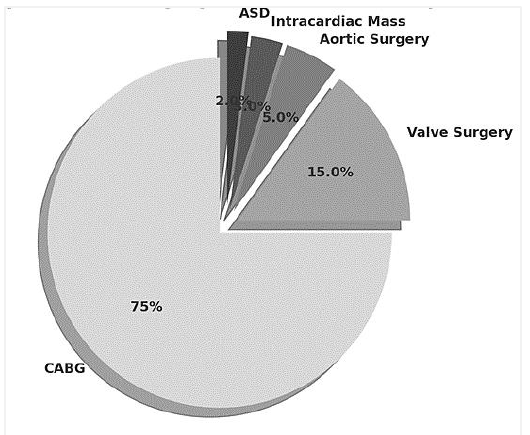
During the study period from January 2014 to December 2024, a total of 1,135 patients underwent open-heart surgery at our center. Of these, 977 patients (86%) underwent coronary artery bypass grafting (CABG), 104 (9.1%) valve surgery, 42 (3.7%) aortic surgery, 7 (0.6%) excision of an intracardiac mass, and 5 (0.4%) atrial septal defect (ASD) repair (Figure 1). Medical records and imaging data of 7 patients who underwent surgery for an intracardiac mass were reviewed.
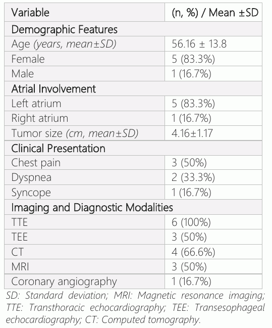
One patient was excluded due to the postoperative pathological diagnosis of thrombus, despite an initial radiological suspicion of myxoma. The remaining six patients, all confirmed histo-pathologically to have cardiac myxoma, were included in the analysis. Thus, cardiac myxomas accounted for 0.52% of all open-heart surgeries performed during the study period. Of the six patients included, 1 (16.7%) was male and 5 (83.3%) were female. The mean age was 56.16 ±13.8 years. In 5 patients (83.3%), the myxoma was located in the left atrium, while in 1 patient (16.7%) it was located in the right atrium. The hospitalization period was 3+0.89 days.
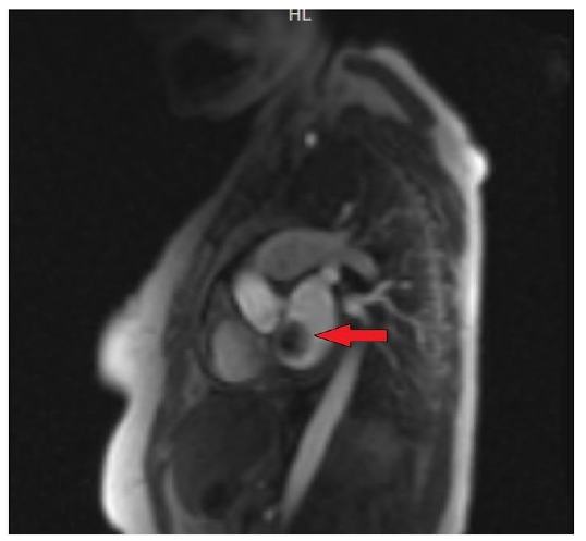
The presenting symptoms varied: 3 patients (50%) presented with chest pain, 2 (33.3%) with dyspnea, and 1 (16.7%) with syncope (Table 1). All patients underwent preoperative evaluation with TTE. In addition, TEE was performed in 3 patients, CT in 4, MRI in 3 (Figure 2), and coronary angiography in 1 patient due to the presence of ischemic risk factors.
Surgical resection of the tumor was performed via median sternotomy under the general anesthesia, using cardiopulmonary bypass established through the aortic and bicaval cannulation. The atrium was opened according to the location of the mass. The tumors were macroscopically visualized and excised entirely along with their pedicle attachments. In 2 patients, ASDs were closed—one identified preoperatively, and the other discovered intraoperatively. Histopathological examination of the resected masses using hematoxylin-eosin staining confirmed the diagnosis of cardiac myxoma in all cases.
The mean follow-up period was 55.16 ± 12.40 months (range: 36–65 months). During follow-up, all patients were regularly monitored for arrhythmias using a 12-lead surface ECG at outpatient visits. In cases with clinical suspicion, 24-hour Holter monitoring was also performed. No cases of atrial fibrillation, atrial tachycardia, or ventricular arrhythmias were observed. Additionally, no tumor recurrence was detected during the follow-up period. Postoperative infective endocarditis developed in one patient, who unfortunately died despite intensive medical treatment. The remaining five patients recovered uneventfully and were discharged in good clinical condition. Due to the small sample size, only descriptive analyses were performed.
Discussion
Primary cardiac tumors are exceedingly rare, with autopsy series reporting an incidence ranging from 0.001% to 0.3% (5). The vast majority of these tumors (75–90%) are benign, with myxomas being the most common among them (6, 7). Recent studies indicate that myxomas occur more frequently in females (65–85%) and are most commonly diagnosed between the ages of 40 and 60 (8, 9). In our series, 83.3% of the patients were female, and the mean age was 56.1±13.8 years, consistent with the literature.
Cardiac myxomas predominantly localize in the left atrium, particularly near the foramen ovale, attached via a pedicle (10, 11). While approximately 80–90% of all myxomas occur in the left atrium, locations in the right atrium or ventricles are less common (12). Similarly, in our series, 5 out of 6 patients (83.3%) had left atrial myxomas, and one patient (16.7%) had a right atrial myxoma. The tumor’s location and size directly influence the type and severity of symptoms (13).
Cardiac myxomas may be asymptomatic or cause symptoms such as chest pain, dyspnea, and syncope, depending on their location. Although dyspnea, constitutional symptoms, and syncope are the most frequently reported symptoms in the literature (14), chest pain (50%) was the most common presenting symptom in our series, followed by dyspnea (33.3%) and syncope (16.7%).
Myxomas located in the left atrium carry a risk of systemic embolism, including cerebral, renal, and splenic emboli. In contrast, right-sided heart myxomas are associated with a higher risk of pulmonary embolism. Recent literature has published that embolic complication rates range from 20% to 30% (15, 16). The absence of embolic complications in our series may be related to early diagnosis and timely surgical treatment. Transthoracic echocardiography is the initial imaging modality of choice with a sensitivity of approximately 95–98% (17). Advanced imaging techniques such as TEE, CT, and cardiac MRI provide more detailed information regarding lesion location, size, and mobility (18, 19). In our study, all patients underwent TTE; TEE (n:3), CT (n:4), MRI (n:3), and preoperative coronary angiography (n=1) were performed as indicated.
Surgical resection remains the gold standard treatment for myxomas, offering an excellent prognosis. Current studies report surgical mortality rates between 1% and 3%, which are influenced by tumor size, location, concomitant cardiac pathologies, and surgical expertise (20). In our series, postoperative mortality due to infective endocarditis occurred in one patient (16.7%), a rate not statistically significant due to the small sample size. Median sternotomy was the surgical approach for all patients, and tumors were excised entirely from their pedicles. Two patients with intraoperatively detected atrial septal defects underwent repair of the defects. Recurrence rates are generally low (1–3%) in sporadic myxomas but can increase to 20–67% in familial cases or those associated with Carney complex (21). No recurrences were observed during the follow-up period in our study; however, the absence of genetic analysis is a limitation in this study. This study has several limitations, including a small sample size that limits generalizability, a lack of genetic testing, particularly relevant to recurrence risk, and its retrospective design. Nevertheless, sharing surgical and clinical experiences with these rare tumors contributes valuable information to the existing literature.
Conclusion
In conclusion, cardiac myxomas are rare tumors that may cause significant morbidity and mortality. Early diagnosis, utilizing effective imaging modalities such as echocardiography, and timely surgical intervention are key to achieving successful outcomes. Careful surgical technique is crucial for preventing recurrences and improving long-term prognosis.
Declarations
- Author contributions (CRediT)
- All authors contributed equally to all 14 CRediT roles (Conceptualization, Data curation, Formal analysis, Funding acquisition, Investigation, Methodology, Project administration, Resources, Software, Supervision, Validation, Visualization, Writing – original draft, Writing – review & editing).
- Funding
-
Funding information was not reported in the published version of this article.
- Conflicts of interest
- The authors reported no conflicts of interest.
- Ethics approval
- Ethics approval details were not reported in the published version of this article.
- Data availability
- A data availability statement was not reported in the published version of this article.
References (21)
4 of 21 references are linked to their DOI.
- Keçeligil HT, Demir Z, Kolbakır F, Demirağ MK, Akar H. Kardiyak Miksoma ve Cerrahi Tedavisi. GKDC Dergisi 1999; 7: 210-216 PubMed Google Scholar
- Crafoord C. Discussion of Glover RP. Late results of mitral commissurotomy. In: Lam CR, ed. Henry Ford Hospital international symposium on cardiovascular surgery; studies in physiology, diagnosis and techniques. Philadelphia: WB Saunders, 1955:202-11. DOI Crossref PubMed Google Scholar
- Sert SS, Kuyumcu MS, İbrişim E. (2024). Efor Dispnesi ile Başvuran Genç Hastada Dev Sol Atriyal Miksoma. Bull Cardiovasc Acad, 2(3), 93-95. PubMed Google Scholar
- Uçar Elalmış Ö, Arslan K. Right Ventricular Myxoma Complicated by Stroke, Cumhuriyet Medical Journal. 2024;46(2): 143-148. DOI Crossref PubMed Google Scholar
- Lins RB, Ribeiro PJ, Franco MM, et al. Cardiac myxoma: a comprehensive systematic review and meta-analysis. Int J Cardiol. 2023;382:19702. PubMed Google Scholar
- Zhang Y, Ma W, Wang X, et al. Surgical treatment and outcomes of cardiac myxoma: a 12-year single-center experience. J Cardiothorac Surg. 2021;16(1):52. PubMed Google Scholar
- Dias RR, Andrade RR, Mady C, et al. Cardiac myxoma: 24-year experience in 196 patients. Rev Bras Cir Cardiovasc. 2019;34(2):166–172. PubMed Google Scholar
- Lee S, Kim JH, Lee HJ, et al. Cardiac myxoma in elderly patients: Surgical outcomes and considerations. Ann Thorac Surg. 2020;109(3):774–779. PubMed Google Scholar
- Sert SS, Kuyumcu MS, İbrişim E. Efor Dispnesi ile Başvuran Genç Hastada Dev Sol Atriyal Miksoma. Bull Cardiovasc Acad. 2024;2(3):93–5. PubMed Google Scholar
- Tsukamoto M, Morimoto R, Okabe H, et al. Characteristics and management of cardiac tumors: 10-year single-center experience. Gen Thorac Cardiovasc Surg. 2023;71(1):41–48. PubMed Google Scholar
- Wang JG, Li YJ, Liu H, Li NN, Zhao J, Xing XM. Clinicopathologic analysis of cardiac myxomas: seven years' experience with 61 patients. J Thorac Dis. 2020;12(4):1121–1129. PubMed Google Scholar
- Randhawa K, Ganame J, Olivieri L. Cardiac MRI in evaluation of cardiac masses. Radiol Clin North Am. 2023;61(3):493–509. PubMed Google Scholar
- Hoffmeier A, Sindermann JR, Scheld HH, Martens S. Cardiac tumors—diagnosis and surgical treatment. Dtsch Arztebl Int. 2022;119(20):345–352. PubMed Google Scholar
- ElBardissi AW, Dearani JA, Daly RC, et al. Survival after resection of primary cardiac tumors: a 48-year experience. Circulation. 2018;138(4):e78–e85. PubMed Google Scholar
- Pinede L, Duhaut P, Loire R. Clinical presentation of left atrial cardiac myxoma: A series of 112 consecutive cases. Medicine (Baltimore). 2001;80(3):159–172. DOI Crossref PubMed Google Scholar
- Uçar Elalmış Ö, Arslan K. Right Ventricular Myxoma Complicated by Stroke. Cumhuriyet Med J. 2024;46(2):143–148. DOI Crossref PubMed Google Scholar
- Araoz PA, Eklund HE, Welch TJ, Breen JF. CT and MR imaging of primary cardiac malignancies. Radio graphics. 2020;40(6):1421–1434. PubMed Google Scholar
- Kassi M, Polsani V, Schutt RC, et al. Cardiac masses on cardiac CT: a review. Curr Cardiol Rep. 2019;21(6):53. Cardiac Myxomas Izzet Emir et al. PubMed Google Scholar
- Bruce CJ. Cardiac tumors: diagnosis and management. Heart. 2021;107(12):985–993. PubMed Google Scholar
- Mkalaluh S, Szczechowicz M, Dib B, et al. Cardiac myxomas: 20 years of experience in 49 patients. Eur J Cardiothorac Surg. 2018;53(5):1034–1039. PubMed Google Scholar
- Carney JA. Carney complex: pathologic features of the major components. Mayo Clin Proc. 2019;94(10):2068– 2078. PubMed Google Scholar
Respond to this article
Comments on a published article are welcome as a Letter to the Editor.
Get an e-mail when new articles in your fields are published
Questions to the authors
Questions are moderated: an editor approves each question, the authors answer, and an editor approves the answer before both appear here.
No answered questions yet.
Ask the authors a question
Discussion
Comments are moderated: they appear after the writer confirms the e-mail address and an editor approves them. Each comment carries a conflict-of-interest statement.
No comments yet.
Write a comment
Tell me when this article is cited
Leave your e-mail address and confirm it. These notices are not being sent yet; your confirmed address is kept for them and used for nothing else.
Read the full text
Open the PDF here
Loading the PDF…
If the PDF does not show here, use the download button above.
How to cite this article
Emir I, Yurt S, Ziyrek U, Onk OA. Cardiac Myxomas: Experience Over Ten Years. Ulutas Med J. 2025;11(3):41-46. doi:10.5455/umj.20250706055401
Emir, I., Yurt, S., Ziyrek, U., & Onk, O. A. (2025). Cardiac Myxomas: Experience Over Ten Years. The Ulutas Medical Journal, 11(3), 41–46. https://doi.org/10.5455/umj.20250706055401
Emir, I. et al. (2025) 'Cardiac Myxomas: Experience Over Ten Years', The Ulutas Medical Journal, 11(3), pp. 41–46. doi: 10.5455/umj.20250706055401.
@article{umj269374,
  title = {Cardiac Myxomas: Experience Over Ten Years},
  author = {Emir, Izzet and Yurt, Serif and Ziyrek, Ugur and Onk, Oruc Alper},
  journal = {The Ulutas Medical Journal},
  year = {2025},
  volume = {11},
  number = {3},
  pages = {41--46},
  doi = {10.5455/umj.20250706055401},
  url = {https://www.ulutasmedicaljournal.com/?mno=269374},
  issn = {2149-388X}
}