The Ulutas Medical Journal · ISSN 2149-0430 (print), 2149-388X (online) · https://www.ulutasmedicaljournal.com/?mno=284199
Case Report · Open access
Traumatic Wound Infection with Graphium
Also available in: العربية · Türkçe · 中文
Abstract
Graphium species are filamentous fungi and belong to the family of Ascomycota. They have been recovered from soil, plant debris, woody substrates, manure, and polluted water, and most of them are plant pathogens. In contrast, human infections caused by Graphium spp are extremely rare. Herein, we reported the first published case of Graphium traumatic wound infection in an immunocompetent patient. Surgery was performed and amphotericin was added into the antimicrobial regimen for better coverage of the fungus. The postoperative course was smooth, and the patient was discharged uneventfully.
Keywords
Full text
Introduction
Graphium species are filamentous fungi and belong to the family of Ascomycota. They have been recovered from soil, plant debris, woody substrates, manure, and polluted water, and most of them are plant pathogens (1). In contrast, human infections caused by Graphium spp are extremely rare (2–4). Thus, the clinical significance of this pathogen remains unclear due to limited knowledge. Herein, we report the first published case of Graphium traumatic wound infection in an immunocompetent patient.
Case Presentation
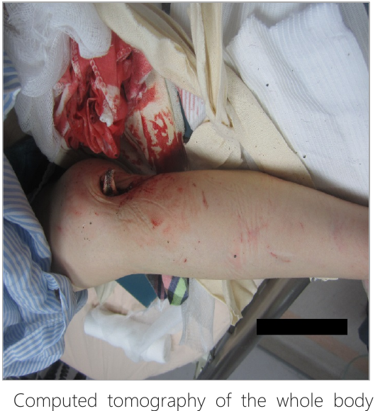
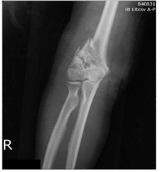
A 38-year-old woman presented with painful deformity of the right elbow and right ankle, and severe abdominal pain after an accidental 3-story fall onto her right side. She denied any history of significant medical illness. Her body temperature on arrival was 37oC, pulse rate 85 beats per minute, respiratory rate 20 per minute, and blood pressure of 90/58 mmHg. Physical examination showed cubitus valgus deformity of the right elbow with an open wound and bone exposure (Figure-1), varus deformity of the right ankle, and tenderness in the lower pelvis. Radiography of the right elbow revealed a comminuted fracture of the right supracondylar humerus, Gustilo type-II (Figure- 2). In addition, radiography of the pelvis and lower limbs disclosed a comminuted fracture of the right femoral inter/sub-trochanter, right bimalleolar fracture, fractures of the bilateral pubic rami, right transverse process of the L5, and left distal tibia, and a displaced fracture of the left middle fibular shaft. Laboratory studies were as follows: white blood cell count, 21.5 x 10⁹ /L with neutrophil predominance (83.8%); serum creatinine, 0.82 mg/dL; alanine aminotransferase, 179 U/L; sodium, 141.3 mmol/L; and C-reactive protein, 103.7 mg/dL (reference value, < 0.8 mg/dL).
revealed liver and spleen lacerations, pelvic fracture and hemoperitoneum. Because of her unstable hemodynamic status and progressive dyspnea, she received aggressive fluid resuscitation, massive blood transfusions and intubation for acute respiratory failure. Emergency angiography showed petechial hemorrhage in the bilateral internal iliac arteries and liver, and she received successful transarterial embolization to stop the bleeding. Empirical antibiotic treatment with flomoxef was prescribed for traumatic wound infection over the right elbow. After her hemodynamic status stabilized, surgical intervention with repeated wound debridement of the right elbow was done.
A sample of the gross turbid discharge with wound tissue was sent for cul-ture, and the wound specimen grew Graphium spp. The fungus which was identified by *MALDI-TOF Mass Spectrometer and had a best identification score of 1.841, which indicated genus-level identification. Thus, amphotericin was added into antimicrobial regimen for better coverage of the fungus. The postoperative course was smooth, and she was discharged uneventfully. Follow-up in the outpatient clinic showed that the wound healed well.
Discussion
The present report demonstrated an unusual fungus - Graphium spp. which caused a traumatic wound infection in an immunocompetent patient. The source of infection could have been the soil where the patient fell. To the best of our knowledge, definite Graphium spp.- associated human infections are rare, and only two human cases have been reported previously. In 2007, Kumar reported a case of Graphium basitruncatum with a presentation of persistent fungemia and skin lesions in a patient with acute leukemia (2). In 2012, El Feghaly reported a case of G. basitruncatum fungemia in a child post stem-cell transplantation.(3) In contrast to previous reports (2, 3), our patient had a traumatic would infection and no significant immunocompromised condition. As seen in this case, Graphium spp. can cause traumatic wound infection in an immunocompetent patient, further expanding the disease spectrum of Graphium spp. infections.
In vitro antifungal susceptibility tests showed the MICs for the isolates in Kumar et al ’s report were amphotericin B, 0.5 µg/ml; itraconazole, >16 µg/ml; voriconazole, 8 µg/ml; caspofungin, 2 µg/ml; fluconazole, >64µg/ml (2), and in El Feghaly et al’s report, were amphotericin B, 8µg/ml; itraconazole, >16 µg/ml; voriconazole, >16 µg/ml; posaconazole >16 µg/ml (3). The patient with leukemia was post chemotherapy, and had clinical improvement with liposomal amphotericin B, but the improvement coincided with recovery of neutrophils and was temporary (2). The stem cell transplantation recipient died under treatment with amphotericin and anidulafungin (3). Our patient had a favorable clinical response to amphotericin B and repeated surgical debridement. In summary, the clinical response was different between our case and previous reports, probably because of the significantly different immune status. Because of limited experience with Graphium spp. infections, the optimal antifungal treatment remains unclear. Therefore, further largescale in vitro and in vivo studies are warranted to establish the appropriate treatment guidelines for this unusual fungus.
Conclusion
In conclusion, we describe the first case of a traumatic wound infection caused by Graphium spp. in an immunocompetent patient. Her clinical condition responded well to antibiotics and wound debridement.
Declarations
- Author contributions (CRediT)
- All authors contributed equally to all 14 CRediT roles (Conceptualization, Data curation, Formal analysis, Funding acquisition, Investigation, Methodology, Project administration, Resources, Software, Supervision, Validation, Visualization, Writing – original draft, Writing – review & editing).
- Funding
-
Funding information was not reported in the published version of this article.
- Conflicts of interest
- A conflict of interest statement was not reported in the published version of this article.
- Ethics approval
- Ethics approval details were not reported in the published version of this article.
- Data availability
- A data availability statement was not reported in the published version of this article.
References (4)
2 of 4 references are linked to their DOI.
- Okada, G. Genus Graphium. J Antibact Antifung Agents 1994; 22: 45-54. PubMed Google Scholar
- Kumar D, Sigler L, Gibas CF, Mohan S, Schuh A, Medeiros BC, et al. Graphium basitruncatum fungemia in a patient with acute leukemia. J Clin Microbiol 2007;45:1644 -1647. DOI Crossref PubMed Google Scholar
- El Feghaly RE, Sutton DA, Thompson EH, Fu J, Wickes BL, Al-Zubeidi D, et al. Graphium basitruncatum fungemia in an immunosuppressed child post stem-cell transplantation. Med Mycol Case Rep 2012;1:35-38. DOI Crossref PubMed Google Scholar
- Apostol JG, Meyer SL. Graphium endophthalmitis. Am J Ophthalmol 1972;73:566-569. PubMed Google Scholar
Respond to this article
Comments on a published article are welcome as a Letter to the Editor.
Get an e-mail when new articles in your fields are published
Questions to the authors
Questions are moderated: an editor approves each question, the authors answer, and an editor approves the answer before both appear here.
No answered questions yet.
Ask the authors a question
Discussion
Comments are moderated: they appear after the writer confirms the e-mail address and an editor approves them. Each comment carries a conflict-of-interest statement.
No comments yet.
Write a comment
Read the full text
Open the PDF here
Loading the PDF…
If the PDF does not show here, use the download button above.
How to cite this article
Wu CL, Chao CM, Lin WT. Traumatic Wound Infection with Graphium. Ulutas Med J. 2018;4(1):44-46. doi:10.5455/umj.20181126062738
Wu, C. L., Chao, C. M., & Lin, W. T. (2018). Traumatic Wound Infection with Graphium. The Ulutas Medical Journal, 4(1), 44–46. https://doi.org/10.5455/umj.20181126062738
Wu, C.L., Chao, C.M. and Lin, W.T. (2018) 'Traumatic Wound Infection with Graphium', The Ulutas Medical Journal, 4(1), pp. 44–46. doi: 10.5455/umj.20181126062738.
@article{umj284199,
  title = {Traumatic Wound Infection with Graphium},
  author = {Wu, Chun-Liang and Chao, Chien-Ming and Lin, Wei-Ting},
  journal = {The Ulutas Medical Journal},
  year = {2018},
  volume = {4},
  number = {1},
  pages = {44--46},
  doi = {10.5455/umj.20181126062738},
  url = {https://www.ulutasmedicaljournal.com/?mno=284199},
  issn = {2149-388X}
}