The Ulutas Medical Journal · ISSN 2149-0430 (print), 2149-388X (online) · https://www.ulutasmedicaljournal.com/?mno=289039
Case Report · Open access
Metastatic Adrenocortical Carcinoma of the Lung In A Four Years Old Child
Also available in: العربية · Türkçe · 中文
- Medical intern at King Saud University, Riyadh, Saudi Arabia ROR
- Associate Consultant at Pediatric Surgery at King Fahad Medical City, Riyadh, Saudi Arabia ROR
- Fellow at Pediatric Surgery department at Qassim University, Qassim, Saudi Arabia ROR
- Histopathologist at King Fahad Medical City, Riyadh, Saudi Arabia. ROR
- Consultant at Pediatric Surgery at King Fahad Medical City, Riyadh, Saudi Arabia ROR
Abstract
- Background
- Studies of adrenocortical carcinoma is an aggressive rare type of endocrine tumors. It possesses a high mortality rate. This type of carcinomas occurs sporadically, while it could be part of hereditary syndromes. Only limited data is found about the recurrence of these tumors after resection and their prognosis is unclear.
- Case Presentation
- We present a case of a four-year-old boy who presented with virilization symptoms and was referred for further investigation of left abdominal mass. He was cushingoid upon examination. The patient was managed by left nephrectomy and chemotherapy. Upon follow up, the residual mass lesion was seen on CT. Re-resection of the residual tumor was done. Adjuvant mitotane therapy and radiotherapy were started, 7 months later the patient was discovered to have left lung metastasis in which he underwent left lung wedge resection.
Keywords
Full text
Introduction
Adrenocortical tumors are rare in children, while adrenocortical carcinoma is even more uncommon in this age group (1). Although ACC is rare but it’s one of the aggressive types of endocrine tumors (2). It has been reported by its tragic high mortality rate(3, 4). The worldwide annual incidence of ACT is only 0.3 - 0.38 per million children below the age of 15 years (5, 6). Commonly adrenocortical carcinoma occurs sporadically. It could also present as part of hereditary syndromes like Li-Fraumeni syndrome and Beckwith-Wiedemann syndrome (7, 8), in children with genetic predisposition (R337H mutation) like in south Africa where its prevalence is more than three times higher than other areas (6).
The peak incidence of ACC is between the fourth and fifth decades, although it can occur in all age groups (2). Females have slight predominance (9). Lung metastasis have been reported with ACC (10).
In this report, we present a case of a 4-yearold boy with metastatic left adrenocortical carcinoma. Lung metastasis have occurred almost 7 months after the primary tumor resection. The study was approved by Institutional Review Board At King Fahad Medical City in Riyadh, Saudi Arabia.
Case Presentation
Four years old Sudanese boy was referred for further management of left abdominal mass. Symptoms started 2 months prior to presentation, when he developed acne mainly in the forehead involving scalp, weight gain, increase in appetite, gradual increase in the abdominal girth, penile enlargement with pubic hair, change in sweat smell (adult odor), fatigability, mild on and off abdominal pain, urinary frequency and behavioral changes. There was no family history of tumors or cancers. The mother underwent thyroidectomy due to goiter with hypothyroidism. On examination, he was vitally stable with cushingoid features, rounded face with pudgy cheeks (Moon face), penile enlargement with pubic hair but no facial hair.
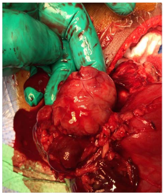
The abdomen was distended without any tenderness. Palpable mass was noted in the left side of the abdomen. Otherwise normal examination with clear chest. CT of abdomen showed a large circumscribed left flank heterogeneous soft tissue lesion measuring 10x9x11 cm, which caused displacement of the surrounding vas-cular structure with no evidence of encasement. There was displacement of the left kidney inferiorly, with no evidence of liver, lung, or osseous metastasis, and no intra-spinal extension. Laboratory work up showed high plasma LDH level (Almost triple the normal), low plasma ACTH level, normal serum cortisol and total testosterone levels, high serum dehydro epiandrosterone sulfate levels. The case was discussed in multidisciplinary tumor board meeting and the plan was operative resection. Up-front surgical resection plus left nephrectomy 26.5.2016 was done with fragile tumor spillage intraopera-tively. Irrigation and suction was done. The tumor was involving the upper pole of the kidney and it was inseparable from the renal parenchyma (Figure-1) so the decision was to do radical nephrectomy in addition to adrenalectomy to reduce the chance of recurrence. Post-operative course was uneventful. Pathologic examination of the primary tumor revealed multinodulated white tan mass measuring 10x8x4 cm replacing most of the kidney parenchyma and reaching to the renal sinus and perinephric fat grossly. No adrenal gland was identified grossly. Tumor was not reaching the vascular margins and no lymph nodes identified in the hilar fat.
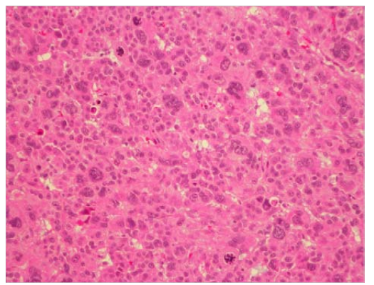
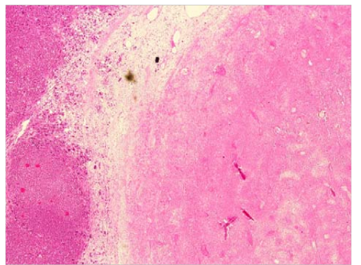
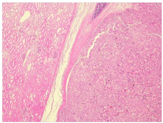
Under microscope, the tumor showed diffused proliferation of highly pleomorphic epithelioid cells seen as sheets and nests with highly pleomorphic and bizzar nuclei with some multinucleated cells. Cells show abundant eosi-nophilic granular cytoplasm. Clear cells change was seen in about 10% of tumor. Mitoses were brisk with many atypical mitoses no-ted (Figure-2). Necrosis was prominent (Figure-3). Vascular invasion near the capsule was also noted. However, the renal vein and arterial vas-cular margins were clear. The tumor appeared to have pushing borders at the tumor-kidney interface and didn’t show any evidence of invasion into the renal parenchyma (Figure-4). On immunohistochemistry, the tumor was positive with Vimentin, Melanen A and Synap-tophysin and was negative with Cytokeratin, EMA (Only focally positive), Inhibin, Calretinin, S100,Chromnogranin,and CD56 (which showed very focal positive). This immunoprofile was in keeping with adrenal cortical tumor. Although negative staining with both inhibin and calretinin is unusual, but it can rarely be seen in adrenal cortical neoplasma. MelanA positivity is helpful for the diagnosis. The diagnosis was agreed on by two independent pathologists. Post-operative follow-up CT scan 10 days after surgery showed interval resection of the large left suprarenal mass and left kidney, small enhancing soft tissue seen in the left paravertebral region, could be residual tumor or peritoneal deposits. With no evidence of chest metastasis. The patient underwent chemotherapy COG (ARAR0332 protocol). He had four cycles with poor response, rather suggestive of tumor progression on chemotherapy with rising DHEA levels.
CT chest, abdomen and pelvis three months after, showed Residual well demarcated predominantly hypodense soft tissue mass lesion no-ted in the left renal/adrenal region measures 3.9x2.3x3cm most likely representing local residual. In the chest satisfactory position of the left central line, no hilar lymphadenopathy. Both lungs are within normal limits with no suspicious pulmonary nodules.
Macroscopic re-resection of the residual tumor was done 4 months after the primary resection, where the mass was identified, separated from medial aspect of IVC and was dissected with no evidence of tumor residual. Pathologic examination showed residual viable tumor, positive regional lymph node, no chemotherapy changes and invasion of fibro-fatty soft tissue (stage 3). Clinically he was doing fine with no virilization symptoms. 11 days post re-resection abdomen/pelvis CT showed, the previously seen left paraspinal left tissue residual mass lesion has been surgically resected. A focal area of enhancing soft tissue is seen in the tumor bed. There is another focal hypodense mass lesion measures 15x21 mm seen in the left prevertebral compartment abutting the posterior wall of the splenic artery. Adjuvant oral mitotane therapy and local radiotherapy were initiated. Follow up CT, 3 months after, revealed subtle reduction in size of the residual mass. The residual soft tissue seen in the left adrenal gland bed is demonstrated showing more enhancing component, it roughly measures 12x10 cm as compared to 22x12 mm done 3 months back. CT chest showed interval development of a lung nodule seen in the lingual measuring 5.6x5.3 mm. No major airspace consolidation or collapse. No pleural effusion or pneumothorax. The major tracheobronchial tree is patent. No size significant axillary, mediastinal or hilar lymph nodes. There’s evidence of a small enhancing nodule close to the posterior left 11th rib measuring 12x6 mm suggestive of another metastatic nodule.
The patient hadmetastatic adrenocortical carcinoma to the left lung, underwent left lung wedge resection. Pathology showed residual microscopic focus of metastatic adrenocarcinoma 5%, Resection margin negative for tumor, he was started on radiotherapy to the left lung surgical bed.
Whole body PET scan, FDG avid left paraaortic nodules as well as left chest well solitary nodule measuring 1.1x1 cm. The rest of the body showed no suspicious FDG uptake. A week after, MRI chest on showed enhanced lesion in 11th intercostal space seen in the post contrast coronal T1- weighted image measures 1.2x0.7 cm. The lesion shows high signal intensity in coronal STIR weighted image and isointensity in coronal T1-weighted image.
Discussion
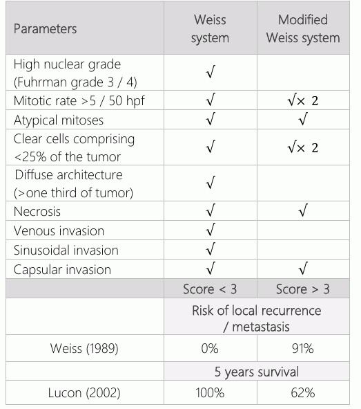
Adrenocortical carcinoma is known by its rarity since it represents only 0.2% of all childhood cancers(7).The diagnosis ofadrenocortical neoplasm in children is usually made as a result of symptoms caused by hormonal production by the cancer cells (11). In this case, the cushingoid features and sexual precocity drove the discovery of the mass, which was investigated and the tumor was found subsequently. Left sided ACC are generally more common than right side, as in our case (12). Carcinoma can’t be distinguished from an adenoma from patient presentation, even in the presence of laboratory findings and imaging studies. Although large tumor size in imaging and heterogeneity can predict carcinomas (13). Categorization of carcinoma from adenoma in adrenocortical pathology is challenging. In 1989 the introduction of Weiss criteria was adopted to differentiate carcinoma from adenoma. This has been modified in 2002 as ‘modified Weiss system’ and is now the preferred modality to be used (14). In this case we used the modified Weiss system (Table-1). Presence of atypical mitoses, clear cells comprising around 10 % of the tumor and necrosis would put our patient in 62% 5 years survival rate. Larger tumor size, high percent necrosis and high mitotic rate are associated with poor outcome (15). Margin status of the tumor, extension of tumor outside the adrenal gland, presence of metastasis and age were associated with worst survival (16, 17). Using TNM staging system (Table-2) (18) prior the re-resection our patient was thought to be stage 2. Afterward, he was discovered to have stage 3 cancer. Post-radical resection of ACC, adjuvant mitotane could lengthen recurrence free survival period (19). In addition, radiotherapy is recommended in cases with high risk of local recurrence (2). Therefore, adjuvant mitotane therapy and local radiotherapy were initiated. Whereas, the 13 institutions study concluded that there’s no association between adjuvant mitotane after resection and improvement of recurrence-free survival or overall survival (20). Our patient developed lung metastasis while on mitotane therapy. In a series study done in Turkey, out of 20 ACC patients, 3 had lung metastasis at presentation in which one of them had concurrence liver metastasis. Furthermore, during mitotane therapy within 2 years’ time, 3 patients developed lung and liver metastasis, while another 2 patients developed lung and bone metastasis. They have stated that (frequent imaging studies and endocrine evaluation was of utmost importance to detect recurrence in long-term follow-up of patients)(10).
Conclusion
Adrenocortical carcinoma is a rare neoplasm in children, it has a high mortality rate. Lung metastasis could occur after tumor resection even after initiation of mitotane and local radiotherapy.
Declarations
- Author contributions (CRediT)
- All authors contributed equally to all 14 CRediT roles (Conceptualization, Data curation, Formal analysis, Funding acquisition, Investigation, Methodology, Project administration, Resources, Software, Supervision, Validation, Visualization, Writing – original draft, Writing – review & editing).
- Funding
-
Funding information was not reported in the published version of this article.
- Conflicts of interest
- A conflict of interest statement was not reported in the published version of this article.
- Ethics approval
- The study was approved by Institutional Review Board At King Fahad Medical City in Riyadh, Saudi Arabia.
- Data availability
- A data availability statement was not reported in the published version of this article.
References (20)
17 of 20 references are linked to their DOI.
- Breidbart E, Cameo T, Garvin JH, Hibshoosh H, Oberfield SE. Pubertal outcome in a female with virilizing adrenocortical carcinoma. J Pediatr Endocrinol Metab. 1 styczeń 2016;29(4): 503–9. DOI Crossref PubMed Google Scholar
- Fassnacht M, Kroiss M, Allolio B. Update in Adrenocortical Carcinoma. J Clin Endocrinol Metab. grudzień 2013;98(12): 4551–64. DOI Crossref PubMed Google Scholar
- Stewart DR, Morris Jones PH, Jolleys A. Carcinoma of the adrenal gland in children. J Pediatr Surg. luty 1974 [cytowane 30 wrzesień 2016];9(1):59–67. DOI Crossref PubMed Google Scholar
- Zaitoon MM, Gary Mackie G. Adrenal cortical tumors in children. Urology. grudzień 1978 [cytowane 30 wrzesień 2016]; 12(6):645–9. DOI Crossref PubMed Google Scholar
- Hisada M, Garber JE, Li FP, Fung CY, Fraumeni JF. Multiple Primary Cancers in Families With Li-Fraumeni Syndrome. JNCI J Natl Cancer Inst. 15 kwiecień 1998;90(8):606–11. DOI Crossref PubMed Google Scholar
- Stiller CA. International variations in the incidence of childhood carcinomas. Cancer Epidemiol Biomarkers; 1994; 3(4):305 LP-310. PubMed Google Scholar
- Ribeiro RC, Figueiredo B. Childhood adrenocortical tumours. Eur J Cancer. maj 2004 [cytowane 30 wrzesień 2016]; 40(8):1117–26. Metastatic Adrenocortical Carcinoma Alobaid, et al. DOI Crossref PubMed Google Scholar
- Wasserman JD, Zambetti GP, Malkin D. Towards an understanding of the role of p53 in adrenocortical carcino genesis. Mol Cell Endocrinol [Internet]. marzec 2012 [cytowane 1 październik 2016];351(1):101–10. DOI Crossref PubMed Google Scholar
- Patil KK, Ransley PG, McCullagh M, Malone M, Spitz L. Functioning adrenocortical neoplasms in children. BJU Int. 9 kwiecień 2002 [cytowane 1 październik 2016];89(6):562–5. DOI Crossref PubMed Google Scholar
- Ciftci BAO, Tanyel FC, Bu N. Adrenocortical Tumors in Children. 2001;36(4):549–54. DOI Crossref PubMed Google Scholar
- Wooten MD, King DK. Adrenal cortical carcinoma. Epidemiology and treatment with mitotane and a review of the literature. Cancer. 1 grudzień 1993;72(11):3145–55. DOI Crossref PubMed Google Scholar
- Srinivas K. Adrenocortical Tumors in Children. J Evol Med Dent Sci. 29 sierpień 2015;4(70):12184–6. DOI Crossref PubMed Google Scholar
- Wolthers OD, Cameron FJ, Scheimberg I, Honour JW, Hindmarsh PC, Savage MO. Androgen secreting adreno cortical tumours. Arch Dis Child. styczeń 1999;80(1):46–50. PubMed Google Scholar
- Lau SK, Weiss LM. The Weiss system for evaluating adrenocortical neoplasms: 25 years later. Hum Pathol. czerwiec 2009;40(6):757–68. DOI Crossref PubMed Google Scholar
- Klein JD, Turner CG, Gray FL, Yu DC, Kozakewich HP, Perez-Atayde AR. Adrenal cortical tumors in children: factors associated with poor outcome. J Pediatr Surg. czerwiec 2011;46(6):1201–7. DOI Crossref PubMed Google Scholar
- Gulack BC, Rialon KL, Englum BR, Kim J, Talbot LJ, Adibe OO. Factors associated with survival in pediatric adrenocortical carcinoma: An analysis of the National Cancer Data Base (NCDB). J Pediatr Surg. styczeń 2016;51(1):172–7. DOI Crossref PubMed Google Scholar
- McAteer JP, Huaco JA, Gow KW. Predictors of survival in pediatric adrenocortical carcinoma: A Surveillance, Epide-miology, and End Results (SEER) program study. J Pediatr Surg. 30 maj 2013;48(5):1025–31. DOI Crossref PubMed Google Scholar
- Union for International Cancer Control. The TNM Classification of Malignant Tumours. 7 th Editio. Gospodaro wicz M, Wittekind Christian, Leslie S, redaktorzy. 2009. PubMed Google Scholar
- Terzolo M, Angeli A, Fassnacht M, Daffara F, Tauchmanova L, Conton PA. Adjuvant mitotane treatment for adrenocortical carcinoma. N Engl J Med. 2007;356. DOI Crossref PubMed Google Scholar
- Postlewait LM, Ethun CG, Tran TB, Prescott JD, Pawlik TM, Wang TS, i in. Outcomes of Adjuvant Mitotane after Resection of Adrenocortical Carcinoma: A 13-Institution Study by the US Adrenocortical Carcinoma Group. J Am Coll Surg. 2016 [cytowane 19 listopad 2016];222(4):480–90. The Ulutas Medical Journal Convenient Online Pdf submission Fast response through peer review No space constraints/color figure charges Immediate publication after acceptance Indexed in CrossREF and Google Scholar To submit your manuscript, please click on https://www.ulutasmedicaljournal.com DOI Crossref PubMed Google Scholar
Respond to this article
Comments on a published article are welcome as a Letter to the Editor.
Get an e-mail when new articles in your fields are published
Questions to the authors
Questions are moderated: an editor approves each question, the authors answer, and an editor approves the answer before both appear here.
No answered questions yet.
Ask the authors a question
Discussion
Comments are moderated: they appear after the writer confirms the e-mail address and an editor approves them. Each comment carries a conflict-of-interest statement.
No comments yet.
Write a comment
Read the full text
Open the PDF here
Loading the PDF…
If the PDF does not show here, use the download button above.
How to cite this article
Alobaid LA, Jafari I, Almosallam O, Rahman MAU, Aljubab A. Metastatic Adrenocortical Carcinoma of the Lung In A Four Years Old Child. Ulutas Med J. 2018;4(1):38-43. doi:10.5455/umj.20180123053002
Alobaid, L. A., Jafari, I., Almosallam, O., Rahman, M. A. U., & Aljubab, A. (2018). Metastatic Adrenocortical Carcinoma of the Lung In A Four Years Old Child. The Ulutas Medical Journal, 4(1), 38–43. https://doi.org/10.5455/umj.20180123053002
Alobaid, L.A. et al. (2018) 'Metastatic Adrenocortical Carcinoma of the Lung In A Four Years Old Child', The Ulutas Medical Journal, 4(1), pp. 38–43. doi: 10.5455/umj.20180123053002.
@article{umj289039,
  title = {Metastatic Adrenocortical Carcinoma of the Lung In A Four Years Old Child},
  author = {Alobaid, Lulu Abdulaziz and Jafari, Ilhama and Almosallam, Osama and Rahman, Muhammed Amin ur and Aljubab, Abdulwahhab},
  journal = {The Ulutas Medical Journal},
  year = {2018},
  volume = {4},
  number = {1},
  pages = {38--43},
  doi = {10.5455/umj.20180123053002},
  url = {https://www.ulutasmedicaljournal.com/?mno=289039},
  issn = {2149-388X}
}