The Ulutas Medical Journal · ISSN 2149-0430 (print), 2149-388X (online) · https://www.ulutasmedicaljournal.com/?mno=37498
Original Article · Open access
Does Varicella Zoster Virus in Children Affect Temporal Artery Structure or Blood Flow?
- Department of Physical Medicine and Rehabilitation, Giresun University, Faculty of Medicine, Giresun, TURKEY ROR
- Department of Pediatrics, Giresun University, Faculty of Medicine, Giresun, TURKEY ROR
- Department of Radiology, Giresun University, Faculty of Medicine, Giresun, TURKEY ROR
Abstract
- Introduction
- Varicella-Zoster Virus (VZV) is a member of the Herpesviridae family of viruses and causes two different clinical conditions. The main objective of our study is not to diagnose GCA but instead, we wanted to assess whether there is a structural or flow change following VZV during childhood using US in temporal artery, which is the most commonly affected artery by GCA in advanced age and to review whether the patients show any clinical symptoms related with GCA.
- Materials and Methods
- The study was designed as a prospective single-blind study and included 30 patients diagnosed with varicella under 16 by the same pediatrician and shows infection characteristics.
- Results
- None of the patients included in the study showed halo signs. Moreover, none of the patients showed compression signs, occlusion, or stenosis results. The mean value for intima-media complex thickness was measured as 0.25mm (0.18 - 0.27mm) in a common superficial temporal artery.
- Conclusion
- Chickenpox patients did not show any ultra sonographic changes in their temporal artery and no clinical symptoms related to GCA, yet we think that the relationship between VZV infection and GCA development warrants further studies with larger series and longer follow-up periods.
Keywords
Full text
Introduction
Varicella-Zoster Virus (VZV) is a member of the Herpesviridae family of viruses and causes two different clinical conditions. Varicella, or colloquially known as chickenpox, is a relatively moderate clinical condition characterized by widespread exanthematous rashes, caused by exposure to the virus in people without immunity to the virus. Shingles, or herpes zoster is the recurrent infection of VZV which lay dormant within the organism after primary infection (1). Chickenpox is easily transmitted from the infectious person through air, droplets, or direct contact (2). Although most varicella infections clear on their own in 7-10 days, complication rates are known to increase with age and cause severe morbidity or even mortality in rare cases.
Giant cell arteritis (GCA) is a systemic type of vasculitis that involves middle-to-large caliber arteries and is usually seen in advanced age. Headache is the most common symptom which can be accompanied by other complaints such as claudication of the jaw, vision problems, and scalp sensitivity. Diagnosis is made by ultra sound (US) and other imaging methods in addition to lab parameters and temporal artery biopsy (3). US was shown to have better sensitivity and specificity in GCA diagnosis when compared to biopsy and it is also proven to be much more cost-effective than biopsy in the literature (4).
VZV complications include post-herpetic neuralgia, meningoencephalitis, myelitis, and vasculopathy (5). Vasculopathies usually affect cerebral arteries and can cause conditions such as transient ischemic attacks or even stroke in some cases (6). Besides, other studies report VZV or VZV vaccine complications in extracranial temporal arteries, ophthalmic and retinal arteries in the form of GCA with its symptoms and results (7, 8). On the other hand, some other studies report no sighting of VZV in the temporal artery and that VZV does not play a role in GCA etiology (9). There are no previous studies that assess whether the temporal artery is affected following varicella infection using the US in the literature. Those contradicting ideas in the literature cause a conundrum whether VZV has a role in GCA etiology. The main objective of our study is not to diagnose GCA but instead, we wanted to assess whether there is a structural or flow change following VZV during childhood using US in temporal artery, which is the most commonly affected artery by GCA in advanced age and to review whether the patients show any clinical symptoms related with GCA.
Materials and Methods
The study was designed as a prospective single-blind study in the University Medical Faculty Departments of Physical Medicine and Rehabilitation, Pediatrics and Radiology. Ethics approval for the study was obtained from the Ethics Committee of the University. All participants and/or their parents signed an informed consent form before participating in the present study.
The study included 30 patients diagnosed with varicella under 16 by the same pediatrician and shows infection characteristics. Varicella characteristics were defined as lesions initially starting as erythematous macules and changing into vesicles with clear liquid inside, halo-formed erythema around vesicles, and that lesions showed polymorphic properties (different staged lesions on the same region)(4). Sociodemographic property of the participants (age, gender,education), varicella immunization history, comorbidity, medications, temporal artery-related symptoms (claudication of jaw, headache, vision issues, earache, sensitivity on temples), whether they received any symptomatic treatment against chickenpox and developed any complications during follow-up (skin infections, CNS involvement, pneumonia, rhabdomyolysis,hepatitis,myocarditis, nephritis, hemorrhagic diathesis) were all recorded in detail. Patients with conditions that might affect vascular structures (vasculitis, hypertension, diabetes mellitus), severe infection, fever and malaise, limited cooperation or mental retardation, malignancies, and open wounds in the procedure site were excluded.
The pediatrician, who made the chickenpox diagnosis by using the criteria stated above, assessed clinical symptoms in those patients such as fever, newly started headaches, and its localization, temporal artery sensitivity and pulse, presence of jaw claudication, vision loss, and pulse changes. Gray-scale and colored Doppler US were used to assess temporal artery during the initial diagnosis and on their 6th-month follow-up visit by a radiologist with 10 years of experience (T. B) who was blind to the patients' initial diagnosis and the reason for their assessment (10). US assessment was done using Toshiba Aplio 500 US device (Toshiba Medical System Corporation, Tokyo, Japan), with a high-frequency linear probe (PLT-1005 BT [4-14 MVZV]) which supports Doppler mode. Doppler frequency was set as 10MVZM, PRF 2-3 kVZV, focus setting at 5mm, and depth setting as 1-2 cm. Common superficial arteries and their frontal and parietal branches were bilaterally reviewed on longitudinal and axial planes. GCA characteristic signs such as halo sign, compression sign, occlusion, and stenosis presence were assessed (4). Moreover, intima-media complex measurement, which is known to show increased values in vessels with vasculitis, was done using Schäfer VS et al.'s study as reference (11). "Compression sign" was defined as artery not being compressed following pressure application by US probe, "occlusion" was defined as temporal artery filled with hypoechoic material despite decreasing PRF and increasing color gain setting and inability to obtain Doppler signal and finally, "stenosis" was defined as the presence of turbulent colored pattern and persisting diastolic filling in Doppler US, accompanied by increased maximum systolic flow in pressure-waved Doppler US, and were all recorded as either "positive" or negative" (11).
Statistical Analysis
The Statistical Package for Social Sciences (SPSS), Version 22.0 (Chicago, IL, USA), was used for the statistical analysis. The descriptive data are presented as means ± standard deviation and medians (minimum-maximum).
Results
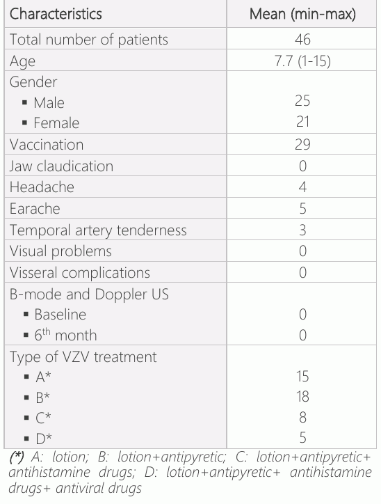
Table-1 shows the demographic values of the patients. None of the 30 patients included in the study showed halo signs. Moreover, none of the patients showed compression signs, occlusion, or stenosis results. The mean value for intima-media complex thickness was measured as 0.25 mm (0.18 - 0.27mm) in a common superficial temporal artery.
Discussion
There are contradicting studies in the literature about the relationship between VZV infection and GCA development. In our study, none of the patients diagnosed with chickenpox showed any clinical or US results that can be attributed to GCA in the short or long term, and that there were no clinical or US connections between childhood varicella infection and GCA development later on in life.
GCA etiology is still unclear in the literature. However, it is thought that the relationship between vessel walls and the immune system can play a role in the pathogenesis of the disease (12). Activated vascular dendritic cells collect CD4+ T cells and macrophages towards the vessel wall. Then those cells penetrate through the tissue from the adventitia layer towards the intima layer by vasa vasorum, causing granuloma formation (14). Since intracellular pathogens also frequently cause similar granulomatous inflammation situations, infections are thought to be a trigger in some of the patients (15). As for VZV, VZV was shown to have the ability to cause vasculopathy and the pathology samples of the affected vessels show multi-core giant cell presence, similar to GCA (16). Some other studies reported detecting VZV antibodies and DNA in a detailed review of temporal artery and aorta specimens of GCA patients with positive and negative biopsy results (17–21). On the contrary, some patients reported no VZV detected in temporal artery specimens of GCA patients (22–28). An additional case-control study reported no relation between VZV and GCA (29), yet another case-control study reported an increased risk of GCA following VZV infections (30). After seeing those contradicting results in the literature, it piqued our curiosity whether we would be able to detect any clinical or US findings correlated with GCA patients in children with VZV infections and if there are any ultrasonographic changes caused by VZV infections, which is a childhood disease in general, on temporal arteries during childhood, even though GCA is a condition seen in elderly patients. The study is a first in that sense, which warrants interest in this subject. The reason that we used US assessment instead of temporal artery biopsy, which is the most used method in GCA diagnosis today, is that our main objective was not to diagnose GCA, but instead, to detect whether there are any ultrasonographic changes in the temporal artery following VZV infections in children and the fact that temporal artery biopsy is an invasive and high-risk procedure with complications and that it only assesses a certain part of the vessels structure and only one single vessel, and finally that US imaging was proven to be a much more cost-effective non-invasive method with higher sensitivity and specificity for GCA diagnosis (4). 4 characteristic pathological signs of GCA can be detected using the US. Those are thickening of the vessel wall (halo sign), inability to compress artery (compression sign), stenosis, and vessel occlusion. Cellular infiltrates and edema can collect on intima and then spread out to media and adventitia layers in GCA. This thickening on the vessel wall shows a hypoechoic view around the vessel and is named as halo sign (30). Even though there are serious studies that show halo sign sensitivity as 55-100% and specificity as 78-100% in the US, this imaging method still did not replace temporal artery biopsy in GCA diagnosis (6). TABUL study, which prospectively compared temporal artery biopsy and colored Doppler US, reported sensitivity as 39% to 54% and specificity as 100% to 81% (10). One of the key points in ultrasonographic assessment is the requirement of an experienced radiologist to make that assessment. In the literature, the majority of the clinicians who perform the US were shown to be incompetent in temporal artery assessment (10). To avoid this obstacle in our study, we employed an experienced radiologist who is experienced in temporal artery US assessment.
We planned to use US for the assessment of temporal artery in VZV patients for the reasons stated above, however, this also caused some limitations to our study. One of the limitations is that even though performed by experienced radiologist, the ultrasonographic assessment method is still extensively studied for use in definitive GCA diagnosis, although it was shown to be a reliable method and is still discussed today. Another limitation is that we were unable to perform temporal artery biopsy in any of the patients, which is the key point in the diagnosis. However, we don't think that it is ethical to perform temporal artery biopsies just to diagnose or detect VZV on the related tissue in patients without GCA clinical symptoms or lab findings. Finally, our last limitation is the short amount of follow-up periods, which was six months. It is possible that those patients included in the study might develop clinical/ultrasonographic signs of GCA in the future and that we might be able to detect those changes during our routine follow-up periods. Therefore; we think further studies on this subject with those aforementioned limitations are warranted.
Conclusions
Chickenpox patients did not show any ultra sonographic changes in their temporal artery and no clinical symptoms related to GCA, yet we think that the relationship between VZV infection and GCA development warrants further studies with larger series and longer follow-up periods.
Declarations
- Author contributions (CRediT)
- All authors contributed equally to all 14 CRediT roles (Conceptualization, Data curation, Formal analysis, Funding acquisition, Investigation, Methodology, Project administration, Resources, Software, Supervision, Validation, Visualization, Writing – original draft, Writing – review & editing).
- Funding
-
Funding information was not reported in the published version of this article.
- Conflicts of interest
- The authors declared no conflict of interest for the present article.
- Ethics approval
- The study was conducted according to the declaration of Helsinki. The study was approved by the Ethic Committee of Giresun University (Date: 07.11.2018; No :5/1/29) and the patients gave informed consent prior to participation.
- Data availability
- A data availability statement was not reported in the published version of this article.
References (30)
27 of 30 references are linked to their DOI.
- Azap A, Kurt H, Varisella Zoster Virüs İnfeksiyonları. Ankara Üniversitesi Tıp Fakültesi Mecmuası, 2001 54.04 PubMed Google Scholar
- Kanra G, Kara A. Varisella zoster virus enfeksiyonları. Çocuk Sağlığı ve Hastalıkları Dergisi 2002;45:260-74 PubMed Google Scholar
- Ulusoy, Hasan, Nebahat Sarıca, and Şule Arslan. "Polymyalgia Rheumatica and Giant Cell Arteritis: A Case Report and Review of Literature." Turkish Journal of Physical Medicine and Rehabilitation 55.1 (2009) PubMed Google Scholar
- Schmidt, Wolfgang A. "Ultrasound in the diagnosis and management of giant cell arteritis." Rheumatology 57.suppl_2 (2018): ii22- ii31 DOI Crossref PubMed Google Scholar
- Gilden D, Cohrs RJ, Mahalingam R, Nagel MA. Varicella zoster virus infection of the nervous system. In: Johnson MV, Adams HP, Fatemi SA, eds. Neurobiology of disease. 2nd ed. New York: Oxford University Press, 2015. In press DOI Crossref PubMed Google Scholar
- Nagel MA, Gilden D. Update on varicella zoster virus vasculopathy. Curr Infect Dis Rep 2014; 407:1–8 DOI Crossref PubMed Google Scholar
- Nagel MA, Khmeleva N, Boyer PJ, Choe A, Bert R, Gilden D. Varicella zoster virus in the temporal artery of a patient with giant cell arteritis. J Neurol Sci 2013; 335:229 –30 DOI Crossref PubMed Google Scholar
- Lotan, I., & Steiner, I. (2017). Giant cell arteritis following varicella zoster vaccination. Journal of the neurological sciences, 375, 158-159 DOI Crossref PubMed Google Scholar
- Muratore, Francesco, et al. "No detection of varicella-zoster virus in temporal arteries of patients with giant cell arteritis." Seminars in arthritis and rheumatism. Vol. 47. No. 2. WB Saunders, 2017 DOI Crossref PubMed Google Scholar
- Luqmani, R. et al. The Role of Ultrasound Compared to Biopsy of Temporal Arteries in the Diagnosis and Treatment of Giant Cell Arteritis (TABUL): a diagnostic accuracy and cost-effectiveness study. Health Technol. Assess. (Rockv). 20, 1–238 (2016) DOI Crossref PubMed Google Scholar
- Schäfer VS, Juche A, Ramiro S, Krause A, Schmidt WA. Ultrasound cut-off values for intima-media thickness of temporal, facial and axillary arteries in giant cell arteritis. Rheumatology 2017;56:1479– 83 DOI Crossref PubMed Google Scholar
- England, B. R., Mikuls, T. R., Xie, F., Yang, S., Chen, L., & Curtis, J. R. (2017). Herpes zoster as a risk factor for incident giant cell arteritis. Arthritis & Rheumatology, 69(12), 2351-2358 DOI Crossref PubMed Google Scholar
- Weyand CM, Goronzy JJ. Immune mechanisms in medium and large-vessel vasculitis. Nat Rev Rheumatol 2013;9:731–40 DOI Crossref PubMed Google Scholar
- Russo MG, Waxman J, Abdoh AA, Serebro LH. Correlation between infection and the onset of the giant cell (temporal) Varicella Zoster Virus in Children Kulaklı et al. arteritis syndrome: a trigger mechanism? Arthritis Rheum 1995;38:374–80 DOI Crossref PubMed Google Scholar
- Nagel MA, Cohrs RJ, Mahalingam R, Wellish MC, Forghani B, Schiller A, et al. The varicella zoster virus vasculopathies: clinical, CSF, imaging, and virologic features. Neurology 2008;70:853–60 DOI Crossref PubMed Google Scholar
- Mitchell BM, Font RL. Detection of varicella zoster virus DNA in some patients with giant cell arteritis. Invest Ophthalmol Vis Sci 2001;42:2572– 7 DOI Crossref PubMed Google Scholar
- Nagel MA, White T, Khmeleva N, Rempel A, Boyer PJ, Bennett JL, et al. Analysis of varicella-zoster virus in temporal arteries biopsy positive and negative for giant cell arteritis. JAMA Neurol 2015;72:1281–7 DOI Crossref PubMed Google Scholar
- Gilden D, White T, Khmeleva N, Katz BJ, Nagel MA. Blinded search for varicella zoster virus in giant cell arteritis (GCA)- positive and GCA-negative temporal arteries. J Neurol Sci 2016;364:141–3 DOI Crossref PubMed Google Scholar
- Gilden D, White T, Khmeleva N, Heintzman A, Choe A, Boyer PJ, et al. Prevalence and distribution of VZV in temporal arteries of patients with giant cell arteritis. Neurology 2015;84:1948– 55 DOI Crossref PubMed Google Scholar
- Gilden D, White T, Boyer PJ, Galetta KM, Hedley -Whyte ET, Frank M, et al. Varicella zoster virus infection in granulomatous arteritis of the aorta. J Infect Dis 2016;213:1866–71 DOI Crossref PubMed Google Scholar
- Kennedy PG, Grinfeld E, Esiri MM. Absence of detection of varicella-zoster virus DNA in temporal artery biopsies obtained from patients with giant cell arteritis. J Neurol Sci 2003;215:27–9 DOI Crossref PubMed Google Scholar
- Nordborg C, Nordborg E, Petursdottir V, LaGuardia J, Mahalingam R, Wellish M, et al. Search for varicella zoster virus in giant cell arteritis. Ann Neurol 1998;44:413–4 DOI Crossref PubMed Google Scholar
- Alvarez-Lafuente R, Fernandez-Gutierrez B, Jover JA, Judez E, Loza E, Clemente D, et al. Human parvovirus B19, varicella zoster virus, and human herpes virus 6 in temporal artery biopsy specimens of patients with giant cell arteritis: analysis with quantitative real time polymerase chain reaction. Ann Rheum Dis 2005;64:780–2 DOI Crossref PubMed Google Scholar
- Helweg-Larsen J, Tarp B, Obel N, Baslund B. No evidence of parvovirus B19, Chlamydia pneumoniae or human herpes virüs infection in temporal artery biopsies in patients with giant cell arteritis. Rheumatology (Oxford) 2002;41:445– 9 DOI Crossref PubMed Google Scholar
- Rodriguez-Pla A, Bosch-Gil JA, Echevarria-Mayo JE, Rossello-Urgell J, Solans-Laque R, Huguet-Redecilla P, et al. No detection of parvovirus B19 or herpesvirus DNA in giant cell arteritis. J Clin Virol 2004;31:11–5 DOI Crossref PubMed Google Scholar
- Solomon, I. H., Docken, W. P., & Padera, R. F. (2019). Giant cell arteritis is not associated with varicella zoster virus in temporal artery biopsies or ascending aortic resections. The Journal of Rheumatology, jrheum-180912 DOI Crossref PubMed Google Scholar
- BuckinghamEM, FoleyMA, Grose C, et al. Identification of herpes zoster-associated temporal arteritis among cases of giant cell arteritis. Am J Ophthalmol. 2018;187:51–60. doi:10.1016/j.ajo.2017.12.017 DOI Crossref PubMed Google Scholar
- Schafer VS, Kermani TA, Crowson CS, Hunder GG, Gabriel SE, Ytterberg SR, et al. Incidence of herpes zoster in patients with giant cell arteritis: a population-based cohort study. Rheumatology (Oxford) 2010;49:2104–8 DOI Crossref PubMed Google Scholar
- Rhee RL, Grayson PC, Merkel PA, Tomasson G. Infections and the risk of incident giant cell arteritis: a population-based, casecontrol study. Ann Rheum Dis 2017;76:1031–5 DOI Crossref PubMed Google Scholar
- Schmidt WA, Kraft HE, Volker L, Vorpahl K, Gromnica-Ihle EJ. Colour Doppler sonography to diagnose temporal arteritis. Lancet 1995;345:866 DOI Crossref PubMed Google Scholar
Respond to this article
Comments on a published article are welcome as a Letter to the Editor.
Get an e-mail when new articles in your fields are published
Questions to the authors
Questions are moderated: an editor approves each question, the authors answer, and an editor approves the answer before both appear here.
No answered questions yet.
Ask the authors a question
Discussion
Comments are moderated: they appear after the writer confirms the e-mail address and an editor approves them. Each comment carries a conflict-of-interest statement.
No comments yet.
Write a comment
Read the full text
Open the PDF here
Loading the PDF…
If the PDF does not show here, use the download button above.
How to cite this article
Kulakli F, Bulut M, Bekci T. Does Varicella Zoster Virus in Children Affect Temporal Artery Structure or Blood Flow?. Ulutas Med J. 2021;7(1):54-59. doi:10.5455/umj.20201229071421
Kulakli, F., Bulut, M., & Bekci, T. (2021). Does Varicella Zoster Virus in Children Affect Temporal Artery Structure or Blood Flow?. The Ulutas Medical Journal, 7(1), 54–59. https://doi.org/10.5455/umj.20201229071421
Kulakli, F., Bulut, M. and Bekci, T. (2021) 'Does Varicella Zoster Virus in Children Affect Temporal Artery Structure or Blood Flow?', The Ulutas Medical Journal, 7(1), pp. 54–59. doi: 10.5455/umj.20201229071421.