The Ulutas Medical Journal · ISSN 2149-0430 (print), 2149-388X (online) · https://www.ulutasmedicaljournal.com/?mno=293537
Original Research · Open access
Association between Presence of Esophageal Varices on Endoscopy and Extreme Degree of F4-Stage Fibrosis on Fibroscan in HCV-Related Chronic Liver Disease
- Rawalpindi Medical University (RMU), Surgical Unit, Rawalpindi ROR
- Rawalpindi medical university, medical unit, Benazir Bhutto hospital Rawalpindi ROR
- CMO, holy family hospital, Rawalpindi ROR
Abstract
- Background
- Hepatitis C Virus (HCV) infection is one of the causes which cause liver fibrosis, cirrhosis and carcinoma if left untreated. Patients who present with acute HCV infection, 50-70% of them develop chronic infection. This study attempted to see an association between F4 fibrosis in liver on fibroscan and esophageal varices on endoscopy in HCV related chronic liver disease (CLD) patients.
- Method
- This study was conducted in Gastroenterology and Hepatology division of holy family hospital, Rawalpindi. Total 117 patients of 10 to 90 years were included. All patients were having HCV related CLD. Patients having F4 fibrosis stage were included while patients having fibrosis stage up to F3 fibrosis stage were excluded. All data regarding age, gender, F4 fibrosis and esophageal varices was entered and analyzed using the statistical package of social sciences while descriptive analysis was performed.
- Results
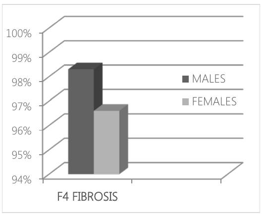
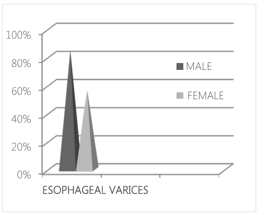
- Amongst 114 patients who presented with F4 fibrosis stage on fibroscan 69.3% (79) of them were diagnosed for esophageal varices on endoscopy and 30.7% (35) were not having esophageal varices on endoscopy. Patients who were having esophageal varices on endoscopy 84.5% (49) of them were males and 55.9% (33) were females. Study participants having F4 fibrosis stage, 98.3% of them were males and 96.6% were females. So, there was not much difference between males and females having an extreme degree of fibrosis on fibroscan.
- Conclusion
- Patients who were having F4 fibrosis stage on fibroscan, 2/3 of them were having esophageal varices on endoscopy. Esophageal varices were more present in males as compared to females.
Keywords
Full text
Introduction
Hepatitis is a disease causing inflammation of the liver. The complications of the disease are fibrosis, liver cancer, and cirrhosis (1). Hepatitis A,B,C,D,Eand G viruses cause liver inflammation and from these viruses hepatitis A, B and C are the most common.Thepatientswho are infected acutely due to Hepatitis C Virus infection, that 50 to 70% of them will develop the chronic liver disease. Chronic infection can lead to cirrhosis, liver cancer and liver failure (2). Almost 71 million people have been infected with chronic hepatitis C across worldwide (3). Prevalence of HCV infection in Pakistan is 4.8% (4). Different studies have been conducted previously to show the fibroscan as the most noninvasive procedure for detection of liver stiffness. One study showed results that Esophageal varices were present in 63.8%, gastric varices in 2.1% and portal gastropathy in 55.3% of patients who have had fibrosis on fibroscan (5). Another study showed results that patients who presented with fibrosis stage F4 (21KP) 92% of them showed esophageal varices (6). Another study showed that liver stiffness measured by fibroscan is the most noninvasive method to detect portal hypertension changes in patients (7). Another study revealed results as 90% of group C patients showing varices 81% of them had a liver stiffness (8). Another study showed that liver stiffness was significantly related to portal hypertension in HCV related liver disease patients (9). An Egyptian study showed results that overall accuracy of fibroscan for the detection of esophageal varices was 96.6% and EV were present with more than 20 KPa pressure (10). A study concluded the strong relationship between liver fibrosis and portal hypertension with p≤0.0001 (r=0.81). It also concluded that patients with F4 stage 63.8% of them showed esophageal varices (11).
Hepatitis C virus infection is one of the most common causes which lead to chronic liver disease. The consequences of CLD are cirrhosis, liver cancer, and liver failure. This study has not been documented in our area. So this could be one of the 1st studies being conducted in Pakistan to find an association between portal hypertension changes (esophageal varices) and liver fibrosis so that noninvasive method can be used pre endoscopically.
Material and Methods
This prospective study was conducted at gastroenterology and Hepatology division of Holy Family Hospital Rawalpindi in 2017. 117 patients of age between 10-90 years were included. All patients were HCV positive and had the chronic liver disease.
Fibroscan was used to see fibrosis stage of the liver in these patients and they all were having F4 fibrosis stage except 3 patients which had F3-F4 fibrosis stage. Patients having hepatitis other than HCV infections were not included. Also, all those patients which were having fibrosis stage up to F3 were excluded for study. Endoscopy was performed in all patients to see esophageal varices whether present or not. This study was attempted after approval from Institutional Research Forum of Rawalpindi University.
Statistical Analysis
WHO sample size calculator, the sample size was calculated with anticipated population proportion 0.926 keeping level of confidence 95 % and absolute precision required 5%. The minimally required sample size was 114 and we included 117 patients. After IRF approval and informed oral consent from patients who fulfill the selection criteria relevant data was taken from the record and shifted to performance. All data were analyzed using Statistical Package of Social sciences version 22. All categorical variables age, esophageal varices, F4 fibrosis stage frequencies along with percentages were calculated.
Results
For all 117 patients included in this study group, 58 (49.6%) were males and 59 (50%) were females. All the study participants were positive for the HCV antibody. Amongst 117 patients 114 of them were having F4 fibrosis stage on fibroscan while 3 patients were having an F3-F4 stage on fibroscan.
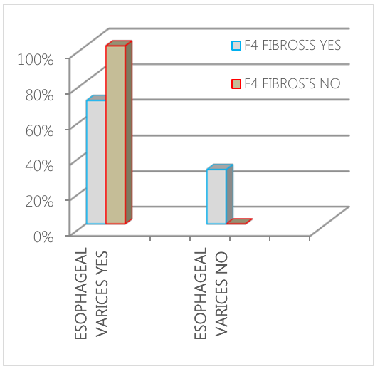
Amongst 114 patients with F4 fibrosis stage on fibroscan, 69.3% (79) were diagnosed for esophageal varices on endoscopy and 30.7% (35) were not having esophageal varices on endoscopy (figure-1). Gender wise distribution of F4 fibrosis is shown in figure-2. In participants having F4 fibrosis stage, 98.3% were males and 96.6% were females. So, there was not much difference between males and females having an extreme degree of fibrosis on fibroscan. Gender wise distribution of esophageal varices is shown in figure-3. It showed that in the patients with esophageal varices on endoscopy, 84.5% (49) were males and 55.9% (33) were females. Distribution of esophageal varices was more in males as compared to females.
The mean age for patients having esophageal varices was 56.6±9.7 years, while patients who did not have esophageal varices mean age for them was 50.4 with a standard deviation of 7.8 years. Mean age for patients having F4 fibrosis was 54.6±9.5 years.
Discussion
Hepatitis is a disease with its complications i.e. liver cirrhosis, liver fibrosis and liver cancer if left untreated (4). This study was attempted to find an association between esophageal varices on endoscopy and F4 fibrosis stage on fibroscan in HCV related CLD so that noninvasive method can be used pre endoscopically (6, 9). When there is a chronic infection due to HCV, it affects the liver and liver fibrosis occurs. After liver fibrosis patients develop portal hypertension changes; esophageal varices, gastric varices, portal gastropathy, and splenomegaly (2). Fibroscan is a noninvasive method to detect liver stiffness and it can be used before endoscopy if the extreme degree of fibrosis is found on fibroscan (1, 7). Our study showed the results that about 2/3 of patients who were having F4 fibrosis stage on fibroscan were diagnosed for esophageal varices on endoscopy which coincides with findings of Vizzutt (5) et al which showed that 63.8% patients with F4 stage showed esophageal varices. Our study is also consistent with findings of Bureau et al (6) which showed the results that high proportion of patients who presented with F4 stage were diagnosed for esophageal varices. A non-invasive method like fibroscan should be used before endoscopy so that portal hypertension changes can be diagnosed and treated at early stage. Our research also showed the results that those patients who presented with esophageal varices they were mostly males as compared to females. This might be due to the reason that in our society mostly males are a drug addict.
Hepatitis C virus transfers due to intravenous injections, shaving, and blood transfusions (11). All these things are more common amongst males in our society. Mostly males are willing for blood donation. But in our study, we only compare esophageal varices with F4 fibrosis stage on fibroscan. What about esophageal varices at F1, F2, and F3 fibrosis stage ? This is a question for future studies. In future studies should be conducted to find an association between F1, F2 and F3 fibrosis stage and other portal hypertension changes so that the disease with its life-threatening complications can be diagnosed at the very early stage and treated accordingly. And in future, there should be studies which also take other tests like liver biopsy and ultrasound to be compared with fibroscan. our study is a first step in the platform of early diagnosis and better treatment of HCV related complications in our region.
Conclusion
Patients who were having F4 fibrosis stage on fibroscan, 2/3 of them were having esophageal varices on endoscopy. Esophageal varices were more present in males as compared to females. Fibroscan can be used as a screening for endoscopy to detect portal hypertension changes in those patients which have an extreme degree of fibrosis.
Declarations
- Author contributions (CRediT)
- All authors contributed equally to all 14 CRediT roles (Conceptualization, Data curation, Formal analysis, Funding acquisition, Investigation, Methodology, Project administration, Resources, Software, Supervision, Validation, Visualization, Writing – original draft, Writing – review & editing).
- Funding
-
Funding information was not reported in the published version of this article.
- Conflicts of interest
- The authors declare that they have no conflict of interest in the current study.
- Ethics approval
- Ethics approval details were not reported in the published version of this article.
- Data availability
- A data availability statement was not reported in the published version of this article.
References (11)
5 of 11 references are linked to their DOI.
- WHO. What is hepatitis?http://www.who.int/features/qa/ 76/en. (Accessed 10 September 2017) PubMed Google Scholar
- Hepatitis (Viral Hepatitis, A, B, C, D, E, G) http://www. medicinenet.com/viral_hepatitis/article.htm. (Accessed 10 September 2017) PubMed Google Scholar
- WHO. Hepatitis C. www.who.int/mediacentre/factsheets/ fs164/en. (Accessed 10 september 2017) PubMed Google Scholar
- Quershi, H; bile, KM; Jooma et al. Eastern Mediterranean Health Journal, suppl. Supplement; Alexandria 16 (Aug 2010): S15-23. PubMed Google Scholar
- Vizzutti F, Arena U, Romanelli RG, Rega L, Foschi M, Colagrande S et al. Liver stiffness measurement predicts severe portal hypertension in patients with HCV-related cirrhosis. Hepatology. May 2007;45 (5):1290-7. DOI Crossref PubMed Google Scholar
- Bureau C, Metivier S, Peron JM, S elves J, Robic MA, Gourroud PA, et al. Transient elastography accurately predict the presence of significant portal hypertension in patients with the chronic liver disease. Alimentary pharmacology and therapeutics. 2008; 27(12):1261-8. DOI Crossref PubMed Google Scholar
- Colecchia A, Montrone N, Scailoi E, Bacchi-Reggiani ML, Colli A, Casazza G, et al. Measurement of Spleen Stiffness to Evaluate Portal Hypertension and the Presence of Esophageal Varices in Patients With HCV-Related Cirrhosis. Gastroenterology 2012;143(3):646-4. DOI Crossref PubMed Google Scholar
- Augustin S, Millan M, Gonzalez A, Martell M, Gelabert A, Segarra A, et al. Detection of early portal hypertension with routine data and liver stiffness in patients with asymptomatic liver disease: A prospective study. Journal of Hepatology. 2014; 60(3):561-9. DOI Crossref PubMed Google Scholar
- Leomine M, Katsahian S, Ziol M, Nahon P, Ganne-Carrie N, Kazemi F, et al. Liver stiffness measurement as a predictive tool of clinically significant portal hypertension in patients with compensated hepatitis C or alcohol -related cirrhosis. Aliment Pharmacol Ther. 2008 Nov 1; 28(9):1102-10. PubMed Google Scholar
- Helmy, Ali AZ, Hamed, et al. Transient elastography (Fibroscan) in the prediction of esophageal varices in hepatitis C virus-related chronic liver disease: an Egyptian experience. Egyptian liver journal 2016; 6(3):43-47. DOI Crossref PubMed Google Scholar
- Vizzutti F, Arena U, Romanelli RG, Rega L, Foschi M, Colagrande S et al. Liver Stiffness Measurement Predicts Severe Portal Hypertension in Patients with HCV-Related Cirrhosis. Hepatology 2005; 45(5):1290-7 PubMed Google Scholar
Read the full text
Open the PDF here
Loading the PDF…
If the PDF does not show here, use the download button above.
How to cite this article
Amin M, Jabbar MA, Ahmad H, SAMAR S. Association between Presence of Esophageal Varices on Endoscopy and Extreme Degree of F4-Stage Fibrosis on Fibroscan in HCV-Related Chronic Liver Disease. Ulutas Med J. 2018;4(2):85-89. doi:10.5455/umj.20180312034434
Amin, M., Jabbar, M. A., Ahmad, H., & SAMAR, S. (2018). Association between Presence of Esophageal Varices on Endoscopy and Extreme Degree of F4-Stage Fibrosis on Fibroscan in HCV-Related Chronic Liver Disease. The Ulutas Medical Journal, 4(2), 85–89. https://doi.org/10.5455/umj.20180312034434
Amin, M. et al. (2018) 'Association between Presence of Esophageal Varices on Endoscopy and Extreme Degree of F4-Stage Fibrosis on Fibroscan in HCV-Related Chronic Liver Disease', The Ulutas Medical Journal, 4(2), pp. 85–89. doi: 10.5455/umj.20180312034434.