The Ulutas Medical Journal · ISSN 2149-0430 (print), 2149-388X (online) · https://www.ulutasmedicaljournal.com/?mno=288793
Case Report · Open access
Giant Cervical Chondrosarcoma
Also available in: العربية · Türkçe · 中文
- Ankara Oncology Training and Research Hospital, Department of Orthopedics, Ankara, Turkey ROR
- Ankara Atatürk Training and Research Hospital, Department of Radiology, Ankara, Turkey ROR
Abstract
- Background
- Chondrosarcoma is the third most common malignant tumor of the bone and most commonly occurs between 30 and 60 years. Primary tumors form directly in bone and soft tissues, whereas secondary tumors develop on existing benign cartilage-containing tissue.
- Case Presentation
- In this case report, we presented a rare case of cervical chondrosarcoma in a patient who admitted to the hospital with a swelling in the neck. There was the history of Multiple Hereditary Exocytosis in the male patient aged 33 years and in his family. On X-ray, MRI and CT scans, there was an expansive mass appearance with dimensions of about 14x6x7.5 cm in the right cervical region. The tumor did not expand toward the spinal canal. The needle biopsy revealed that the patient had a chondrosarcoma, thus extensive resection was performed. Malign tumors of the cervical spine have a good prognosis if en bloc resection is possible.
Keywords
Full text
Introduction
Osteochondroma is the most common skeletal neoplasm. It is probably a develop-mental malformation, consisting of a bone-derived body, and a cartilaginous cap. Hereditary Multiple Osteochondromatosis is an autosomal dominant disorder, and the rate of malignancy is unknown but is thought to be as low as 3-5% (1, 2).
Chondrosarcoma is the third most common malignant tumor of the bone and most commonly occurs between 30 and 60 years of age (3, 4). It divides into two as primary and secondary. Primary tumors form directly in bone and soft tissues, whereas secondary tumors develop on existing benign cartilage-containing tissue. Secondary chondrosarcomas are seen in younger age groups (1).
Less than 10% of the chondrosarcomas are located in the vertebrae and are most common in the thoracic vertebrae, while rarely seen in the cervical vertebrae (4). In this case report, we present a chondrosarcoma located in the cervical vertebrae and developed on the ground of multiple hereditary exostoses.
Case Report
Our case is a 33-year-old male patient diagnosed with multiple hereditary exostoses and also there are similar lesions on both two siblings and his mother. The patient had previously undergone total excision operations at various sites (left hand 2. finger, distal of the left femur, proximal of the right tibia and pelvis) in different centers. The patient was asked if data concerning the case could be submitted for publication, and he consented.
Since the patient underwent operations and all of them were benign tumors, he has neglected the growing mass in the right neck region. Eventually, the patient applied to our clinic because of the mass in the neck becoming increasingly bigger.
On the clinical examination, in the right neck region, he has a mass of approximately 10 cm and the mass were immobile, rigid, and restricting the neck movements. Besides, there were some osteochondroma-related small protrusions and scars associated with previous operations in various parts of the body.
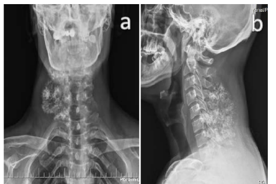
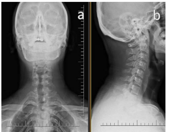
On the anteroposterior and lateral cervical spine radiograph; at the lower cervical vertebra level, there was a view of a large, cartilaginous and destructive mass which was mostly composed of soft tissue and containing diffuse popcorn-like calcifications, and of which borders could not clearly distinguish with posterior arches of adjacent vertebrae (Figure 1a and 1b).
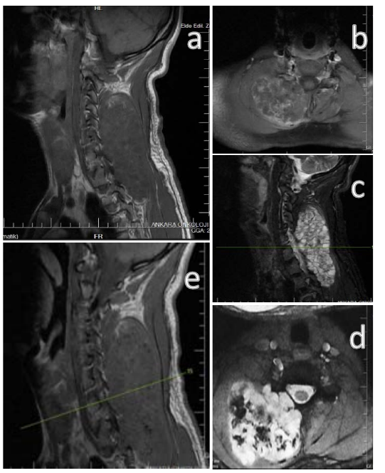
When magnetic resonance images are examined, on the sagittal T1A, T2A, axial T1A, T2A and fat-suppressed T2A sections, at C3-T1 level was seen that the lesion expanded to adjacent intervertebral foramen and occasionally included lamina and transverse processes of adjacent right vertebrae. On the T1A images, it was seen that a giant solid mass, which was isointense with muscle and the signal voids originating from calcification. In addition, on the T2A, and on the fat-suppressed, T2A images the lesion was hyperintense. The tumor did not have an invasion of the spinal canal (Figure 2a, 2b, 2c, 2d, 2e).
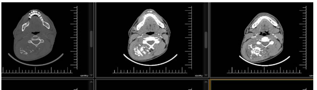
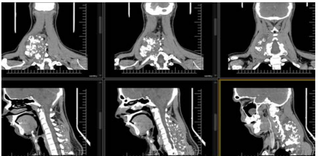
On the computerized tomography images, there was a large solid hypodense lesion (compared to muscle) which located at the C3- T1 level and pushing the adjacent muscles to the right and causing scalloping in the transverse processes and lamina of adjacent vertebrae. In addition, there were popcorn-like chondroid calcifications (Figure3a, 3b).
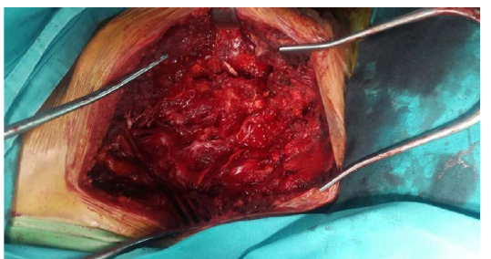
The needle biopsy performed. The pathology report was a grade-2 chondrosarcoma originating in an osteochondroma. After general anesthesia, the patient was placed in the prone position and sterile inking and covering procedures applied. Later, the lesion resected with wide borders by entering with a longitudinal incision. After hemostasis achieved, the tumor bed was closed by placing a hemovac drain. When the resection material examined macroscopically, it was a gray-brown colored, 15x10.5x8 cm sized, irregularly shaped, clear density tissue with small nodules on the surface (Figure 4 and 5).
On the first postoperative day, anteroposterior and lateral radiographs were taken and on the second postoperative day, the hemovac drain was removed and the patient was discharged (Figure 6). The patient was followed up for the daily dressing and on the 15th postoperative day, sutures were removed.
Discussion
Osteochondroma is the most common skeletal neoplasm and is mostly asymptomatic and usually diagnosed incidentally in childhood. The tumor grows up with skeletal maturation and usually is noticed at this stage, and the growth stops when the skeletal maturation ends (1, 5).
In about 90% of cases, osteochondroma is a single lesion and most commonly in the metaphysis of long bones, especially in the proximal tibia, proximal humerus or distal femur (1). The patient, who was followed by for his lesions, had tumors that were located bilaterally in the femur, proximal tibia, and proximal humerus.
Hereditary Multiple Osteochondromatosis is an autosomal dominant disorder.It may contain both sessile and pediculated lesions. In our case, the family story was positive.
In adults, growing osteochondroma suggests malignant transformation.The male gender and being older than 40 years is a malignancy criterion for being a primary chondrosarcoma, however, secondary chondrosarcomas which originated in Ollier Disease or Multiple Hereditary Osteochondromatosis are seen in the earlier age (1, 6). In terms of age and gender, our case compatible with the cases reported in the literature.
In the literature, malignant transformation risk is reported higher in osteochondromas located within the pelvis, shoulder, and hipbone. Some other criteria of malignancy are the larger tumor than 6 cm, soft tissue component, and calcifications seen on the radiograph (1).
A direct radiography is generally sufficient for diagnosing osteochondromas. The existence of a bony tissue, integrated with the origin and covered with hyaline cartilage, is diagnostic. On the MRI, the malignant transformation should be considered if the thickness of the cartilage surface is thicker than 2 cm in the adult and thicker than 3 cm in the children. Radiologically, cervicalchondrosarcomas appear as destructive bony lesions in the cervical spinal column or as paraspinal masses with calcification (7).
Surgeryis recommended for osteochondroma if the patient has pain, neurovascular pressure, abnormal growth pattern, the risk of malign transformation, or limitation of joint movement. The main symptom in central chondrosarcomas is pain, while the main symptom of peripheral lesions is a growing mass (swelling), however, the pain is very rare in the peripheral lesions(1). In our patient, all of the surgical indications mentioned earlier, except pain, were present at the time of admission.
Chondrosarcomas divides into two groups as primary (75%) and secondary (25%). Primary chondrosarcomas directly originate in bone and soft tissues, while secondary chondrosarcomas develop from pre-existing benign cartilage containing tissue (enchondroma, osteochondroma). They can be central or peripheral depending on their location. Primary chondrosarcomas are most central, whereas secondary chondrosarcomas are mostly peripheral. Our patient had a secondary peripheral chondrosarcoma.
Chondrosarcomas are more common in pelvic, thoracic, shoulders and hip bones. Less than 10% of chondrosarcomas is located in the vertebrae while they most frequently located in the thoracic vertebrae and followed by cervical and lumbar vertebrae (3, 6). Spinal canal extension is rarely in the spinal chondrosarcomas, but the chondrosarcomas involving both intradural and extradural components have also reported in the literature (8). There was no tumor enlargement into the spinal canal in our case.
Mostly classical chondrosarcomas are of low to moderate grade and grow slowly, whereas peripheral chondrosarcomas grow much slower.In addition,peripheral chondrosarcomas usually do not metastasize, but recurrences occur frequently. After the intralesional excision recurrence rate is 70%, but after wide resection, it is 10% (1).
Prognosis of the spinal chondrosarcoma is relatively good with a 5-year survival for grade 1 tumors is 90%, for grade 2 tumors is 81% and for grade 3 tumors is 43% (3). Like the other borderline and malignant tumors, the most successful treatment forspinal chondrosarcoma is a complete en bloc resection of the tumor (7, 9–11). This often requires a spinal reconstruction involving a multidisciplinary team of orthopedic, plastic and neurosurgery specialists.Chondrosarcomasare chemotherapy and radiotherapy resistant tumors. If en bloc resection is not possible, partial removal of the tumor after the radiotherapy may provide palliation of pain and improve neurological deficits(6, 9). The surgeons in other departments did not need because of the absence of spinal canal extension and skin reconstruction in our case. Chemotherapy and radiotherapy didn't perform after wide resection of the tumor.
Conclusion
Chondrosarcomas located in the cervical spine is a rare entity and preoperatively the relationship between tumors and vital neurovascular structures should be assessed properly. The most successful treatment is resection of the mass with large borders, but chemotherapy and radiotherapy are ineffective.
Declarations
- Author contributions (CRediT)
- All authors contributed equally to all 14 CRediT roles (Conceptualization, Data curation, Formal analysis, Funding acquisition, Investigation, Methodology, Project administration, Resources, Software, Supervision, Validation, Visualization, Writing – original draft, Writing – review & editing).
- Funding
-
There is no funding source.
- Conflicts of interest
- The authors declare that they have no conflict of interest in the current study.
- Ethics approval
- Ethics approval details were not reported in the published version of this article.
- Data availability
- A data availability statement was not reported in the published version of this article.
References (11)
7 of 11 references are linked to their DOI.
- Rodop O. osteochondroma. Dabak N, editor. in: Multidisipliner Yaklaşımla Kemik ve Yumuşak Doku Tümörleri (1. ed). Ankara Bayt Yayıncılık; 2013: 117-21. PubMed Google Scholar
- Ozturk R, Aydın M, Arıkan ŞM,Şimşek MA, Toptaş E, Güngör BŞ. Belirsiz diferansiasyonlu ekstremite yumuşak doku sarkomları: 60 hastalık seride klinik prezentasyon, tedavi ve sonuçları. Bakırköy Tıp Dergisi PubMed Google Scholar
- Merchant S, Mohiyuddin SM, Rudrappa S, Deo RP, A S, Menon LR. Cervical chondrosarcoma- rare malignancy: a case report. Indian J Surg Oncol. 2014 Dec;5(4):293-6. DOI Crossref PubMed Google Scholar
- Matsumoto Y, Takahashi Y, Harimaya K, Nakagawa T, Kawaguchi K, Okada S. Dedifferentiated chondrosarcoma of the cervical spine: a case report. World J Surg Oncol. 2013 Feb 2;11:32. DOI Crossref PubMed Google Scholar
- Ozturk R, Karakoç Y. Arthroscopic treatment of benign tumors and tumor-like lesions located in and around the knee joint. Ulutas Med J DOI Crossref PubMed Google Scholar
- Strike SA, McCarthy EF. Chondrosarcoma of the spine: a series of 16 cases and a review of the literature. Iowa Orthop J. 2011;31:154-9. PubMed Google Scholar
- Sundaresan N, Rosen G, Boriani S. Primary Malignant Tumors of the Spine. Orthop Clin North Am.2009 Jan; 40(1):21– 36. DOI Crossref PubMed Google Scholar
- Hekmatnia, A; Ghazavi, A; Aminmansour, B; Mahzouni, P.A Case of Chondrosarcoma That Primarily Developed in the Cervical Spine. Iran J Radiol. 2012 Mar;9 (1):57-59 DOI Crossref PubMed Google Scholar
- Boriani S, De Iure F, Bandiera S, Campanacci L, Biagini R, Di Fiore M, Bandello L, Picci P, Bacchini P. Chondrosarcoma of the mobile spine. Spine. 2000 Apr;25(7):804–12. DOI Crossref PubMed Google Scholar
- Ozturk R., Arıkan ŞM, Şimşek MA,Özanlağan E, Güngör BŞ. Management of solitary fibrous tumors localized in extremity: case series and a review of the literature. Eklem Hastalik Cerrahisi 2017;28(2):121-7 doi: 10.5606/ehc.2017.52092 DOI Crossref PubMed Google Scholar
- Toğral G, Arıkan M, Aktaş E, Öztürk R, Güven O, Eksioğlu F. Surgical Management Of Bone Metastases From Urological Malignancies: An Analysis Of 70 Cases. Acta Orthop Traumatol Turc 2015;49(6):634– 640. PubMed Google Scholar
Respond to this article
Comments on a published article are welcome as a Letter to the Editor.
Get an e-mail when new articles in your fields are published
Questions to the authors
Questions are moderated: an editor approves each question, the authors answer, and an editor approves the answer before both appear here.
No answered questions yet.
Ask the authors a question
Discussion
Comments are moderated: they appear after the writer confirms the e-mail address and an editor approves them. Each comment carries a conflict-of-interest statement.
No comments yet.
Write a comment
Tell me when this article is cited
Leave your e-mail address and confirm it. These notices are not being sent yet; your confirmed address is kept for them and used for nothing else.
Read the full text
Open the PDF here
Loading the PDF…
If the PDF does not show here, use the download button above.
How to cite this article
Beltir G, Arikan SM, Ates OF, Gungor BS. Giant Cervical Chondrosarcoma. Ulutas Med J. 2018;4(2):118-123. doi:10.5455/umj.20180119064908
Beltir, G., Arikan, S. M., Ates, O. F., & Gungor, B. S. (2018). Giant Cervical Chondrosarcoma. The Ulutas Medical Journal, 4(2), 118–123. https://doi.org/10.5455/umj.20180119064908
Beltir, G. et al. (2018) 'Giant Cervical Chondrosarcoma', The Ulutas Medical Journal, 4(2), pp. 118–123. doi: 10.5455/umj.20180119064908.
@article{umj288793,
  title = {Giant Cervical Chondrosarcoma},
  author = {Beltir, Galip and Arikan, Sefik Murat and Ates, Omer Faruk and Gungor, Bedii Safak},
  journal = {The Ulutas Medical Journal},
  year = {2018},
  volume = {4},
  number = {2},
  pages = {118--123},
  doi = {10.5455/umj.20180119064908},
  url = {https://www.ulutasmedicaljournal.com/?mno=288793},
  issn = {2149-388X}
}