The Ulutas Medical Journal · ISSN 2149-0430 (print), 2149-388X (online) · https://www.ulutasmedicaljournal.com/?mno=283167
Case Report · Open access
Lymphoma Mimicking Pancreatic Adenocarcinoma: A Case Report
Also available in: العربية · Türkçe · 中文
Abstract
- Background
- Pancreatic lymphoma is very rare and occurs below <1%. Because clinical signs and symptoms are similar to pancreatic adenocarcinoma, diagnosis of pancreatic lymphoma is usually very difficult. We aimed to present Magnetic Resonance (MR) findings of a pancreatic lymphoma mimicking pancreatic adenocarcinoma.
- Case Presentation
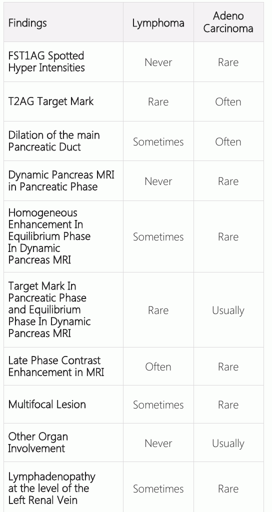
- The case was pathologically diagnosed with Diffuse B-cell lymphoma and there were no jaundice findings. Obstruction was not present in the structure other than being pushed into the veins. There was a diffusion restriction without necrosis with low ADC values parallel to increased cellularity. Apart from these, the table in the distinction between lymphoma and adenocarcinoma is helpful in other findings.
- Conclusion
- It is useful to know that MRI and diffuse MRI in pancreatic lymphoma may provide useful information as well as a diffuse restriction in both due to adenocarcinoma and lymphoma elevated cellularity
Keywords
Full text
Introduction
Malignant tumors of the pancreas are largely adenocarcinomas. Non-Hodgkin's lymphoma (NHL) is often caused by the lymphatic system. In the NHL, the gastrointestinal tract is the area of extranodal involvement (1). Pancreatic lymphoma is very rare and occurs below <1%. Because clinical signs and symptoms are similar to pancreatic adenocarcinoma, diagnosis of pancreatic lymphoma is usually very difficult (2). We aimed to present Magnetic Resonance (MR) findings of a pancreatic lymphoma case micking pancreatic adenocarcinoma.
Case Presentation
Magnetic Resonance Imaging (MRI) was performed for the differential diagnosis of a massive lesion with no contrast enhancement, which was hypodense compared to pancreas parenchyma in dynamic liver CT uncontrolled and post contrast all phases. With MRI, T1A and T2A coronal images of the patient were obtained, and diffusion and IV contrast postdynamic images were obtained.
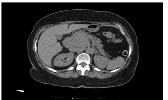
Dynamic pancreatic MR examinations were performed with the diagnosis of abdominal pain and mass on the abdomen, and a lobular contoured mass irregularly limited hypodense lesion (Figure-1) in the CT examination.
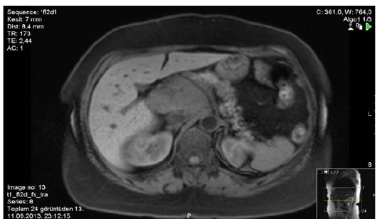
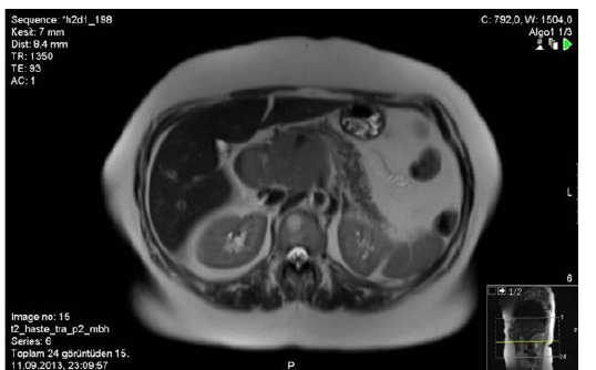
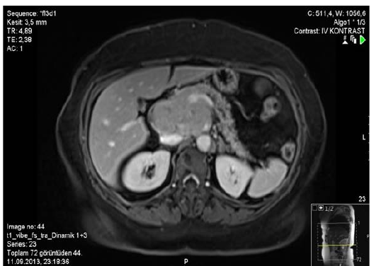
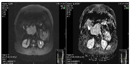
T2x hyper intense (Figure-3), T2A hyper intense (Figure-3), duodenum compression and adjacent vascular structures surrounding the main portal vein and celiac trunks, and heterogeneous contrast at late phase in post contrast study with lobulated contour showing involvement (Figure-4). The described lesion included areas showing diffuse restriction in diffusion imaging (Figure-5).
Discussion
Pancreas-secreting lymphoma is rare, with an extranodal involvement of less than 1% (2). There are various pathologic subtypes such as primary pancreatic lymphoma follicular lymphoma, diffuse large B-cell lymphoma, mucosal-associated lymphoma and T-cell lymphoma. Clinical signs and symptoms are very similar, but it is an important finding that the pancreatic lymphoma is uneventful without jaundice (3). Our case was pathologically diagnosed as Diffuse B-cell lymphoma and there were no jaundice findings. Hypodense is seen as shomogen solid lesions in the noncontrasted sections. MRI is also seen in two types. In T1AGs, the signal intensity is a welldefined homogeneous mass with low signal intensity and heterogeneous signal increases in mild to moderate T2AG. In the second type, low signal intensities are seen on T1W and T1W T2W according to the pancreatic gland and mild moderate contrasting is seen on contrastenhanced examinations (1). The current case was more compatible with the first type and showed moderate contrast enhancement in late venous phase. Adenocarcinoma should be considered primarily in the differential diagnosis of pancreatic lymphoma. In a single primer pancreatic lymphoma the mass is usually larger than a typical adenocarcinoma. Adeno carcinoma rarely causes vessel stenosis or occlusion (1). In our case, obstruction was not present in the stricture other than being pushed into the veins. Diffusion-weighted imaging may vary with the cognitive necrotic areas it contains in pancreatic adenocarcinoma. Low ADC values depend on the fibrotic component and high cellularity it contains and cause restriction in the diffusion. Necrotic areas are a more common finding in angina pancreatic adenocarcinoma (4). In our case, there was diffusion restriction without necrosis with low ADC values parallel to increased cellularity. Apart from these, the table in the distinction between lymphoma and adenocarcinoma is helpful in other findings (3).
Conclusion
Although pancreatic lymphoma is very rare, it should be kept in mind in differential diagnosis in patients with a limited number of welldefined homogeneous internal structures and late phase contrast enhancement, vascular versus ductal obstruction, and clinical absence of jaundice. Biopsy is necessary for definitive diagnosis. It is useful to know that MRI and diffuse MRI in pancreatic lymphoma may provide useful information as well as diffuse restriction in both due to adenocarcinoma and lymphoma elevated cellularity.
Declarations
- Author contributions (CRediT)
- All authors contributed equally to all 14 CRediT roles (Conceptualization, Data curation, Formal analysis, Funding acquisition, Investigation, Methodology, Project administration, Resources, Software, Supervision, Validation, Visualization, Writing – original draft, Writing – review & editing).
- Funding
-
Funding information was not reported in the published version of this article.
- Conflicts of interest
- The authors declare that they have no conflict of interest in the current study.
- Ethics approval
- Ethics approval details were not reported in the published version of this article.
- Data availability
- A data availability statement was not reported in the published version of this article.
References (4)
3 of 4 references are linked to their DOI.
- Haşıloğlu IZ, Havan N, Duymuş R et al. Pankreatik Lenfoma Olgusunda Cok Kesitli Bilgisayarlı Tomografi ve Pozitron Emisyon Tomografi; Bilgisayarlı Tomografi Bulguları, Bakırköy Tıp Dergisi, Cilt 8, Sayı 2, 2012/ Medical Journal of Bakırköy, Volume 8, Number 2, 2012. DOI Crossref PubMed Google Scholar
- Sağlam M, Yılmaz MI, Mas MR et al. Pankreatik Burkitt lenfoma olgusu: radyolojik bulgular. Diagn Interv Radiol 2009; 15:39-42 PubMed Google Scholar
- Fujinaga Y, Lall C, Patel A. et al. MR Features of primary and secondary malignant lymphoma of the pancreas: a pictoral review, Insights Imaging 2013;4:321-329 DOI Crossref PubMed Google Scholar
- Fattahi R, Balci CN, Perman WH et al. Pancreatic Diffusion: Weighted İmaging (DWI): Comparison Between Mass-Forming Focal Pancreatitis, Pancreatic Cancer and Normal Pancreas DOI Crossref PubMed Google Scholar
Respond to this article
Comments on a published article are welcome as a Letter to the Editor.
Get an e-mail when new articles in your fields are published
Questions to the authors
Questions are moderated: an editor approves each question, the authors answer, and an editor approves the answer before both appear here.
No answered questions yet.
Ask the authors a question
Discussion
Comments are moderated: they appear after the writer confirms the e-mail address and an editor approves them. Each comment carries a conflict-of-interest statement.
No comments yet.
Write a comment
Read the full text
Open the PDF here
Loading the PDF…
If the PDF does not show here, use the download button above.
How to cite this article
UNLU S, KAHRAMAN AS, ONER S. Lymphoma Mimicking Pancreatic Adenocarcinoma: A Case Report. Ulutas Med J. 2018;4(2):108-111. doi:10.5455/umj.20171112112954
UNLU, S., KAHRAMAN, A. S., & ONER, S. (2018). Lymphoma Mimicking Pancreatic Adenocarcinoma: A Case Report. The Ulutas Medical Journal, 4(2), 108–111. https://doi.org/10.5455/umj.20171112112954
UNLU, S., KAHRAMAN, A.S. and ONER, S. (2018) 'Lymphoma Mimicking Pancreatic Adenocarcinoma: A Case Report', The Ulutas Medical Journal, 4(2), pp. 108–111. doi: 10.5455/umj.20171112112954.
@article{umj283167,
  title = {Lymphoma Mimicking Pancreatic Adenocarcinoma: A Case Report},
  author = {UNLU, Serkan and KAHRAMAN, Aysegul SAGIR and ONER, Serkan},
  journal = {The Ulutas Medical Journal},
  year = {2018},
  volume = {4},
  number = {2},
  pages = {108--111},
  doi = {10.5455/umj.20171112112954},
  url = {https://www.ulutasmedicaljournal.com/?mno=283167},
  issn = {2149-388X}
}