The Ulutas Medical Journal · ISSN 2149-0430 (print), 2149-388X (online) · https://www.ulutasmedicaljournal.com/?mno=282591
Original Research · Open access
A Blocking Barrier During Reduction of the Elbow Dislocation in Children: Incarcerated Medial Epicondyle Fracture
- Orthopaedics and Traumatology, Ankara Numune Training and Research Hospital, Ankara, Turkey ROR
Abstract
- Background
- In children, an incarcerated medial epicondyle fracture of the distal humerus associated with elbow dislocation, may be missed on conventional radiographs. Other reliable radiological criteria are imperative for the diagnosis of these fractures. The purpose of the study was to investigate the clinical results of the operative treatment of incarcerated medial epicondyle fractures after elbow dislocation in children and define new radiological criteria in the diagnosis.
- Materials and Methods
- A sea wave-like shaped parallel double line was seen on anterior-posterior elbow radiographs of non-fractured pediatric patients. The disruption of the sea wave-like sign was defined as a new diagnostic radiographic criterion in incarcerated medial epicondyle fractures after elbow dislocation in children. Seven children with an incarcerated medial epicondyle fracture.
- Results
- The mean age was 12.14±2.1 years (range: 9-15 years) and the mean follow-up period was 29.1±13.6 months (range: 12-48 months). The mean Mayo elbow score was 92.8±4.8 (range: 85-100) at the final follow-up. The new diagnostic radiological criterion was identified in all incarcerated medial epicondyle fractured patients.
- Conclusions
- The results of this study suggest that open reduction internal fixation of incarcerated medial epicondyle fractures after elbow dislocation resulted in satisfactory motion and function. The new diagnostic radiological criterion defined in this study is useful, does not incur any additional cost and may solve the diagnostic problems of incarcerated medial epicondyle fractures of the distal humerus after elbow dislocation in the pediatric population.
Keywords
Full text
Introduction
Medial epicondyle fractures cover up to 20% of pediatric elbow fractures (1). Management of isolated, displaced fractures of the medial humeral epicondyle has been still controversial. Both plaster cast immobilization and surgical approaches have been advocated. Good outcomes have been reported with nonoperative treatment, despite a high rate of nonunion (2–5). In patients with displaced fractures, surgical approaches provide stability and functional range of motion.
Occasionally, the fractured fragment may occupy the retro-epitrochlear groove. The presence of dysesthesias in the territory of the ulnar nerve requires urgent open reduction of the incarcerated fragment. In children, an incarcerated medial epicondyle fracture of the distal humerus associated with elbow dislocation may be missed on diagnosed on conventional radiographs. Other reliable radiological criteria are imperative for the diagnosis of these fractures.
The purpose of the study was to investigate the clinical results of the operative treatment of incarcerated medial epicondyle fractures after elbow dislocation in children and to define a new radiological criterion in the diagnosis.
Materials and Methods
A total of 30 patients with displaced medial epicondyle fracture managed with the open reduction and fixation at our institution between 2007-2015. A retrospective case review was conducted of 7 consecutive children with an incarcerated medial epicondyle fracture after elbow dislocation. The study was approved by the Institutional Review Board. The inclusion criteria of this study were that patients were younger than 16 years, and had an incarcerated medial epicondyle fracture with at least a 12-month follow-up period. Patients with any bony deformity of the ipsilateral upper extremity or previous surgery of the elbow were excluded from the study.
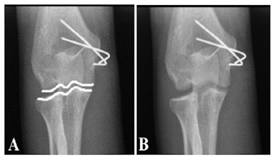
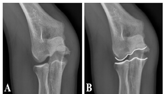
All patients were evaluated with conventional anterior-posterior and lateral radiographs. To facilitate the diagnosis of incarcerated medial epicondyle fractures, we defined new diagnostic radiological criteria. A parallel double the line was seen as a ‘sea wave-like sign on the anterior-posterior elbow radiographs of non-fractured or reduced pediatric patients (Figure- 1). This parallelism was seen to be disrupted in incarcerated medial epicondyle fractures. The incarcerated fragment distracted in the joint space in the medial compartment of the elbow between the trochlea of the humerus and coracoid of the ulna leads to disruption of this parallel double line. We have defined this disruption of the sea wave-like sign as a new diagnostic radiographic criterion for incarcerated medial epicondyle fractures after elbow dislocation in children (Figure-2).
All patients underwent surgery using a similar technique. The patients were positioned supine. A pneumatic tourniquet was used. The patients were prepared and draped in a standard fashion. The affected arm was externally rotated with the elbow in a 90° - 100° flexed position. A skin incision of approximately 5 cm was made over the medial epicondyle. Deep dissection was applied and the ulnar nerve was located. The medial epicondyle was also found. The fragment was reduced with the arm in flexion and fixed with two Kirschner wires. Radiographs were used to confirm reduction and Kirschner wire positions. Elbow stability was assessed. Standard skin closure was applied.
Post-operatively, a long-arm plaster cast was used for 3 weeks. The range of motion of the the elbow was started after cast removal. The Kirschner wires were removed after 4-6 weeks. Physiotherapy was applied only if there was a marked reduction in elbow mobility 6 weeks after cast removal. All patients were in regular follow-up at 3, 6, 12, 24 and 36 months post-operatively. Standard anterior-posterior and lateral radiographs of the elbow were taken at follow-up examination and complications, such as wound problems, were evaluated.
Results
The patients were 6 males and 1 female with a mean age of 12.14±2.193 years (range: 9-15 years) and the mean follow-up period was 29.14±13.61 months (range: 12-48 months). The mean elbow range of motion was 130˚±10˚ (range: 120˚-150˚). All patients had forearm rotation from 90° supination to 90° pronation. The mean Mayo elbow score was 92.86±4.88 (range: 85-100).
All patients had clinical and radiographic signs of healing at the final follow-up. There was no radiographic evidence of loss of reduction at intervals or at the final follow-up. There were no cases of residual deformity or instability. None of the patients had ulnar nerve symptoms post-operatively. The new diagnostic radiological criterion was identified in all the incarcerated medial epicondyle fracture patients.
Discussion
The mechanism of injury for medial epicondyle fractures of the distal humerus has been suggested to be a fall on an outstretched arm with the elbow in extension and the wrist in supination and extension, which forces the elbow into valgus stress. The resultant force leads to avulsion of the medial epicondyle (6, 7). Approximately 60% of cases of medial epicondyle fractures have been reported to be associated with dislocation of the elbow (8). The medial epicondyle is incarcerated in the joint in 15% to 25% of cases (5, 9). Sometimes the elbow may spontaneously relocate and valgus stress radiograph may be helpful in diagnosing concomitant injuries (8, 9).
The treatment of medial epicondyle fractures in the distal humerus is controversial. However, the surgical approach is clearly indicated in cases of intra-articular entrapment of the fragment, on suspicion of entrapment of the ulnar nerve, or in cases of the marked instability of the elbow (10). It has been reported in the literature that union rates are higher in surgically fixated medial epicondyle fractures compared to conservatively treated fractures. However, in a meta-analysis by Kamath et al, no significant difference was determined in the functional outcome between surgically or conservatively treated patients. Interestingly, there was less pain in nonsurgically treated patients despite lower union rates. Therefore, it was accepted that the union of the medial epicondyle did not correlate with the functional outcome (11). Although healing was observed radiographically in all the patients in the current study at the final follow-up, this was not the goal of the treatment. In this study, good outcomes were achieved with operative treatment for incarcerated medial epicondyle fractures of the distal humerus. The findings of the study were comparable with current literature (9, 12, 13).
Multiple surgical interventions have been recorded in the literature such as suture repair, Kirschner wire fixation, screw fixation and excision of the fragment and suturing of the soft tissues of the medial elbow (3–5, 14). In the current study, Kirschner wires were used for fixation and this was found to be an effective fixation method. Authors who advocate screw fixation as the fixation method has stated that with screw fixation early mobilization can be applied postoperatively. However, they also used a splint or cast for two weeks after the operation to protect the fracture displacement (1). Kirschner wire is easy and relatively less complicated fixation material and no complications related to the fixation method developed in the current study.
Surgically treated medial epicondyle fractures have been reported to be hypertrophic, with a deformity of the medial epicondyle and with an “ulnar sulcus,” in many cases (15). None of the patients in the current study had ulnar sulcus deformity at the end of the follow-up period. Although the diagnosis of medial epicondyle fractures is difficult when the diagnosis is missed, delayed ulnar neuropathies have been reported in the literature (10, 16, 17). The missed diagnosis of the intra-articular entrapment of the medial epicondyle can result in a significant restriction of mobility and increased risk of ulnar neuropathy. Early intervention is necessary as prognosis worsens with the passing of time, due to the poorer recovery of the ulnar nerve restriction.
Medial epicondyle fracture associated with elbow dislocation in children may be missed on conventional radiographs because of the small size of the fragment, it can be mistaken for ossification center and the position may be concealed behind the distal humerus (10). If the distal humerus appears symmetrical on an anterior-posterior radiograph, or if the epicondyle is visible on the lateral radiograph, then the epicondyle must be displaced (18, 19). Incarcerated medial epicondyle fractures after elbow dislocation in children is a rare entity, but because of the associated risk of injury to the ulnar nerve and serious restriction of elbow motion, prompt diagnosis and treatment of this fracture is very important. Due to the difficulties experienced during the diagnosis of these fractures, we defined a new radiographic sign, which can be easily noticed by orthopedic surgeons after elbow dislocation in children. The weak point of the study was the small number of cases and that the study was retrospective.
The results of this study suggest that open reduction internal fixation of the incarcerated medial epicondyle fractures after elbow dislocation resulted in a satisfactory motion and function. In the pediatric population, fracture may not be easily diagnosed on conventional radiographs. This new radiological, diagnostic criterion defined in this study is useful, does not lead to any additional cost and may solve the diagnostic problems of the incarcerated medial epicondyle fractures of the distal humerus after elbow dislocation in the pediatric population.
Declarations
- Author contributions (CRediT)
- All authors contributed equally to all 14 CRediT roles (Conceptualization, Data curation, Formal analysis, Funding acquisition, Investigation, Methodology, Project administration, Resources, Software, Supervision, Validation, Visualization, Writing – original draft, Writing – review & editing).
- Funding
-
Funding information was not reported in the published version of this article.
- Conflicts of interest
- The authors declare that they have no conflict of interest in the current study.
- Ethics approval
- The study was approved by the Institutional Review Board.
- Data availability
- A data availability statement was not reported in the published version of this article.
References (19)
13 of 19 references are linked to their DOI.
- Gottschalk HP, Eisner E, Hosalkar HS. Medial epicondyle fractures in the pediatric population. J Am Acad Orthop Surg 2012;20:223– 32. DOI Crossref PubMed Google Scholar
- Kobayashi Y, Oka Y, Ikeda M, Munesada S. Avulsion fracture of the medial and lateral epicondyles of the humerus. J Shoulder Elbow Surg 2000;9(1):59-64. DOI Crossref PubMed Google Scholar
- Fowles JV, Slimane N, Kassab MT. Elbow dislocation with avulsion of the medial hu meral epicondyle. J Bone Joint Surg Br 1990;72(1):102-4. DOI Crossref PubMed Google Scholar
- Farsetti P, Potenza V, Caterini R, Ippolito E. Long-term results of treatment of frac tures of the medial humeral epicondyle in children. J Bone Joint Surg Am 2001;83(9):1299-305. DOI Crossref PubMed Google Scholar
- Wilson NI, Ingram R, Rymaszewski L, Miller JH. Treatment of fractures of the medial epicondyle of the humerus. Injury 1988;19(5):342-4. DOI Crossref PubMed Google Scholar
- Beaty JH, Kasser JR (2010) The elbow: physeal fractures, apophyseal injuries of the distal humerus, osteonecrosis of the trochlea, and T-condylar fractures. In: Beaty JH, Kasser JR (eds) Rockwood and Wilkins’ fractures in children, 7t h ed. Lippincott Williams & Wilkins, Philadelphia, pp 566–77. PubMed Google Scholar
- Smith BG, Pierz KA (2011) Open reduction and internal fixation of fractures of the me dial epicondyle. In: Flynn JM, Wiesel SW (eds) Operative techniques in orthop edic surgery, vol:2. Lippincott Williams&Wilkins, Philadelphia, pp 1042–5. PubMed Google Scholar
- Lindham S, Hugosson C. The significance of associated lesions including dislocation in fractures of the neck of the radius in children. Acta Orthop Scand 1979;50(1):79-83. DOI Crossref PubMed Google Scholar
- Lee HH, Shen HC, Chang JH, Lee CH, Wu SS. Operative treatment of displaced medial epicondyle fractures in children and adolescents. J Shoulder Elbow Surg 2005;14 (2):178-85. DOI Crossref PubMed Google Scholar
- Lima S, Correia JF, Ribeiro RP, Martins RM, Alegrete N, Coutinho J, Costa GM. A rare case of elbow dislocation associated with unrecognized fracture of medial epicondyle and delayed ulnar neuropathy in pediatric age. J Shoulder Elbow Surg 2013;22(3):e9–e11. DOI Crossref PubMed Google Scholar
- Kamath AF, Baldwin K, Horneff J, Hosalkar HS. Operative versus nonoperative management of pediatric medial epicondyle fractures: A systematic review. J Child Orthop 2009;3(5):345-57. DOI Crossref PubMed Google Scholar
- Ip D, Tsang WL. Medial humeral epicondylar fracture in children and adolescents. J Orthop Surg (Hong Kong) 2007;15(2):170-3. DOI Crossref PubMed Google Scholar
- Hines RF, Herndon WA, Evans JP. Operative treatment of medial epicondyle fractures in children. Clin Orthop Relat Res 1987;223:170-4. DOI Crossref PubMed Google Scholar
- Gilchrist AD, McKee MD. Valgus instability of the elbow due to medial epicondyle nonunion: Treatment by fragment excision and ligament repair. A report of 5 cases. J Shoulder Elbow Surg 2002;11(5):493-7. DOI Crossref PubMed Google Scholar
- Skak SV, Grossmann E, Wagn P. Deformity after internal fixation of fracture separation of the medial epicondyle of the humerus. J Bone Joint Surg Br 1994;76(2):297-302. DOI Crossref PubMed Google Scholar
- Haflah NHM, Ibrahim S, Sapuan J, Abdullah S. An elbow dislocationin a child with missed medial epicondyle fracture and late ulnar nerve palsy. J Pediatr Orthop B 2010;19:459-61. PubMed Google Scholar
- Rasool MN. Dislocation of the elbow in children. J Bone Joint Surg Br 2004;86- B:1050-8. PubMed Google Scholar
- Patrick J. Fracture of the medial epicondyle with displacement into the elbow joint. J Bone Joint Surg 1946;28:143-7. PubMed Google Scholar
- Martini M, Hallaj N, Daoud A, Descamps L. Les luxations traumatiques recentes du coude: a propos de 94 observations. Acta Orthop Belg 1978;44:542-54. PubMed Google Scholar
Read the full text
Open the PDF here
Loading the PDF…
If the PDF does not show here, use the download button above.
How to cite this article
Ozdemir G, Deveci A, Bingol O, Kilic E, Ceyhan E, Turan S. A Blocking Barrier During Reduction of the Elbow Dislocation in Children: Incarcerated Medial Epicondyle Fracture. Ulutas Med J. 2018;4(2):103-107. doi:10.5455/umj.20171107113200
Ozdemir, G., Deveci, A., Bingol, O., Kilic, E., Ceyhan, E., & Turan, S. (2018). A Blocking Barrier During Reduction of the Elbow Dislocation in Children: Incarcerated Medial Epicondyle Fracture. The Ulutas Medical Journal, 4(2), 103–107. https://doi.org/10.5455/umj.20171107113200
Ozdemir, G. et al. (2018) 'A Blocking Barrier During Reduction of the Elbow Dislocation in Children: Incarcerated Medial Epicondyle Fracture', The Ulutas Medical Journal, 4(2), pp. 103–107. doi: 10.5455/umj.20171107113200.