The Ulutas Medical Journal · ISSN 2149-0430 (print), 2149-388X (online) · https://www.ulutasmedicaljournal.com/?mno=283352
Case Report · Open access
Tumoral Calcinosis Around the Elbow: Case Report
- Department of Radiology, Malatya State Hospital, Malatya, Turkey ROR
- Department of Radiology, Inonu University School of Medicine, Malatya, Turkey ROR
Abstract
Tumoral calcinosis is a rare ectopic calcification seen around large joints. Large joints are seen in the hip, shoulder and elbow, and less frequently in the foot and ankle. It is unclear why the first two decades of life are caused by familial predisposing aphakia, as well as being seen more frequently. We presented Magnetic Resonance Imaging findings of the patient who presented with tumoral calcinosis elbow pain clinic. Tumoral calcinosis should be kept in mind in differential diagnosis when a mass with soft tissue periarticular calcification is seen in the vicinity of large joints.
Keywords
Full text
Introduction
Tumoral calcinosis (TC) is a rare ectopic calcification seen around large joints (1). Large joints are seen in the hip, shoulder and elbow, and less frequently in the foot and ankle (2). It is unclear why the first two decades of life are caused by familial predisposing aphakia, as well as being seen more frequently (3). We presented Magnetic Resonance Imaging (MRI) findings of the patient who presented with TC elbow pain clinic.
Case Presentation
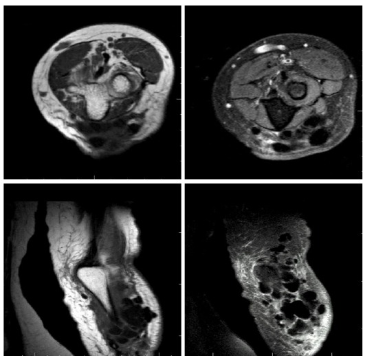
T1AG Sagittal T2AG Sagittal, PDW Coronal, and PDW SPIR Axial sequences were obtained in the patient who complained of elbow pain and who applied to the radiology department for MR extraction.
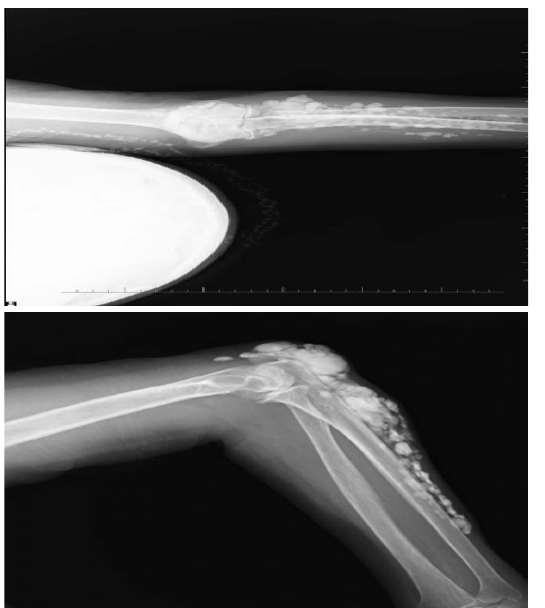
Calcification foci (Figs. 1, 2, 3 and 4) were observed in a number of different sizes, with T1AG axial and coronal, PDW coronal and PDW SPIR coronal sequences clustered together in a multitude of hypo intense periarticular assemblies. On direct radiographs it was confir- med that these lesions were the calcification center (Figs. 5 and 6).
Discussion
TC shows periarticular localization, especially in regions under compression, in the form of masses that are not directly related to the neighboring joint space. There are white sand, bruise color and texture or calcification in the tissues (4). This calcification pathogenesis in the TC is due to trauma-dystrophy, or traumatism (5, 6). Clinical symptoms such as pain and skin erosion can be seen in the TC (7).
The idiopathic form of TC is the family. Another form of TC is due to the high calciumphosphorus ratio seen in chronic renal failure (8). Diagnosis on CT can be made with radiographic findings and the medical story of the patient. Radio graphically; both TC and idiopathic renal insufficiency are secondary to calcification sites that cause multiple well-defined pavement stones. MRI is seen as hypo intense lesion in all sequences. Additionally, edema due to inflammation around pathological calcification is seen as a hyper intense signal in T2AG and Inversion Recovery (IR) sequences (8, 9). Heterotopic ossification, calcific hemangiomas or lymphangiomas, calcified soft tissue calcification, paraosteal osteosarcoma and synovial osteochondromatosis. In synovial osteo chondromatosis loose body bodies may be calcified, but they are intraarticularly located. Clinical history of the disease and location of lesion are of great importance in differential diagnosis (7, 10). TC should be kept in mind in differential diagnosis when a mass with soft tissue periarticular calcification is seen in the vicinity of large joints.
Declarations
- Author contributions (CRediT)
- All authors contributed equally to all 14 CRediT roles (Conceptualization, Data curation, Formal analysis, Funding acquisition, Investigation, Methodology, Project administration, Resources, Software, Supervision, Validation, Visualization, Writing – original draft, Writing – review & editing).
- Funding
-
Funding information was not reported in the published version of this article.
- Conflicts of interest
- A conflict of interest statement was not reported in the published version of this article.
- Ethics approval
- Ethics approval details were not reported in the published version of this article.
- Data availability
- A data availability statement was not reported in the published version of this article.
References (10)
4 of 10 references are linked to their DOI.
- Oeggerli P, Maurer R, Kistler H. Tumoral calcinosis with superinfection and sepsis Schweiz Med Wochenschr 1991; 121 (38): 1387-139 PubMed Google Scholar
- Prahinski JR, Schaefer RA. Tumoral calcinosis of the foot. Foot Ankle Int 2001; 22: 911-913 DOI Crossref PubMed Google Scholar
- Sledz K., et al., Tumoral Calcinosis of the Temporomandibular Joint: CT and MR Findings, AJNR 16: 782-785, Apr 1995 0195-6108 / 95 / 1604-0782 PubMed Google Scholar
- Yurdoğlu C., Özbaydar MU., Adaş M. et al. Familial tumoral calcinosis in three patients in the same family, Acta Orthop Traumatol Turc 2007; 41 (3): 244-248. PubMed Google Scholar
- Slavin RE, Wen J, Kumar D, Evans EB. Familial tumoral calcinosis. A clinical, histopathologic, and ultrastructural study with an analysis of its calcifying process and pathogenesis. Am J Surg Pathol 1993; 17: 788-802 DOI Crossref PubMed Google Scholar
- Boskey AL, Vigorita VJ, Sencer O, Stuchin SA, Lane JM Chemical, Microscopic, and ultrastructural characterization of the mineral deposits in tumoral calcinosis. Clin Orthop Relat Res 1983; (178): 258-69. DOI Crossref PubMed Google Scholar
- Kumara, Singh P, Dobhal H. Case of Recurrent Tumoral Calsinosis in a Young Female, World Journal of Endocrine Surgery, January-April 2013; 5 (1): 18-20 PubMed Google Scholar
- Steinbach LS et al., Tumoral calcinosis: radiologic-pathologic correlation, Skeletal Radiol (1995) 24: 573-578 DOI Crossref PubMed Google Scholar
- Farzan M, Farhood AR., Tumoral Calcinosis: What is the treathment? Report of two cases of different types and review of the literature, Am J Orthop. 2011; 40 (9): E170-E176. PubMed Google Scholar
- Sledz K, Ortiz O, Wax M, Bouguot J, Tumoral Calcinosis of the Temporomandibular Joint: CT and MR Findings, AJNR: 16, April 1995 PubMed Google Scholar
Respond to this article
Comments on a published article are welcome as a Letter to the Editor.
Get an e-mail when new articles in your fields are published
Questions to the authors
Questions are moderated: an editor approves each question, the authors answer, and an editor approves the answer before both appear here.
No answered questions yet.
Ask the authors a question
Discussion
Comments are moderated: they appear after the writer confirms the e-mail address and an editor approves them. Each comment carries a conflict-of-interest statement.
No comments yet.
Write a comment
Read the full text
Open the PDF here
Loading the PDF…
If the PDF does not show here, use the download button above.
How to cite this article
Unlu S, Sanli B, Yildirim IO, Oner S, Dogan M. Tumoral Calcinosis Around the Elbow: Case Report. Ulutas Med J. 2017;3(4):85-87. doi:10.5455/umj.20171114024323
Unlu, S., Sanli, B., Yildirim, I. O., Oner, S., & Dogan, M. (2017). Tumoral Calcinosis Around the Elbow: Case Report. The Ulutas Medical Journal, 3(4), 85–87. https://doi.org/10.5455/umj.20171114024323
Unlu, S. et al. (2017) 'Tumoral Calcinosis Around the Elbow: Case Report', The Ulutas Medical Journal, 3(4), pp. 85–87. doi: 10.5455/umj.20171114024323.