The Ulutas Medical Journal · ISSN 2149-0430 (print), 2149-388X (online) · https://www.ulutasmedicaljournal.com/?mno=225554
Case Report · Open access
The Role of Radiologic and Scintigraphic Imaging in Differentiating Thoracic Pyogenic Spondylodiscitis from Metastasis
Also available in: العربية · Türkçe · 中文
- Department of Nuclear Medicine, Mersin State Hospital, Mersin, Turkey ROR
- Department of Nuclear Medicine, Hacettepe University, Kastamonu School of Medicine and Dışkapı Yıldırım Beyazıt Training and Research Hospital, Ankara, Turkey ROR
- Department of Radiology, Dışkapı Yıldırım Beyazıt Training and Research Hospital, Ankara, Turkey ROR
- Department of Radiology, Turgut Özal University Faculty of Medicine, Ankara, Turkey ROR
Abstract
- Background
- Physical examination, basic laboratory tests and plain radiographs can give important data in diagnosis of the patients presenting with complaints of back pain. However, advanced imaging tools may be necessary for further evaluation of complicated processes in geriatric patients.
- Case presentation
- We presented plain radiography, magnetic resonance imaging (MRI) and bone scintigraphy (BS) findings of thoracic pyogenic spondylodiscitis in a 72-year-old male patient which resembles metastatic disease.
- Conclusion
- Multimodal imaging, particularly MRI and BS can provide useful data in the initial diagnosis and follow-up of geriatric patients with a complicated vertebral pathology. Spondylodiscitis can mimic metastatic disease.
Keywords
Full text
Introduction
Secondary vertebral malignancies such as metastasis from endometrial cancer can resemble spondylodiscitis in some cases (1). However, spondylodiscitis can also mimic metastastatic disease such as metastases from prostate cancer (2) or urothelial carcinoma (3). Malignant neoplastic disease of the spine in geriatric patients is common, and its most frequently seen form is secondary metastatic disease (4). An increase in incidence of spondylodiscitis has been reported in recent years (5).Though routine physical examination, laboratory tests and plain radiographs provide significant information, it may be difficult to differentiate these two entities and advanced imaging tools may be required especially in complicated geriatric cases presenting with severe back pain. The role of magnetic resonance imaging (MRI) and bone scintigraphy (BS) in bone metastases and bone infections is well defined (6). Here we present the contribution of plain radiography, MRI and BS in the management of thoracic pyogenic spondylodiscitis which resembles metastatic disease in a geriatric case.
Case Presentation
A 72-year-old male was referred with complaints of severe pain in his upper back which has been lasting for two months. All the procedures were performed according to the World Medical Association Declaration of Helsinki (revised in 2000, Edinburgh).
The patient was informed about procedures and consent was obtained from him. Patient’s history revealed that he had experienced several interventions for dilatation of urethral stricture, a transurethral resection of a bladder mass and frequent urinary tract infections. Physical examination findings were limitation in body movements, pain with lumbar extension, a relatively hard prostate gland and subfebrile fever (37.7 oC). Patient’s reflexes, muscle strength and sensory examinations were normal. In laboratory tests, C-reactive protein (CRP) was 26.5 mg/L and erythrocyte sedimentation rate (ESR) was 51 mm/h.
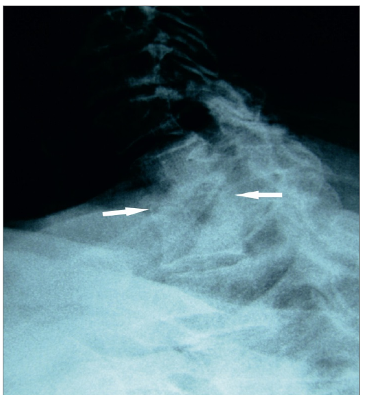
His urine appeared blurred, in which abundant leukocyturia (82 leukocytes in each field) and (++) gram negative bacilli were detected. His serum prostate-specific antigen level was in normal range. Cystoscopy was unsuccessful because of urethral stricture. On his lateral upper thoracic plain radiograph, narrowing in T1–2 intervertebral disc space and sclerotic appearance in the end-plates surrounding the disc space were detected (Figure-1).
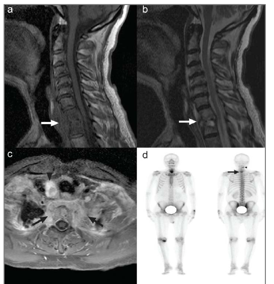
On thoracic MRI, T1 and T2 vertebra corpuses and adjacent paravertebral soft tissue mass showed hypointensity on T1- weighted images (Figure-2a) and slight, heterogenous hyper-intensity on T2-weighted images (Figure-2b). Signal intensity of narrowed, irregular inter-vertebral disc space was similar to that of neighbouring vertebral end-plates. Contrast-enhanced, fat-saturated T1-weighted axial images revealed pathologic enhancement in the vertebral corpuses, pedicles and para-vertebral soft tissues, besides a hyperintense nodular lesion in the right lobe of thyroid gland (Figure-2c). In whole-body BS, increased focal uptake in soft tissue of middle cervical region on the right side and a diffuse, patho-logical, increased uptake in T1–2 vertebrae were observed (Figure-2d). On ultrasono-graphy (US) of the neck, a thyroid nodule having calcifications in the right lobe of thyroid gland was detected and US-guided fine needle aspiration biopsy of the nodule revealed benign cytology.
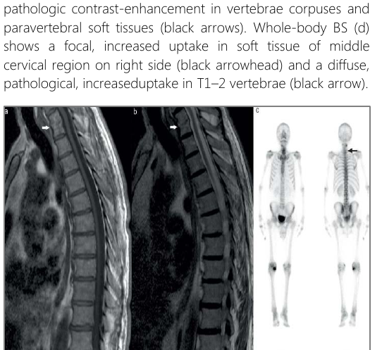
In differential diagnosis, malignant metastatic disease of thoracic vertebrae was considered, but the diagnosis of pyogenic spondylo discitis was eventually made following the evaluation of clinical, laboratory and imaging findings. After application of medical therapy with antibiotics and anti-inflammatory drugs besides immobilisation, patient was revaluated six months later. A significant decrease in pain level and in limitation of movements was observed. Serum CRP and serum ESR values were lower than the initial ones, which were recorded as 11.6 mg/L and 34 mm/h, respectively. Urine tests were normal. In control thoracic MRI and BS (Figure 3a–c), significant regression in vertebral lesions was detected.
Discussion
Common reasons for back pain in geriatric age group are primary or metastatic tumours, fractures, degenerative or infectious disc diseases. Spine is a major site for bone metastases (6) particularly in geriatric cancer patients (4). In the presence of discogenic sclerosis, great care should be taken to exclude neoplastic disease.
Recurrent urinary system infection or surgery can pave the way for an infection to reach the vertebral column through the venous system (5, 7). The most common predisposing factors are genitourinary, soft tissue and gastrointestinal infections, where pyogenic bacteria (staphylococcus aureus, streptococcus species and gram negative bacilli) are the most commonly determined pathogens (6, 8, 9). Microorganisms can spread into the vertebral end-plate or the intervertebral disc region (6, 10, 11).
Plain radiography has a role in diagnosis of vertebral osteomyelitis and metastases, in which narrowing of disc space can be the earliest radiographic sign in osteomyelitis and ostelytic/osteoblastic lesions can represent metastases (6). However, MRI can be preferred to evaluate both infectious and malignant processes (4, 5, 12). Non-specific Modic type- 1 alteration in the early stage of vertebral osteomyelitis is quite common (7). In most of the cases with spondylodiscitis, hypointensity on T1-weighted images, hyperintensity on T2- weighted images in vertebral corpuses and intervertebral discs are demonstrated.
Infection typically surrounds disc space (6) and intervertebral disc involvement is more frequent finding for infectious spondylodiscitis, but these are very rare in malignant disease of spine (4, 13). In the present case, besides vertebral corpuses, T1–2 intervertebral disc involvement was prominent, which helped us in making the diagnosis of spondylodiscitis rather than malignant disease of spine, either primary or metastatic. In evaluating the spinal infections, obtaining contrast-enhanced, fat-saturated images besides routine sequences is recommended. In differentiation of pyogenic and tuberculousspondylitis, contrast-enhanced MRI images can be useful.
A thin, smooth and enhancing abscess wall, a well-defined paraspinal abnormal signal, subligamentous spread to three or more vertebral levels and multiple vertebral or entire body involvement were more suggestive of tuberculous than pyogenic spondylitis (10). In the present case, clinical and laboratory findings besides treatment results after six months were in favour of pyogenic etiology. In suspicious cases without a known primary tumour, a whole-body BS with 99mTc-labeled biphos-phonates is often preferred for the initial evaluation of skeletal system, as we did in the present case. In our case, initial and control BS revealed that the pathology was rather localized unlike most bone metastases and therefore helped us in making the diagnosis.
Though spondylodiscitis can be differentiated from vertebral metastases even in complicated cases including the patients who underwent previous spinal irradiation (12), it may be challenging to differentiate these two entities in patients presenting with the involvement of several contiguous vertebrae on MRI (14). In such cases, short-term MRI follow-up of the patients who are on antibiotic treatment, and computed tomographyguided biopsy will help in differentiation of them (14, 15).
In the present case, we did not need to perform biopsy because by using MRI and BS images we could be able to demonstrate rather active and progressive phase of pyogenic spondylodiscitis at the time of initial diagnosis and also regression of the pathology after the treatment. It was stated that in vertebral lesions of unknown origin, spondylodiscitis or new primary lesion should be taken into consideration rather than metastatic disease in differential diagnosis, even though the patient gives a history of a former malignancy (15). We consider that it will be appropriate to decide about the best time for control imaging after evaluating the initial clinical, laboratory and imaging findings.
Conclusion
Multimodal imaging, particularly MRI and BS can provide useful data in the initial diagnosis and follow-up of a geriatric patient with a complicated vertebral pathology, as it was in our case.
Declarations
- Author contributions (CRediT)
- All authors contributed equally to all 14 CRediT roles (Conceptualization, Data curation, Formal analysis, Funding acquisition, Investigation, Methodology, Project administration, Resources, Software, Supervision, Validation, Visualization, Writing – original draft, Writing – review & editing).
- Funding
-
Funding information was not reported in the published version of this article.
- Conflicts of interest
- The authors declared no conflicts of interest.
- Ethics approval
- Ethics approval details were not reported in the published version of this article.
- Data availability
- A data availability statement was not reported in the published version of this article.
References (15)
10 of 15 references are linked to their DOI.
- Bayraktutan U, Kantarci M, Yuce I, Sade R, Ogul H, Karaca L. Endometrial cancer metastasis mimicking spondylodiscitis and psoas abscess. Spine J 2016;16(1):e9–e10. xx Multimodal Imaging in Pyogenic Spondylodiscitis Sevin AYAZ, et al. DOI Crossref PubMed Google Scholar
- Alexiou E, Georgoulias P, Valotassiou V, Georgiou E, Fezoulidis I, Vlychou M. Multifocal septic osteomyelitis mimicking skeletal metastatic disease in a patient with prostate cancer. Hell J Nucl Med 2015;18(1):77–78. PubMed Google Scholar
- Kawamura N, Okumi M, Hirai T, et al. A case report of pyogenic spondylodiscitis mimicking spinal metastasis of urothelial carcinoma. Hinyokika Kiyo 2010;56(11):635–7. PubMed Google Scholar
- Guillevin R, Vallee JN, Lafitte F, Menuel C, Duverneuil NM, Chiras J. Spine metastasis imaging: review of the literature. J Neuroradiol 2007;34:311– 21. DOI Crossref PubMed Google Scholar
- Bruyn GA, Tondu PR. Back pain and condensation of the eighth thoracic vertebra: is it always a metastatic disease? Clin Exp Rheumatol 2006;24:89– 92. PubMed Google Scholar
- Dahnert W. Radiology Review Manual. 6th ed. Philadelphia, Lippincott Williams and Wilkins 2007:120, 217, 219, 1084, 1087– 9. PubMed Google Scholar
- Ritchie DA. Commentary on the MRI appearances of early osteomyelitis and discitis. Clin Radiol 2010;65:982–3. DOI Crossref PubMed Google Scholar
- Hadjipavlou AG, Mader JT, Necessary JT, Muffoletto AJ. Hematogenous pyogenic spinal infections and their surgical management. Spine 2000;25:1668–79. DOI Crossref PubMed Google Scholar
- Ozuna RM, Delamarter RB. Pyogenic vertebral osteomyelitis and postsurgical disc space infections. Orthop Clin North 1996;27:87 -94 DOI Crossref PubMed Google Scholar
- Jung NY, Jee WH, Ha KY, Park CK, Byun JY. Discrimination of tuberculous spondylitis from pyogenic spondylitis on MRI. AJR Am J Roentgenol 2004;182:1405–10. DOI Crossref PubMed Google Scholar
- Turpin S, Lambert R. Role of scintigraphy in musculoskeletal and spinal infections. Radiol Clin North Am 2001;39:169–89. DOI Crossref PubMed Google Scholar
- Rades D, Bremer M, Goehde S, Joergensen M, Karstens JH. Spondylodiscitis in patients with spinal cord compression: a possible pitfall in radiation oncology. Radiother Oncol 2001;59(3):307–9. DOI Crossref PubMed Google Scholar
- Kakitsubata Y, Theodorou DJ, Theodorou SJ,Nabeshima K, Tamura S. Metastatic disease involving the discovertebral junction of the spine. Joint Bone Spine 2009;76:50–56. DOI Crossref PubMed Google Scholar
- Lee CM, Lee S, Bae J. Contiguous spinal metastasis mimicking infectious spondylodiscitis. J Korean Soc Radiol 2015;73(6):408–12. DOI Crossref PubMed Google Scholar
- Gasbarrini A, Boriani L, Salvadori C, et al. Biopsy for suspected spondylodiscitis. Eur Rev Med Pharmacol Sci 2012;16 Suppl 2:26–34. To submit your manuscript, please click on http://ulutasmedicaljournal.com PubMed Google Scholar
Respond to this article
Comments on a published article are welcome as a Letter to the Editor.
Get an e-mail when new articles in your fields are published
Questions to the authors
Questions are moderated: an editor approves each question, the authors answer, and an editor approves the answer before both appear here.
No answered questions yet.
Ask the authors a question
Discussion
Comments are moderated: they appear after the writer confirms the e-mail address and an editor approves them. Each comment carries a conflict-of-interest statement.
No comments yet.
Write a comment
Tell me when this article is cited
Leave your e-mail address and confirm it. These notices are not being sent yet; your confirmed address is kept for them and used for nothing else.
Read the full text
Open the PDF here
Loading the PDF…
If the PDF does not show here, use the download button above.
How to cite this article
Ayaz S, GUltekin SS, Dilli A, Teber MA. The Role of Radiologic and Scintigraphic Imaging in Differentiating Thoracic Pyogenic Spondylodiscitis from Metastasis. Ulutas Med J. 2016;2(2):117-121. doi:10.5455/umj.20160421120053
Ayaz, S., GUltekin, S. S., Dilli, A., & Teber, M. A. (2016). The Role of Radiologic and Scintigraphic Imaging in Differentiating Thoracic Pyogenic Spondylodiscitis from Metastasis. The Ulutas Medical Journal, 2(2), 117–121. https://doi.org/10.5455/umj.20160421120053
Ayaz, S. et al. (2016) 'The Role of Radiologic and Scintigraphic Imaging in Differentiating Thoracic Pyogenic Spondylodiscitis from Metastasis', The Ulutas Medical Journal, 2(2), pp. 117–121. doi: 10.5455/umj.20160421120053.
@article{umj225554,
  title = {The Role of Radiologic and Scintigraphic Imaging in Differentiating Thoracic Pyogenic Spondylodiscitis from Metastasis},
  author = {Ayaz, Sevin and GUltekin, Salih Sinan and Dilli, Alper and Teber, Mehmet Akif},
  journal = {The Ulutas Medical Journal},
  year = {2016},
  volume = {2},
  number = {2},
  pages = {117--121},
  doi = {10.5455/umj.20160421120053},
  url = {https://www.ulutasmedicaljournal.com/?mno=225554},
  issn = {2149-388X}
}