The Ulutas Medical Journal · ISSN 2149-0430 (print), 2149-388X (online) · https://www.ulutasmedicaljournal.com/?mno=192673
Original Article · Open access
Cholesteatoma Surgery With Three Different Methods: A Clinical Study For Recidivism Research
- Department of Otorhinolaryngology, MMT Tarsus American Hospital, Mersin, Turkey ROR
- Department of Otorhinolaryngology, Osh State University, Osh, Kyrgyzstan ROR
Abstract
- Introduction
- The study aimed to determine the rates of recidivistic disease after primary surgery, localization of cholesteatoma, pathologic findings detected during the operation, and the additional surgical procedures performed in patients with chronic otitis media with cholesteatoma.
- Materials and Methods
- Three hundred forty-five patients suspected of chronic otitis media with cholesteatoma searched their files and operation notes in Cukurova University Hospital between January 2010 and January 2020 retrospectively. Three surgical techniques were applied: Canal Wall Down (CWDM), Canal Wall Up (CWUM), and Radical (RM) mastoidectomy. Cholesteatoma localization, ossicular system status, condition of the facial canal, semicircular canal defects, tegmen defect, tympanoplasty, and whether hearing reconstruction is performed were investigated. In follow-up, revision surgeries were performed on 61 patients with suspicions of recidivistic disease. The revision surgery region was where cholesteatoma was detected, and the condition of the ossicles was investigated. Patients' demographic characteristics, follow-up time, the recidivism rate in surgical method, cholesteatoma localization in the first surgery, and cholesteatoma localization in revision surgery were compared.
- Results
- 27.2% of 345 patients were CWUM, 23.5% were CWDM, and 49% were RM. Cholesteatoma localization was 60.9% of them in mastoid cavities, 59,4% of them in the attic and epitympanum, 56,2% of them in the middle ear, and 12,8% of them in only sinus tympani and facial recesses. Patients were followed for a mean of 36 months. In the follow-up period, revision mastoidectomy was performed for ten patients with recidivistic disease. Revision rates after CWUM were 62,3%, revision rates after CWDM were 9,8%, and revision rates after RM was 27,9%. Conclusions. The choice of surgical procedure is an essential parameter in forming a recidivistic disease. Especially in open technical surgeries (CWDM and RM), the risk of recidivistic disease is significantly lower than that of closed technical surgeries (CWUM). Keywords: Cholesteatoma, recidivistic disease, ossicle erosion, radical mastoidectomy
Keywords
Full text
Introduction
Otitis media is one of the most common diseases in children after viral and bacterial infections. It can cause many complications such as acute or chronic perforation, middle ear atelectasis, tympanosclerosis, adhesive otitis media, ossicle chain erosion or fixation, Petrous apicitis, chronic otomastoiditis, cholesteatoma, labyrinthitis, facial paralysis and intracranial complications (1).
Chronic suppurative otitis media is a disease characterized by the middle ear cavity and the chronic inflammation of the mastoid cells, together with continuous or recurrent ear discharge and ear membrane perforation. Chronic otitis media has the following types: allergic or secretory tubulotympanicit, chronic mucosal otitis media, chronic mucosa and bone necrosis otitis media, particular type chronic otitis media (tuberculosis and syphilitic otitis), chronic otitis media with cholesteatoma. The one that causes most complications is chronic otitis media with cholesteatoma (2).
Cholesteatoma is a cystic lesion developed by keratinized stratum squamous epithelium. This entity is divided into two groups: congenital and acquired (3). Acquired cholesteatoma is divided into two groups. Cholesteatoma treatment is done surgically. The primary goal of the surgery is cholesteatoma eradication and prevention of recurrence (1). In the surgical treatment, three essential techniques are performed: Canal Wall Down (CWDM), Canal Wall Up (CWUM), and Radical Mastoidectomy (RM).
Lesions of cholesteatoma re-detected after surgical treatment are divided into residual cholesteatoma and recurrence cholesteatoma. The recidivistic disease is formed by evaluating both residual and recurrence cholesteatoma (4). In the closed technique (CWUM), external ear canal will not be lowered. After full cortical mastoidectomy, posterior tympanotomy is performed through facial recess to enter the middle ear.Tympanoplasty with mastoidectomy is in this group. The open technique (CWDM and RM) is an operation in which the posterior wall of the external auditory canal is lowered, and the middle ear and mastoid are made into a single cavity. In cases of middle ear cholesteatoma and osteitis, an open technique may be applied if cholesteatoma is very common. It can also be used for small sclerotic mastoids without cholesteatoma. Radical mastoidectomy is the surgical method to be applied if there is residual or recurrent cholesteatoma after tympanoplasty or if the cholesteatoma is not entirely removed in patients who initially undergo cholesteatoma surgery and the hearing function is also insufficient. Radical mastoidectomy is the surgical method to be applied if there is residual or recurrent cholesteatoma after tympanoplasty or in cases where cholesteatoma surgery was first performed and cholesteatoma was not removed entirely and if, in these patients, hearing is not enough.
The aim of this study is to demonstrate and compare recidivism rates of cholesteatoma after three different surgery techniques. The secondary objective is to show the rates of cholesteatoma localization, ossicular chain erosion, whether mastoid obliteration was applied or not, condition of the facial canal (exposed/intact), semicircular canal defects, tegmen defect and whether tympanoplasty and hearing reconstruction were performed or not in chronic otitis media with cholesteatoma.
Materials and Methods
In this study, 345 patients with chronic otitis media with cholesteatoma were operated on between January 2010 and January 2020 at Cukurova University, and patients’ files and surgery notes were investigated retrospectively. Among these patients, patients who underwent revision surgery were also noted.
In patients undergoing first and revision surgery, the type of the surgical procedure, the location of the lesion, ossicular chain erosion, semicircular canal dehiscence (Yes/No), hearing reconstruction (Yes/No),hearing reconstruction, applied tympanoplasty (Yes/No), the type of tympanoplasty, mastoid obliteration (Yes/No), complications and additional problems were noted. All patients were operated by highly experienced surgeons in the field of otology. During the operation, the region where the cholesteatoma was detected. According to the localization of cholesteatoma, the bridge was lowered during the revision. Regular monthly clinic control was applied after the operation. Cholesteatoma was detected during surgical exploration in patients suspected of having otoscope findings during policlinic control or suspicious cholesteatoma detected on radiological examinations.
Surgical techniques were evaluated in three main groups: canal wall down, canal wall up, and radical mastoidectomy. To find the recidivism rate, cholesteatoma localization in the first surgery and cholesteatoma localization in revision surgery were statistically compared. In addition, the findings and complications encountered during the first and revision surgeries were statistically compared.
Statistical Analysis
Standard deviation, mean, median, lowest, highest, frequency, and ratio values were utilized in the descriptive statistics of the data. Age, gender, surgery technique, cholesteatoma localization, condition of the facial canal, mastoid obliteration, tympanoplasty, tegmen defect andhearing reconstructionwere assessed with descriptive statistics. The Kolmogorov- Smirnov test measured the distribution of the variables. Mann-Whitney u test was used for quantitative analysis. The comparison of the ages of the patients with and without revision surgery was evaluated using the Mann-Whitney U test. The chi-square test was used to analyze qualitative data, and the Fischer test was used when chi-square test conditions were not met. The comparison of gender distribution and ear side of the patients with and without revision surgery was analyzed with the Chi-square test. The chi-square test was also used to assess cholesteatoma localization, the comparison of surgery technique, condition of the facial canal, mastoid obliteration, tympanoplasty, tegmen defect, and hearing reconstruction in the patients with and without revision surgery. SPSS 22.0 program was used in the analyses.
Results
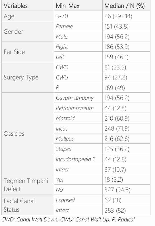
In this study, operative notes of patients with chronic otitis media with cholesteatoma at Cukurova University were investigated. A total of 345 patients were included in the study. One hundred fifty-one were women (43.8%), and 194 were male. The minimum age was 3, and the maximum was 70 (median age 26). 186 (53.9%) patients operated from the right ear, and 159 (46.1%) operated from the left ear. CWDM operation was applied to 81 patients (23.5%), CWUM operation was applied to 94 patients (27.2%), and RM operation was applied to 169 patients (49%). In these patients, cholesteatoma localization was detected in attic and epitympanum 205 (59.4%), cavum timpani (mesotympanum, hypotympanum) 194 (56.2%), only retrotympanum (sinus tympani, facial recess) 44 (12.8%), and 210 (60.9%) in mastoid antrum. In the ossicular system of these patients was found in Lysis of incus 248 patients (71.9%), malleus 216 (62.6%), stapes 125 (36.2%), and only 44 cases (12.8%) in the incudostapedial joint. 37 (10.7%) patients' ossicular systems were intact, and no lysis was observed. There were 18 (5.2%) patients with tegmen tympani defects, while 327 (94.8%) patients had no tegmen tympani defects. In 62 (%18) patients, the facial canal was open on the tympanic segment. 283 (82%) were intact (Table 1).
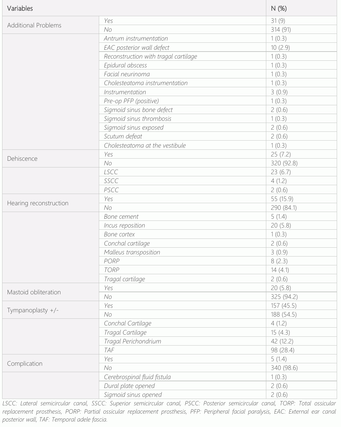
Hearing reconstruction was performed in 55 (15.9%) of all patients. In those patients, bone cement 5 (9%), incus interposition 20 (36.36%), bone cortex 1 (1.8%), conchal cartilage 2 (3.6%), malleus transposition 3 (5.4%), partial ossicular replacement prosthesis (PORP) 8 (14.5%), total ossicular replacement prosthesis (TORP) 14 (25.4%), tragal cartilage2(3.6%) was performed. Twenty (5.8%) underwent a surgery for mastoid obliteration. Twenty-five (7.2%) patients were diagnosed with SSC dehiscence. 23 (6.7%) were found to have lateral (horizontal) SSC, 4 (1.2%) had superior SSC, and 2 (0.6%) had posterior SSC. Tympanoplasty was performed in 157 patients (45.5%). In 98 patients (28.4%) TMF (temporal muscle facia), in 42 patients (12.2%) tragal cartilage perichondrium, in 15 patients (4.3%) tragal cartilage, in 4 patients (1.2%) conchal cartilage were used. 5 (1.4%) of the patients had complications. Two (0.6%) patients had dural plate defects, and 2 (0.6%) patients had sigmoid sinus defects. The complications were repaired with temporal jugular fascia. Additionally, in evaluation of cholesteatoma per-operatively before intervention 1 (0.3%) antral otoevidman, 10 (2.9%) external auditory canal posterior wall defects, 1 (0.3%) epidural abscess, 1 (0.3%) facial neurinoma, 4 (1.2%) extensive mastoid otoevidman, 3(0.9%) sigmoid plate defects, 1(0.3%) sigmoid sinus thrombosis, 2 (0.6%) scutum defects, 1 (0.3%) vestibular cholesteatoma were found (Table 2).
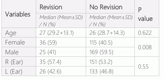
As a result of regular follow-ups, patients with suspicious otoscopic findings or patients who were found to have recidivistic disease after diffusion MRI investigation had revision surgeries. A total of 61 patients underwent revision mastoidectomy. Revision operation rate was found to be 17.6%. The average age of patients with revision surgery was 27. The patients' age difference was insignificant (p> 0.05) in the revised and non-revision groups. Of the patients, 36 (59%) were female and 25 (41%) were male. The rate of female patients in the revision group was significantly higher than the non-revision (p<0.05). 35 patients (57.4%) were operated on the right ear, and 26 patients (42.6%) on the left ear. In the revision and non-revision groups, the distribution of the sides did not differ significantly (p>0.05) (Table 3).
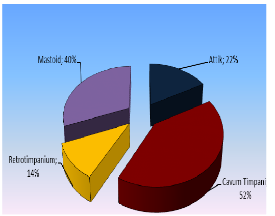
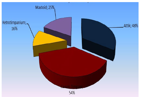
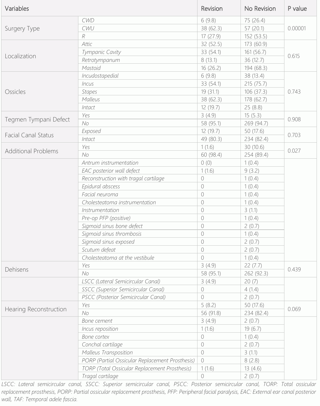
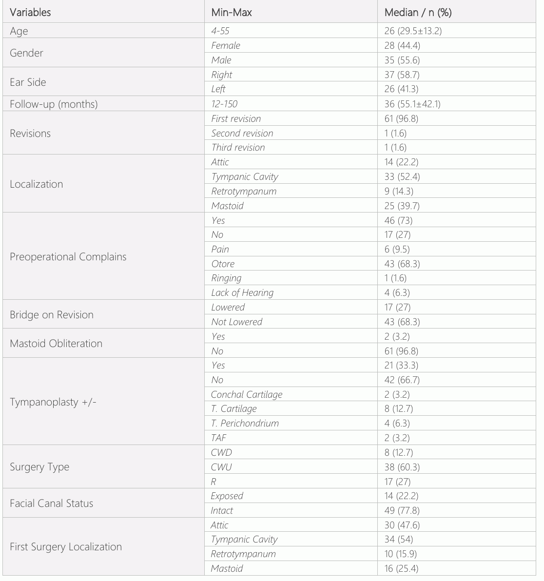
Revision mastoidectomy was performed in 6/81 (7.4%) of the patients who underwent CWDM surgery, 38/94 (40.4%) in patients who underwent CWUM surgery, and 17/169 (10%) in patients who underwent RM. The revision rate after CWUM surgery was significantly higher (p <0.05) (Table 4). Tegmen tympani defect rate was insignificant (p>0.05) in patients with and without revision surgery. The distribution of facial canal defects was not significant (p>0.05) in patients with and without revision surgery (Table 4). In the revision group, the additional problem rate was found to be significantly lower (p<0.05) than in the non-revision group (Table 4). The reason is that the first operation of the disease with the additional problem (common cholesteatoma) is the RM operation. Therefore, it can be said the rate of recidivism is higher in CWUM/CWDM-operated patients (Table 4). In the group with and without revision surgery, the SSC dehiscence ratio did not differ. In patients with and without revision surgery, complication rate was not different (Table 5). The follow-up period of patients with revision surgery ranged from 12 to 150 months (mean 36). One of the patients (1.6%) underwent the second revision operation due to recidivism, and the third revision operation was performed in 1 patient (1.6%). Cholesteatoma localization in patients who underwent revision surgery was detected in the attic and epitympanum 14 (22.2%), cavum tympani (mesotympanum, hypotympanum) 33 (52.4%), retro tympanum (sinus tympani, facial recess) 9 (14.3%), and 25 (39.7%) in mastoid cavity and antrum. Preoperative complaints of patients with revision surgery were determined as otorrhea (ear drain) 43(68.3%), ear pain 6(9.5%), hearing loss 4 (6.3%), and ear tinnitus 1 (1,6%). Bridges (external ear way posterior wall) of 17/63 patients who underwent revision surgeries were lowered. The patients who underwent revision operation had first operation localization in 30/63 (47.6%) attic and epitympanum, 34/63 (54%) cavum tympani (mesotympanum, hypotympanum, forehead), 10/63 (15.9%) retrotympanum sinus tympani, facial recess), 16/63 (25.4%) mastoid cavity and antrum (Table 5). Compared with the cholesteatoma placement in the first operation and the cholesteatoma placement in the revision surgery, recidivism is observed, especially in the mastoid cavity (40%) (Figures 1 and 2).
Discussion
Chronic otitis media (COM) is characterized by eardrum perforation, occasional suppurative discharge from the external auditory canal, and conductive hearing loss. One of the most critical problems of COM is developing cholesteatoma. Cholesteatoma in the middle ear is benign, but clinically, it shows a malign pattern and has a high lytic effect (5). In a study, the authors demonstrated a cholesteatoma in 36% of patients with COM with perforated eardrum and 4% with a nonperforated eardrum (6). Opposite ear involvement in 10-17% of patients with cholesteatoma was reported (7). Surgical experience, patient age, cholesteatoma size and localization, and outgrowth of disease in areas not accessible during surgery were found to be related to recidivism (8, 9). Furthermore, in a study by Visvanathan et al., a high facial bridge and a lack of external ear canal plasticity increased the risk of cholesteatoma recidivism (10). In our country, these patients usually complain of malodorous ear drains. Another study detected 85% of the complaints as ear drains (11). In our research, the most frequent complaint in patients who underwent revision surgery was ear drain (68.3%). In a (Gaillardin et al.) study performed on 113 patients with cholesteatomas, the rate of ossicular lysis was 58%. The lysis mainly occurred in the long arm of the incus (12). In a study by Egeli et al. on patients who underwent RM, malleus showed 65% partial and/or total lysis, incus showed 72% partial and/or total lysis and stape showed 63% partial and/or total lysis (13). In our study, the rate of ossicular lysis was found to be 89.2%, withmostly incus lysis71.9%, malleus lysis 62.6%, stapes lysis 36.2%, and incudostapedial joint lysis 12.8%. These results are consistent with the literature. In a study by Tanyeri et al. involving 56 patients, the total recidivism rate was 7%. One of the important causes of cholesteatoma recurrence is the surgical approach (23, 24). The significant predictors of recidivism are age, mastoid involvement, incus erosion, stapes erosion and type of surgical technique, and sinus cholesteatoma (25–29). The study of Diom et al. showed that the total recidivism rate was 13% in chronic otitis media patients with cholesteatomas in the following 15 years after surgery. In another study, records of 132 patients were retrospectively investigated, and the patients were followed up for six years. As a result, the recidivism rate in this study was lower in CWDM than in CWUM (16). According to Prasad et al., 40 children underwent CWUM surgery, 39 patients underwent revision surgery due to recurrence or residual disease. (11). Although studies show no change in recidivism rates between the two methods, the general belief is that the recidivism rates are higher in the CWUM method.
Göçmen et al. examined 114 patients who were operated for pediatric cholesteatoma (17). The selected surgical procedure in this study was CWUM in 31% of the patients and CWDM in 69%. Reasons for the more frequent selection of the CWDM method: most patients are likely to have follow-up problems due to low socioeconomic status, widespread illness, and the more straightforward implementation of the CWDM method by experienced surgeons. In Gocmen’s study, despite the higher residence rate in patients with CWUM, there was no significant difference between hearing outcome and recidivism rates between CWUM and CWDM patients. In our study, recidivism rates were 9.8% in patients with CWDM surgery, 40.8% in patients with CWUM surgery, and 10.5% in those with RM.
The advantages of CWUM are; rapid healing, easy post-operative care, no need to avoid water, and more options for hearing aids if necessary. The disadvantages are; the inability to evaluate epitympanum and mastoid cavity in the post-operative examination, inability to access the supratubal region unless the malleus head and incus are removed, excessive recurrence, frequent formation of retraction pouches on eardrum and excessive surgical intervention for complete eradication of the disease (18, 19). Regardless of the technique applied to the patient who underwent surgery for cholesteatoma, the essential consideration is post-operative follow-up. Diffusion MRI and HRCT (High-resolution computed tomography) are essential. A study conducted by Tierney et al. detected HRCT sensitivity as 43%, specificity 42-51%, and predictive value 28% in cases of residual and recurrent cholesteatoma (20). In a study conducted on 23 patients who had primer cholesteatoma surgery and secondary surgery, preparation residual cholesteatoma or recurrence cholesteatoma was detected up to 2mm's HRCT, sensitivity was %86, specificity was 87%. In addition, 20 of 23 patients had preoperative HRCT imaging showing soft tissue in the middle ear (21). In a study by Vecruysse et al., Preoperative diffusion MR was applied to 100 patients with 45 residual cholesteatomas and 55 primer cholesteatomas. The sensitivity was reported as 81%.
Regardless of the surgical technique used to reduce the risk of residual disease in the cholesteatoma, the surgeon must thoroughly clean the surrounding area of cholesteatoma, including the cholesteatoma matrix. However, the cholesteatoma on the facial nerve should be treated more carefully. New surgical methods can be applied in areas that are difficult to reach.
Conclusion
As a result, discussing the advantages and disadvantages of the closed technique (CWUM) and the open technique (CWDM and RM) with the patient when deciding on the surgery technique may be wiser. For some patients, it is crucial to protect their hearing aids and keep their hands free from water. This may be more important for some patients to prevent their illness from recurring. Patient compliance (whether to regularly follow-up, smoking, etc.) is essential. At this point, it is thought that it is helpful to give detailed information to the patient before the operation and to tell the patient the next step after the operation. It may be appropriate to consider the option of treatment with open cavity mastoidectomy in patients with cholesteatoma, considering the low risk of recidivistic disease, socioeconomic status of the patient, and the rare need for second session surgery.
Declarations
- Author contributions (CRediT)
- All authors contributed equally to all 14 CRediT roles (Conceptualization, Data curation, Formal analysis, Funding acquisition, Investigation, Methodology, Project administration, Resources, Software, Supervision, Validation, Visualization, Writing – original draft, Writing – review & editing).
- Funding
-
Funding information was not reported in the published version of this article.
- Conflicts of interest
- A conflict of interest statement was not reported in the published version of this article.
- Ethics approval
- The present study was approved by the Ethical committee of Cukurova University, Faculty of Medicine (ID: 04/09/2015- T45-K5).
- Data availability
- A data availability statement was not reported in the published version of this article.
References (29)
12 of 29 references are linked to their DOI.
- Bluestone CD, Klein OJ. Otitis Media In Infants And Children. Third Edition. Philadelphia, W.B Sounders Company. 2001:2-7. PubMed Google Scholar
- Akyıldız N. Kulak hastalıkları ve Mikrocerrahi, Bilimsel Tıp Kıtapları, 1998:419-451. PubMed Google Scholar
- Gerçeker M. KBB Hastalıkları ve Baş Boyun Cerrahisi, Ankara, MN medikal ve Nobel Tıp Kitapevi. 2009:69. PubMed Google Scholar
- Neudert M, Lailach S, Lasurashvili N, Kemper M, Beleites T, Zahnert T. Cholesteatoma recidivism: comparison of three different surgical techniques. Otol Neurotol. 2014; 35:1801-8. PubMed Google Scholar
- Ferlito A, Devaney KO, Rinaldo A, Milroy CM, Wenig BM, Iurato S, McCabe BF. Clinicopathological consultation. Ear cholesteatoma versus cholesterol granuloma. Ann Otol Rhinol Laryngol. 1997;106:79-85. DOI Crossref PubMed Google Scholar
- Cummings C, Fredrickson J, Harker L, Krause CJ. Otolaryngology -Head and Neck SurgerySecond Edition. Mosby-year Book, Inc. Missouri, 1993:2823-2832. PubMed Google Scholar
- Olszewska E, Wagner M, Bernal-Sprekelsen M, Ebmeyer J, Dazert S, Hildmann H, Sudhoff H. Etiopathogenesis of cholesteatoma. Eur Arch Otorhinolaryngol. 2004;261:6-24. PubMed Google Scholar
- Schraff SA, Strasnick B. Pediatric cholesteatoma: a retrospective review. Int J Pediatr Otorhinolaryngol. 2006;70:385-93. DOI Crossref PubMed Google Scholar
- Soldati D, Mudry A. Cholesteatoma in children: techniques and results. Int J Pediatr Otorhinolaryngol. 2000;52:269-76. DOI Crossref PubMed Google Scholar
- Visvanathan V, Kubba H, Morrissey MS. Cholesteatoma surgery in children: 10-year retrospective review, J Laryngol Otol. 2012;126:450-3. DOI Crossref PubMed Google Scholar
- Prasad SC, Melia CLA, Medina M, Vincenti V, Bacciu A, Bacciu S, Pasanisi E. Long-term surgical and functional outcomes of the intact canal wall technique for middle ear cho lesteatoma in the paediatric population. ACTA otorhinolaryngologica italica 2014;34:354-361 PubMed Google Scholar
- Gaillardin L, Lescanne E, Morinière S, Cottier JP, Robier A. Residual cholesteatoma: Prevalence and location. Follow-up strategy in adults, European Annals of Otorhinolaryngology, Head and Neck Diseases. 2012;129:136-140. DOI Crossref PubMed Google Scholar
- Egeli Yücel Z, Yazıcı MF. Radikal Mastoidektomi Uygulanan Hastalarda Patolojik Değerlendirme. Türk Otolarengoloji Arşivi. 1998;36:41-44. PubMed Google Scholar
- Y, Fieflen T, Koyuncu M, Tekat A, Ünal R, Elhami R. Kolesteatoma cerrahisinde teknikler ve sonuçlarımız. Turk Arch Otolaryngol. 1998;36:77-81. PubMed Google Scholar
- Diom ES, Cisse Z, Tall A, Ndiaye M, Pegbessou E, Ndiaye IC, Diallo BK, Diouf R, Diop EM. Management of acquired cholesteatoma in children: a 15 year review in ENT service of CHNU de FANN Dakar., Int J Pediatr Otorhinolaryngol. 2013;77:1998-2003. DOI Crossref PubMed Google Scholar
- Deniz M, Uslu C, KoldaĢ C, Deniz B. Which technique is better for cholesteatoma surgery? B-ENT. 2015;11:109-15. PubMed Google Scholar
- Göçmen H, Kiliç R, Ozdek A, Kizilkaya Z, Safak MA, Samim E. Surgical treatment of cholesteatoma in children. Int J Pediatr Otorhinolaryngol. 2003;67:867-72. DOI Crossref PubMed Google Scholar
- Yanagihara N. Surgical treatment of choleastaotoma problems: indications and technique. In: Sade J, editor. Cholesteatoma and mastoid surgery. 1st ed. Amsterdam: Kugler Publications. 1982:97-9. PubMed Google Scholar
- Chole RA, Brodie HA. Surgery of the mastoid and petrosa. In: Bailey BJ, editor. Head and neck surgery-otolaryngology. 2nd ed. Philadelphia: JB Lippincott. 1998:2053-72. PubMed Google Scholar
- Tierney PA, Pracy P, Blaney SP, Bowdler DA. An assessment of the value of the preoperative computed tomography scans prior to otoendoscopic „second look‟ in intact canal wall mastoid surgery. Clin Otolaryngol Allied Sci. 1999;24:274-276. DOI Crossref PubMed Google Scholar
- Dündar A. Diffusion MRI in Residual Cholesteatoma, Int Adv Otol. 2015;11:58-62. PubMed Google Scholar
- Vercruysse JP, De Foer B, Pouillon M, Somers T, Casselman J, Offeciers E. The value ofdiffusion-weighted MR imaging in the diagnosis of primary acquired and residual cholesteatoma: a surgical verifi ed study of 100 patients. Eur Radiol. 2006;16:1461-1467. DOI Crossref PubMed Google Scholar
- Nyrop M, Bonding P. Extensive cholesteatoma: long-term results of three surgical techniques. J Laryngol Otol 1997;111:521–6 DOI Crossref PubMed Google Scholar
- Darrouzet V, Duclos JY, Portmann D, Bebear JP. Preference for the closed technique in the management of cholesteatoma of the middle ear in children: a retrospective study of 215 consecutive patients treated over 10 years. Am J Otol 2000;21:474–81 PubMed Google Scholar
- Djurhuus BD, Faber CE, Skytthe A. Decreasing incidence rate for surgically treated middle ear cholesteatoma in Denmark 1977–2007. Dan Med Bull 2010;57:A4186 PubMed Google Scholar
- Rosenfeld RM, Moura RL, Bluestone CD. Predictors of residual-recurrent cholesteatoma in children. Arch Otolaryngol Head Neck Surg 1992;118:384–91 DOI Crossref PubMed Google Scholar
- Roger G, Denoyelle F, Chauvin P, Schlegel-Stuhl N, Garabedian EN. Predictive risk factors of residual cholesteatoma in children: a study of 256 cases. Am J Otol 1997;18:550–8 PubMed Google Scholar
- Gristwood RE, Venables WN. Factors influencing the probability of residual cholesteatoma. Ann Otol Rhinol Laryngol 1990; 99:120–3 25 77212121 Cholesteatoma Surgery Huseyin Keskin DOI Crossref PubMed Google Scholar
- Ann Otol Rhinol Laryngol 2010;119:304–12 26 De CE, Marchese MR, Scarano E, Paludetti G. Aural acquired cholesteatoma in children: surgical findings, recurrence and functional results. Int J Pediatr Otorhinolaryngol 2006;70: 1269– PubMed Google Scholar
Read the full text
Open the PDF here
Loading the PDF…
If the PDF does not show here, use the download button above.
How to cite this article
Keskin H, Guvenmez O. Cholesteatoma Surgery With Three Different Methods: A Clinical Study For Recidivism Research. Ulutas Med J. 2024;10(1):15-25. doi:10.5455/umj.20240229070431
Keskin, H., & Guvenmez, O. (2024). Cholesteatoma Surgery With Three Different Methods: A Clinical Study For Recidivism Research. The Ulutas Medical Journal, 10(1), 15–25. https://doi.org/10.5455/umj.20240229070431
Keskin, H. and Guvenmez, O. (2024) 'Cholesteatoma Surgery With Three Different Methods: A Clinical Study For Recidivism Research', The Ulutas Medical Journal, 10(1), pp. 15–25. doi: 10.5455/umj.20240229070431.