The Ulutas Medical Journal · ISSN 2149-0430 (print), 2149-388X (online) · https://www.ulutasmedicaljournal.com/?mno=24159
Original Article · Open access
Dermatophytosis in Bhairahawa, Nepal: Prevalence and Resistance Pattern of Dermatophyte Species
Also available in: العربية · Türkçe · 中文
- Department of Microbiology, Universal College of Medical Sciences, Bhairahawa, Nepal ROR
- Department of Pharmacology, Universal College of Medical Sciences, Bhairahawa, Nepal ROR
- Department of Dermatology, Universal College of Medical Sciences, Bhairahawa, Nepal ROR
Abstract
- Introduction
- Dermatophytosis is colonization by dermatophytic fungus of the keratinized tissues like hair, nails and skin. They are considered important as a public health problem. The study was aimed to isolate, identify, and detect the in-vitro antifungal sensitivity pattern of various dermatophytes isolates from clinically diagnosed cases of dermatophytosis.
- Materials and Methods
- One hundred and sixty patients of all age groups and both sexes and clinically diagnosed with dermatophytosis were recruited in this study. The specimens included skin scales, hair pluckings, and nail clippings. Identification and isolation were done by microscopic examination, culture, and biochemical analysis.
- Results
- Dermatophytosis was more common in males (60.62%) than females (39.37%). Tinea corporis (31.25%) was the most common clinical presentation followed by Tinea faciei (25%). Trichophyton rubrum (36.19%) was the most common isolate followed by Trichophyton mentagrophytes (15.23%). Out of four antifungal drugs used, fluconazole was found most resistant while Itraconazole was the most effective drug.
- Conclusion
- The epidemiology of dermatophyte infections may change with time. Antifungal susceptibility testing will aid the clinician in initiating prompt and appropriate antifungal therapy and prevent the emergence of resistance.
Keywords
Full text
Introduction
Dermatophytes are keratinophilic hyaline molds that can cause disease in keratinized tissues like hair, skin, and nail (1). The members of this dermatophytic group include Trichophyton, Microsporum and Epidermophyton (2). Based on the reservoir and route of transmission, dermatophytes may be of anthropophilic (human), zoophilic (animals), or geophilic (soil) origin. These organisms are named according to the affected body site: Tinea capitis (head), T. corporis (trunk), T. cruris (perianal area), T. pedis (foot and interdigital area), and T. unguium (nail) (3). The most common etiological agents are Trichophyton rubrum, T. mentagraphytes, T. interdigitale, T. tonsurans, and Microsporum canis. T. rubrum is the most frequently isolated agent in clinics (1). Nepal is such a country where a wide variation in climate, socio - economic status, religion and customs is quite prevalent in different parts of the country. In developing countries, other than hot and humid climatic conditions, low hygiene, poor access to water, overcrowding contact also plays significant etiological role for dermatophytosis (4–9).
Treatment options for dermatophytosis are topical as well as systemic antifungal drugs. But during course of time dermatophytes have also evolved drug resistance for single as well as multiple drug simultaneously. Studies conducted worldwide show that resistance among dermatophytes is not uncommon (10, 11). Due to high temperature and increased humidity, there are increased cases of dermato phytosis and other fungal infections especially in terrain and hilly region of Western Nepal. Since there was increased incidence of drug resistance observed over a period of time to the antimycotic drugs commonly used for the treatment i.e., fluconazole, terbinafine and clotrimazole, the need for testing of antifungal susceptibilities of dermatophytes has become apparent. Recently CLSI (Clinical and Laboratory Standards Institute) has approved a micro broth dilution method for antifungal susceptibility testing of molds, but these tests are cumbersome and difficult to be performed in routine laboratory setup. The agar-based disc diffusion (ABDD) is an easy method to determine the antifungal susceptibility of dermatophytes, but data regarding these methods are scarce and not standardized (2, 12, 13). The application of in vitro antifungal susceptibility testing for guidance of antifungal drug therapy has been limited due to uncertain correlation between in vitro and in vivo action of drugs (2, 14).
This study was planned to determine the prevalence of dermatophytes infection in Bhairahawa, Nepal as well as the resistance of the recovered dermatophyte species to antifungal drugs. So far, skin fungal infection is empirically treated and fungal culture and sensitivity is not routine recommended in our region; therefore, only handful of data is available regarding incidence of skin infection and drug resistance. Therefore, this study was planned to find out the same.
Materials and Methods
A hospital-based prospective observational study was conducted at Universal College of Medical Sciences Teaching Hospital (UCMS-TH) Bhairahawa from March 2019 to October 2019.
Ethical Statement
Ethical approval was taken from the institutional review Committee (IRC) of UCMS-TH prior to the sample collection (I.R.C. Reg. No. UCMS/IRC /036/019). A total of 160 patients of all age group and both sexes attending Dermatology outpatient and clinically diagnosed with dermatophytosis were recruited in this study after informed consent. Patients with surface infections, accidental and surgical cases and also patients who were already on antifungal treatment were excluded from the study. A detailed history of selected cases was recorded that included name, age, sex, address, duration of illness and other complaints. All the clinically diagnosed 160 cases were subjected to mycological work. The specimens included skin scales, hair and nails. The site of the lesions was cleaned with 70% alcohol, samples were collected in a sterile paper folds and labelled with details of patients. All the samples were subjected to direct microscopy and culture. One part of the specimen was directly observed under microscope by potassium hydroxide (KOH) mount using 10% for skin and 40% for hair and nail samples. Another part of the sample was inoculated on slants of Sabouraud’s dextrose agar (SDA) with chloramphenicol (0.05 mg/ml) and cycloheximide (0.5mg/ml). Culture tubes were examined thrice weekly for appearance of growth, cultures were incubated for 1 month before discarding them as negative. Cultures yielding growth were evaluated to species level-based colony morphology, microscopic properties in Lacto phenol cotton blue (LPCB) mount and urease test. The LPCB was obtained from Hi-Media Laboratories Pvt. Ltd., Mumbai, India. The isolates were subjected to the agar-based disc diffusion method to the study of sensitivity pattern of antifungals using antifungal drugs as described by Nweze et al, (12) and Prabhat Kiran Khatri et al.(15). All the dermatophytes were sub cultured on potato dextrose agar and incubated at 280C to enhance sporulation for 1 week. Following growth, conidia were harvested in sterile saline and conidial suspension was adjusted to between 1.0×10⁶ applied over MHA plates, after which the plates were incubated at 280 C for 5-7 days.
Trichophyton mentagrophytes ATCC 9533 and Trichophyton rubrum ATCC 28188 strains served as control. After the colonies grew, the zones of inhibition around the disc were measured in millimeters and recorded as sensitive, intermediate or resistant (9, 12, 13). Control plates with fungus inoculum and without antifungal disc were also tested.
All the data from cases was fed in MS Excel (Microsoft office 2018) and then analyzed by Statistical Package for Social Service (SPSS) for window version; SPSS 22, Inc., Chicago, IL). All data were expressed in terms of percentage.
Results
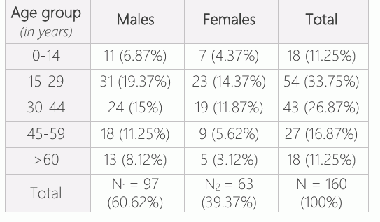
Out of 160 clinically diagnosed cases of dermatophytosis, males (60.62%) were more affected than females (39.37%) with male: female ratio 1.54:1. Most of the affected patients belonged to the age group of 15-29 years (33.75%) followed by 30-44 years (26.87%) which is shown in Table-1. Majority of the affected patients belonged to low socio economic status and were involved in active physical works like manual laborer, farmers, spores/ml by microscopic carpenter, tailor, domestic help etc.
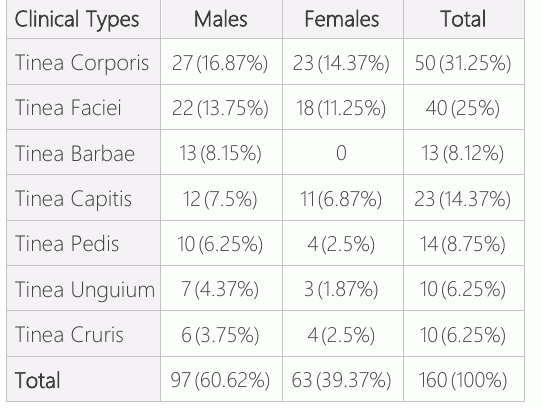
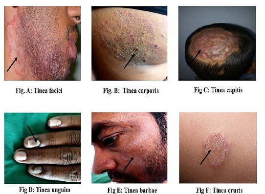
and 5×10⁶ enumeration with cell counting hemocytometer (Neubauer chamber) (16). Four antifungal drugs were tested against dermato phytes isolates. The following commercially available antifungal drugs were obtained from HiMedia Laboratory Pvt. Ltd., Mumbai, India; fluconazole (25 µg), itraconazole (10 µg) and ketoconazole (10µg). Plates of non-supplemented Muller Hinton Agar (MHA) were streaked evenly in three directions with a sterile cotton swab dipped into the standardized inoculums suspension. Plates were allowed to dry then antifungal disc were Tinea corporis (31.25%) was the most common clinical presentation followed by Tinea faciei (25%) and Tinea capitis (14.37%). There was higher incidence of Tinea corporis and Tinea faciei in males compared to females i.e. 27 (16.87%), 22(13.75%) respectively which is shown in Table-2.
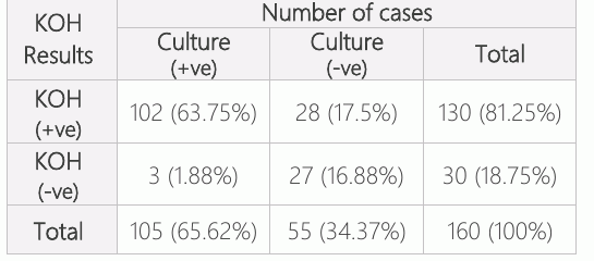
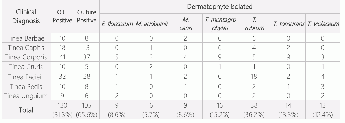
Out of 160 samples processed, 130 (81.3%) were positive for KOH mount while 105 (65.6%) were culture positive. Out of 130 KOH positive samples, 102 (63.8%) were both KOH positive and culture positive, rest were culture negative which is elucidated in Table-3.
Samples from patient with Tinea cruris resulted 100% KOH positivity followed by those from cases of Tinea capitis which showed 78.26% KOH positivity. Highest cultural positivity was observed in cases of Tinea corporis (74%) followed by Tinea faciei (70%) and Tinea barbae (61.5%). Trichophyton rubrum (36.2%) was the most common isolate followed by Trichophyton mentagrophytes(15.2%),Trichophyton tonsurans (13.3%) and Trichophyton violaceum (12.4%). Trichophyton rubrum was the most common dermatophyte isolated from 38 clinical types of dermatophytosis. All four isolated dermatophyte species were recovered from Tinea corporis, the most common clinical presentation which is shown in Table-4.
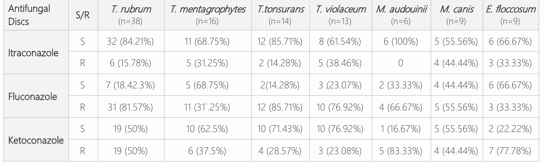
Antifungal susceptibility testing showed itraconazole as the most sensitive antifungal agent, while ketoconazole was the least sensitive. Among the dermatophyte isolates, M. audouinii showed 100% sensitivity against Itraconazole followed by T. rubrum (84.21%) whereas the least sensitivity was shown by M. canis (55.56%). T. mentagrophytes showed 68.75% sensitivity against fluconazole. Similarly, T. violaceum showed highest sensitivity i.e., 76.92 % against ketoconazole followed by T. tonsurans (71.43%) and T. mentagrophytes (62.5%) which is shown in Table-5.
Discussion
Identification of species responsible for the dermatophytoses and their sensitivity pattern is of great importance not for epidemiology but also for therapeutic point of view. Our study site bears tropical climate where high level of humidity and high temperature favor the growth of fungi causing dermatophytoses. In our present study about 33.75% of dermato phytes were isolated from patient belonging to the age groups 15-29 years age. Our results are similar to other studies (17–20) who also reported higher infections in young adults. The higher prevalence is mainly due to the physical activity, hot humidity and high temperature in the region. This leads suitable wet condition for dermatophytes to grow. In this study, out of 160 patients, 97 (60.62%) were males and 63 (39.37%) were females, with male to female ratio being 1.54:1. Male dominance is reported in many places of South Asia (21–23). High prevalence of dermatophytes in males is due to frequent interaction with the society.
The predominant of clinical manifestations of dermatophytoses vary considerably to different studies in literature. In this study tinea corporis was the most dominant clinical manifestation involving 31.25%. Our findings are in accordance with the study by Balakumar S and et al, (24) who also reported Tinea corporis as the dominant clinical diagnosis. High rates of Tinea corporis could be attributed to its symptomatic nature (pruritus) which leads the patient to seek medical advice (5).Whereas study by Hemendra Kumar Sharma et al, (25) showed Tinea unguium as the dominant clinical diagnosis. This variation observed in the clinical type of dermato phytoses is due to varied climate conditions, livelihood, type of occupations, type of occupation, pathogen and host relationship. In the study, out of 160 clinical samples, 130 (81.25%) samples were positive by direct microscopy by KOH mount and 105 (65.62%) samples were culture positive. Out of 130 KOH positive samples, 102 (63.75%) samples were both KOH positive and culture positive, while the rest 28 (17.5%) were culture negative. The direct microscopy and culture findings of present study are relatively in agreement with study done by Basak P et al, (26) (71.1% KOH positive and 59.8% culture positive), Dhyaneswari GP et al, (27) (72.6% KOH positive) and Mahale RP et al, (28) (61.01% culture positive). There is a difference between KOH positivity rate and culture positivity rate in our present study this is because fungal elements were seen under direct microscopy but samples failed to grow on culture which might be due to various factors like unsatisfactory collection of samples containing dead fungal hyphae (29, 30). In this study, some specimens did not show any fungal elements when seen under direct microscopy but showed growth on culture. This might be due to presence of scanty fungal elements which were missed during direct microscopic examination or due to fungal elements in inactive sporulating form, which could not be visualized under microscopy (30). In this study genus Trichophyton represented 77.14% of the isolates of dermatophytes, followed by Epidermophyton (8.57%) and Microsporum (14.28%). The most isolated was Trichophyton rubrum (36.19%) followed by Trichophyton mentagrophytes (15.23%) Trichophyton tonsurans (13.33%), Trichophyton violaceum (12.38%), Microsporum canis (8.57%). The other species isolated was Microsporum audouinii, and Epidermophyton floccosum. Our findings are in accordance with study by Dhyaneswari GP et al, (27) (Trichophyton rubrum 59.6%, Trichophyton mentagrophytes 26%), Walke HR et al, (31) (Trichophyton rubrum 56.37%, Trichophyton mentagrophytes 19.39%), R.K Agarwal et al, (32) (Trichophyton rubrum 42.63%, Trichophyton mentagrophytes 41.81%), and Basak P et al, (26) who have reported dermatophyte Trichophyton rubrum as the dominant species. However, there are studies such as by Hemendra Kumar Sharma et al, (25) who has reported Trichophyton mentagro phytes as most common species isolated.
The determination of in-vitro antifungal susceptibility was reported to be important for the ability to eradicate pathogenic dermato phytes. Most clinical types of dermatophytoses respond well to topical antifungal therapy, while Tinea unguium, Tinea capitis and extensive type of dermatophytoses require systemic therapy. Recently, there has been a rise in antifungal resistant strains of fungi. Therefore, early initiation of correct antifungal therapy is essential for proper treatment and prevention of spread of disease. In the present study, antifungal susceptibility testing by agar-based disc diffusion method (12, 32) was performed for five antifungal drugs: ketoconazole, fluco nazole, itraconazole and nystatin. Itraconazole (76.19 %) was the most sensitive followed by nystatin (63.8%) and ketoconazole (54.28%) and fluconazole (27.61%) was the least sensitive. Present study findings are almost similar with the findings of Basak P et al, (26) itraconazole (97.9%) was the most sensitive antifungal drug while fluconazole (2.7%) was least sensitive). Our findings about poor susceptibility of dermatophytes to fluconazole is compatible with the studies by Hemendra Kumar Sharma et al (25), Basak P et al, (26) and EI Damaty et al (33). The higher resistance to fluconazole may be due to its availability at pharmacies, self-medication by patients due to its over the counter (OTC) availability and rampant practice of irrational prescription by compounder.
In this study, out of 105 isolates, 80(76.19%) were sensitive to Itraconazole, while 20.83% were resistant. Itraconazole is a much more affordable antifungal drug. Our study was in according to the Basak P et al, (31) and EI Damaty et al, who also showed Itraconazole as the effective drug. It has effectiveness against dermatophytes; hence, it must be a preferred treatment option for better outcome in patients suffering from dermatophytoses. In this study out of 105 dermatophyte isolates, 54.28% were sensitive to Ketoconazole while 45.71% were resistant. We have observed average sensitivity to Ketoconazole which is in agree ment with study by Hemendra Kumar Sharma et al,(25) which has suggested Ketoconazole as an average choice for the treatment of dermato phytosis. Our work suggests that disk diffusion antifungal susceptibility testing is simple, inexpensive, and does not require high cost equipment. It allows for a comparison between different antifungal agents and may help optimize the therapy for treating patients with dermatophytosis.
Conclusion
This report documents the emergence and occurrence of dermatophytoses and its agent in Western part of Nepal. Males are more affected than female with dermatophytoses infection. KOH examination is shown to be more sensitive than culture. Majority of the cases were Tinea corporis followed by Tinea pedis and the Tinea faciei and the commonest mycological isolate with Trichophyton taking the lead, among them the commonest species was Trichophyton rubrum. The fungal infections can be treated by a proper dose of itraconazole than other antifungal drug therapy. MIC values should be determined by broth microdilution test to determine the proper dose.
Recommendation
Present study has highlighted the frequency of dermatophytosis in tertiary care hospital which also reflects the overall similar picture in other part of our country. On the basis of the study, it has made following recommendation: Any clinical diagnosis needs to be supported by laboratory diagnosis. Since microscopy and culture are easy to perform, cost effective and this should be done in all suspected cases of dermatophytosis. As antifungal susceptibility testing facilities are now available for dermato phytes, every isolate should be tested against antifungal drugs so that increasing resistance among dermatophytes can be reduced. This may help in surveillance and epidemiological study of resistant strains.
Declarations
- Author contributions (CRediT)
- All authors contributed equally to all 14 CRediT roles (Conceptualization, Data curation, Formal analysis, Funding acquisition, Investigation, Methodology, Project administration, Resources, Software, Supervision, Validation, Visualization, Writing – original draft, Writing – review & editing).
- Funding
-
Funding information was not reported in the published version of this article.
- Conflicts of interest
- The authors declared no conflict of Interest in the present study.
- Ethics approval
- Ethical approval was taken from the institutional review Committee (IRC) of UCMS-TH prior to the sample collection (I.R.C. Reg. No. UCMS/IRC /036/019). A total of 160 patients of all age group and both sexes attending Dermatology outpatient and clinically diagnosed with dermatophytosis were recruited in this study after informed consent.
- Data availability
- A data availability statement was not reported in the published version of this article.
References (33)
17 of 33 references are linked to their DOI.
- Reiss HE, Shadomy H, Lyon M. Mycoses of implantation. Fundamental medical mycology: Wiley-Blackwell, Hoboken; 2012. p. 479-92 DOI Crossref PubMed Google Scholar
- Rippon JW. The changing epidemiology and emerging patterns of dermatophyte species. Current topics in medical mycology. 1985:208-34 DOI Crossref PubMed Google Scholar
- Nenoff P, Krüger C, Ginter‐Hanselmayer G, Tietz HJ. Mycology–an update. Part 1: Dermatomycoses: causative agents, epidemiology and pathogenesis. JDDG: Journal der Deutschen Dermatologischen Gesellschaft. 2014;12(3):188-210 DOI Crossref PubMed Google Scholar
- Suganthi M. Pathogenesis and clinical significance of dermatophytes: A comprehensive review. Innovations in Pharmaceuticals and Pharmacotherapy. 2017;5(1):62-70 DOI Crossref PubMed Google Scholar
- Karim MT, Ghosh S, Chougale R, Vatkar V, Deshkar D, Narute J. Study of Dermatophytes and their Biofilm Production. Journal of Advanced Medical and Dental Sciences Research. 2017;5(11):56-9 PubMed Google Scholar
- Adimi P, Hashemi SJ, Mahmoudi M, Mirhendi H, Shidfar MR, Emmami M, et al. In-vitro activity of 10 antifungal agents against 320 dermatophyte strains using microdilution method in Tehran. Iranian journal of pharmaceutical research: IJPR. 2013;12(3):537 PubMed Google Scholar
- Hanafy AM. In vitro antifungal drug susceptibility of dermatophytes isolated from patients in Al-Medina, Saudi Arabia. Egypt J Exp Biol (Bot). 2012;8(2):245-50 PubMed Google Scholar
- De Sarro A, La Camera E, Fera M. New and investigational triazole agents for the treatment of invasive fungal infections. Journal of Chemotherapy. 2008;20(6):661-71 DOI Crossref PubMed Google Scholar
- Pakshir K, BAHA AL, Rezaei Z, Sodaifi M, Zomorodian K. In vitro activity of six antifungal drugs against clinically important dermatophytes. 2009 DOI Crossref PubMed Google Scholar
- Azambuja CVdA, Pimmel LA, Klafke GB, Xavier MO. Onychomycosis: clinical, mycological and in vitro susceptibility testing of isolates of Trichophyton rubrum. Anais brasileiros de dermatologia. 2014;89(4):581-6 DOI Crossref PubMed Google Scholar
- Jha B, Mahadevamurthy S, Sudisha J, Bora A. Isolation, identification and antifungal susceptibility test of dermato phytes from the patients with onychomycosis in central Nepal. Am J Dermatol Venereol. 2015;4(3):30-6 PubMed Google Scholar
- Nweze E, Mukherjee P, Ghannoum M. Agar-based disk diffusion assay for susceptibility testing of dermatophytes. Journal of clinical microbiology. 2010;48(10):3750-2 DOI Crossref PubMed Google Scholar
- Agarwal R, Gupta S, Mittal G, Khan F, Roy S, Agarwal A. Antifungal susceptibility testing of dermatophytes by agar-based disk diffusion method. Int J Curr Microbiol Appl Sci. 2015;4(3):430-6 PubMed Google Scholar
- Rippon JW. Medical mycology; the pathogenic fungi and the pathogenic actinomycetes: Eastbourne, UK; WB Saunders Company; 1982 PubMed Google Scholar
- Khatri PK, Kachhawa D, Maurya V, Meena S, Bora A, Rathore L, et al. Antifungal resistance pattern among dermatophytes in Western Rajasthan. Int J Curr Microbiol App Sci. 2017;6: 499-509 DOI Crossref PubMed Google Scholar
- Indira G. In vitro antifungal susceptibility testing of 5 antifungal agents against dermatophytic species by CLSI (M38-A) micro dilution method. Clin Microbial. 2014;3(3) DOI Crossref PubMed Google Scholar
- Tartor YH, Abo Hashem ME, Enany S. Towards a rapid identification and a novel proteomic analysis for dermatophytes from human and animal dermatophytosis. Mycoses. 2019;62(12):1116-26 DOI Crossref PubMed Google Scholar
- Janagond AB, Rajendran T, Acharya S, Vithiya G, Ramesh A, Charles J. Spectrum of dermatophytes causing tinea corporis and possible risk factors in rural patients of madurai region, South India. Natl J Lab Med. 2016;5 PubMed Google Scholar
- Ramaraj V, Vijayaraman RS, Rangarajan S, Kindo AJ. Incidence and prevalence of dermatophytosis in and around Chennai, Tamilnadu, India. Int J Res Med Sci. 2016;4(3):695-700 DOI Crossref PubMed Google Scholar
- Janardhan B, Vani G. Clinico mycological study of dermatophytosis. Int J Res Med Sci. 2017;5(1):31-9 DOI Crossref PubMed Google Scholar
- Verenkar M, Pinto M, Rodrigues S, Roque W, Singh I. Clinico-microbiological study of dermatophytoses. Indian journal of pathology & microbiology. 1991;34(3):186-92 PubMed Google Scholar
- Sumathi S, Mariraj J, Shafiyabi S, Ramesh R, Krishna S. Clinicomycological study of dermatophytes. Int J Pharm Biomed Res. 2013;4(2):132-4 PubMed Google Scholar
- Gupta B, Kumar S, Kumar R, Khurana S. Mycological aspects of dermatomycosis in Ludhiana. Indian Journal of Pathology & Microbiology. 1993;36(3):233-7 PubMed Google Scholar
- Balakumar S, Rajan S, Thirunalasundari T, Jeeva S. Epidemiology of dermatophytosis in and around Tiruchirapalli, Tamilnadu, India. Asian Pacific Journal of Tropical Disease. 2012;2(4):286-9 DOI Crossref PubMed Google Scholar
- Hemendra Kumar Sharma NV. In vitro antifungal Susceptibility testing of Dermatophytes isolated from clinical samples in tertiary care hospital around Jaipur, Rajasthan. International journal of medical and health research. 2019(3):40-5 PubMed Google Scholar
- Priyam Basak BM, Swetalona Pattanaik. Prevalence of dermatophytic infections including antifungal susceptibility pattern of dermatophytes in a tertiary care hospita. International Journal of Research in Medical Sciences 2019: 699-705 DOI Crossref PubMed Google Scholar
- Dhyaneswari GP MV, Bhore AV. Clinicomycological profile of dermatophytosis in a tertiary care hospital in Western India. SAS J Med. 2015;1(4):160-5 PubMed Google Scholar
- Mahale RP RM, Tejashree A, Deepashree R, Kulkarni M. Clinicomycological profile of dermatophytosis in a teaching hospital. Int J Pharmaceut Sci Invent. 2014;3(8):43-6 PubMed Google Scholar
- Lavanya V SS. Clinico-mycological study of Dermatophytosis in a tertiary care centre in Bagalkot. Int J Med Health Res. 2015;1(2):63-6 Dermatophytosis Karn et al. PubMed Google Scholar
- Lyngdoh CJ, Lyngdoh WV, Choudhury B, Sangma KA, Bora I, Khyriem AB. Clinico-mycological profile of dermatophytosis in Meghalaya. International Journal of Medicine and Public Health. 2013;3(4) DOI Crossref PubMed Google Scholar
- Walke HR, Gaikwad AA, Palekar SS. Clinico-mycological profile of dermatophytosis in patients attending dermatology OPD in tertiary care hospital, India. Int J Curr Microbiol App Sci. 2014;3(10):432-40 PubMed Google Scholar
- Agarwal R, Gupta S, Mittal G, Khan F, Roy S, Agarwal A. Antifungal susceptibility testing of dermatophytes by agar-based disk diffusion method. Int J Curr Microbiol Appl Sci. 2015;4:430-6 PubMed Google Scholar
- El Damaty HM, Tartor YH, Mahmmod YS. Species identification, strain differentiation, and antifungal susceptibility of dermatophyte species isolated from clinically infected Arabian horses. Journal of Equine Veterinary Science. 2017;59:26-33 DOI Crossref PubMed Google Scholar
Respond to this article
Comments on a published article are welcome as a Letter to the Editor.
Get an e-mail when new articles in your fields are published
Questions to the authors
Questions are moderated: an editor approves each question, the authors answer, and an editor approves the answer before both appear here.
No answered questions yet.
Ask the authors a question
Discussion
Comments are moderated: they appear after the writer confirms the e-mail address and an editor approves them. Each comment carries a conflict-of-interest statement.
No comments yet.
Write a comment
Read the full text
Open the PDF here
Loading the PDF…
If the PDF does not show here, use the download button above.
How to cite this article
Karn SL, Gurung A, Shrivastava AK, Poudel SK, Adhikari SR, Sah CM. Dermatophytosis in Bhairahawa, Nepal: Prevalence and Resistance Pattern of Dermatophyte Species. Ulutas Med J. 2021;7(2):115-123. doi:10.5455/umj.20201208055254
Karn, S. L., Gurung, A., Shrivastava, A. K., Poudel, S. K., Adhikari, S. R., & Sah, C. M. (2021). Dermatophytosis in Bhairahawa, Nepal: Prevalence and Resistance Pattern of Dermatophyte Species. The Ulutas Medical Journal, 7(2), 115–123. https://doi.org/10.5455/umj.20201208055254
Karn, S.L. et al. (2021) 'Dermatophytosis in Bhairahawa, Nepal: Prevalence and Resistance Pattern of Dermatophyte Species', The Ulutas Medical Journal, 7(2), pp. 115–123. doi: 10.5455/umj.20201208055254.
@article{umj24159,
  title = {Dermatophytosis in Bhairahawa, Nepal: Prevalence and Resistance Pattern of Dermatophyte Species},
  author = {Karn, Subhash Lal and Gurung, Ajay and Shrivastava, Amit Kumar and Poudel, Sulochana Khatiwada and Adhikari, Shristi Raut and Sah, Chandra Mohan},
  journal = {The Ulutas Medical Journal},
  year = {2021},
  volume = {7},
  number = {2},
  pages = {115--123},
  doi = {10.5455/umj.20201208055254},
  url = {https://www.ulutasmedicaljournal.com/?mno=24159},
  issn = {2149-388X}
}