The Ulutas Medical Journal · ISSN 2149-0430 (print), 2149-388X (online) · https://www.ulutasmedicaljournal.com/?mno=78740
Original Article · Open access
Evaluation of Biomarkers in Patients with Sepsis Diagnosis in Pediatric Intensive Care Unit
- Department of Pediatric Disease, Silopi State Hospital, Ministry of Health, Turkey ROR
- Department of Pediatric Infectious Diseases, Dicle University School of Medicine, Turkey ROR
- Department of Pediatric Intensive Care Unit, Dicle University School of Medicine, Turkey ROR
- Department of Pediatric Allergy and Immunology, Dicle University School of Medicine, Turkey ROR
Abstract
- Introduction
- Sepsis is one of the leading causes of mortality and morbidity in intensive care units. In this study, we aimed to investigate the etiological cause, focus of infection, culture sample results, and inflammatory markers among patients treated for sepsis at pediatric intensive care units (PICUs).
- Materials and Methods
- We retrospectively reviewed the medical records of 70 patients aged 1 month to 18 years who were treated for sepsis at PICU between January 2014 and May 2019.
- Results
- The median age of the patients was 37 months. The most common underlying etiology was respiratory failure (70%). The most common site of infection causing sepsis was the respiratory system (n:40, 57%). The most commonly isolated agents were Proteus mirabilis and Acinetobacter baumannii. Whereas C reactive protein (CRP) was normal at the time of the diagnosis of sepsis in 28.5% (n=20) of the patients, procalcitonin (PCT) was elevated in all of them. A comparison of the laboratory parameters in the first 24 hours after the diagnosis of sepsis and at the end of the treatment revealed a significant difference between White blood cell (WBC) count, neutrophil-lymphocyte ratio (NLR), the levels of C reactive protein (CRP) and Procalcitonin (p<0.05). In addition, positive correlations were detected between NLR, CRP and PCT (p=0.036, p=0.012/ r=0.251, r:0.299, respectively).
- Conclusion
- We believe that PCT, CRP, and NLR can be used as biomarkers for monitoring patients with sepsis.
Keywords
Full text
Introduction
Sepsis is one of the leading causes of mortality and morbidity in intensive care unit. Multiorgan failure and death may occur unless the infection causing sepsis is effectively treated. Thus, it is of vital importance to have a thorough knowledge of sepsis and start appropriate treatment as quickly as possible (1, 2).
Sepsis is a clinical syndrome characterized by systemic inflammatory response syndrome, which is associated with infection-mediated immune system abnormalities, microcirculatory disorders, and end-organ failure. It has complex pathophysiology mediated by cytokine release, and it results from the effects of the circulating products of bacteria originating from constant bacteremia. There are some clues that the pathophysiology of pediatric sepsis is multi factorial, thus no single pathogen, mediator, or pathway is the sole culprit (3–6).
The introduction of advanced pediatric intensive care procedures, guidelines for early diagnosis and treatment, and support programs have all contributed to lower sepsis-related pediatric mortality, particularly in developed countries (7). Many laboratory parameters are used to diagnose inflammatory diseases and to monitor immune system response. Several specific laboratory tests determine the type and severity of an ongoing infection; however, only a few parameters exist for monitoring critically ill patients and assessing their treatment response (8–13).
In the present study, we aimed to investigate the effects of the etiological cause, focus of infection, culture sample results, and inflammatory markers on monitoring patients treated for sepsis at Dicle University Faculty of Medicine, Department of Pediatrics, Pediatric Intensive Care Units between 2014 and 2019.
Materials and Methods
This study enrolled 70 patients of any sex and age between 1 month and 18 years who were treated for sepsis at Dicle University Faculty of Medicine, Pediatric Intensive Care Unit (PICU) between January 2014 and May 2019. The patients’ clinical and laboratory data were retrospectively obtained from the medical records and available digital media. We recorded full blood count parameters, C-reactive protein (CRP), procalcitonin, culture samples, neutrophil-lymphocyte ratio (NLR), thrombocyte lymphocyte ratio (PLR), and other laboratory parameters, which were studied in the first 24 hours after the diagnosis of sepsis and immediately after the treatment for sepsis was completed at the Pediatric Intensive Care Unit. NLR and PLR were calculated from the full blood count data.
Full blood count samples were collected in tubes containing 1-2 ml EDTA and studied with CELL DYN 3700 Hematology Analyzer device. Procalcitonin was studied with a RADIOMETER AQT90 device using the immune assay method; a level above 0.1 mg/dl according to the reference value was considered to be elevated. CRP was studied with nephelometer (SIEMENS) NnII model device using the nephelometry technique, with the normal reference range being 0-0.5 mg/dl and levels above >0.5 mg/dl being considered high. Blood cultures were obtained at a volume of at least 3 ml under sterile conditions. The samples were studied with the BACTEC FX fully automatic blood culture system at the microbiology laboratory. Aerobic blood culture samples were monitored in the device for 5 days. A sample was prepared from a culture sample signaling proliferation, and a preliminary microbiological identification was performed with Gram staining of that sample. The colonies that grew in the growth medium were identified by genus and/or species with Maldi Biotyper 3.1 (Bruker, Germany) system using the MALFU TOF mass spectrometry technique. Among hematological parameters, leucocyte count was evaluated by age. The values below the lower limit of normal by age were defined as leukopenia, and those above the lower limit of normal by age were defined as leukocytosis. Body temperature above 38.5 °C was defined as hyperthermia and below 36.0 °C as hypothermia.
Patients with the following features were excluded: contaminated blood culture receiving chemotherapy/glucocorticoids, HIV infection, underlying immune deficiency syndrome.
Sepsis was diagnosed based on the criteria established by the International Pediatric Sepsis Consensus Conference dated 2005 (7). These criteria are as follows; SIRS: The presence of at least two of the following four criteria, one of which must be abnormal temperature or leukocyte count: 1. A core temperature of 38.5°C or 36°C. 2. Tachycardia, defined as a mean heart rate 2 SD above normal for age in the absence of external stimulus, chronic drugs, or painful stimuli; or otherwise unexplained persistent elevation over a 0.5- to a 4-hr time 3. Mean respiratory rate 2 SD above normal for age or mechanical ventilation for an acute process not related to underlying neuromuscular disease or the receipt of general anesthesia.
4. Leukocyte count elevated or depressed for age (not secondary to chemotherapy-induced leukopenia) or 10% immature neutrophils Sepsis: SIRS in the presence of or as a result of suspected or proven infection.
Ethical statement
The study was conducted in compliance with the criteria of the Helsinki Declaration. All the participants (family members) accepted the informed/written consent. It was approved by the Dicle University Faculty of Medicine Institutional Ethics Committee (Date: 23.05.2019 No: 200).
Statistical Method
Statistical analysis of the data was done using the IBM Statistics Version 22 package program with 95% confidence. Chi-Square test, Fisher's exact Chi-Square test, and Chi-Square Trend were used for comparison of categorical data between groups, and Mann-Whitney U statistical analyzes were used for comparison of continuous variables between groups since the data were not suitable for normal distribution. The power of NLR values to predict bacteremia with ROC analysis; The survival times according to bacteremia, medical/surgical status, and NLR groups were evaluated by Kaplan Meier survival analysis, p<0.05 was considered significant.
Results
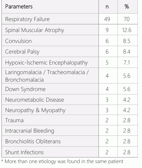
Out of 70 patients enrolled in the study, 36 (51.4%) were male, and 34 (48.6%) were female (p>0.05). The median age of the patients was 37 (min 3-max 188) months. The age range of the study population was 0-5 years for 49 patients (70%); 5-10 years for 18 (25.7%); and 10-18 years for 3 (4.3%). The most common indication for PICU admission was respiratory failure(70%). The indications for PICU admission were shown in Table 1, with some patients having more than one indication.
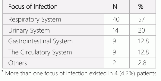
The most common foci of infection-causing sepsis in decreasing frequency were respiratory infections (n: 40, 57%), urinary system infections (n:14, 20%), circulatory system infections (n:9, 12.8%), and gastrointestinal system infections (n:9, 12.4%) (Table 2).
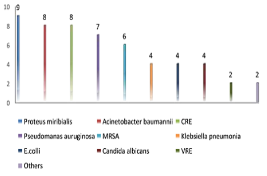
A microorganism proliferated in the culture samples of 52 (74.2%) of 70 patients while no proliferation occurred in the samples of 18 (25.8%) patients. Culture positivity was detected in the tracheal aspirate fluid in 44.2% of the patients, urine samples in 27%, blood samples in 17.3%, stool samples in 17.3%, and other samples in 2%. An analysis of microorganism proliferation in culture samples showed that Proteus mirabilis proliferated in 9 (17.3%) patients, Acinetobacter baumannii in 8 (15.3%), Carbapenem-resistant enterococci (CRE) in 8 (15.3%), Pseudomonas aeruginosa in 7 (13.4%), Methicillin-resistant staphylococcus aureus (MRSA) in 6 (11.5%), Klebsiella pneumoniae in 4 (7.7%), E.coli in 4 (7.7.%), Candida albicans in 4 (7.7%), Vancomycin-resistant enterococci (VRE) in 2 (3.8%), and other microorganisms in 2 (3.8%) (Figure 1).
Two patients were found to have multiple simultaneous proliferation in different culture samples. An analysis of the acute phase reactants in septic patients in the first 24 hours after the diagnosis of sepsis showed that 20 (28.5%) patients had CRP levels within the normal reference range and 50 (71.5%) patients had CRP levels above the normal reference range. Procalcitonin level was high in all patients (100%).
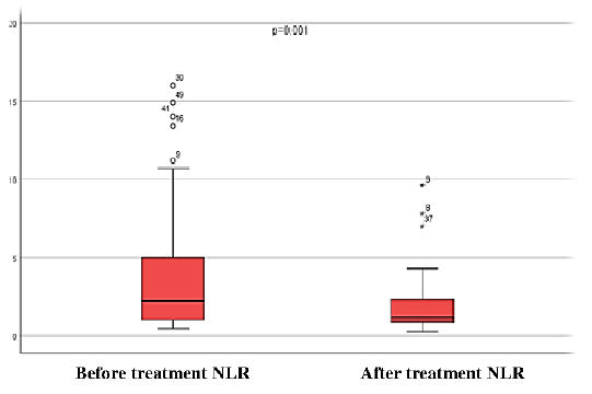
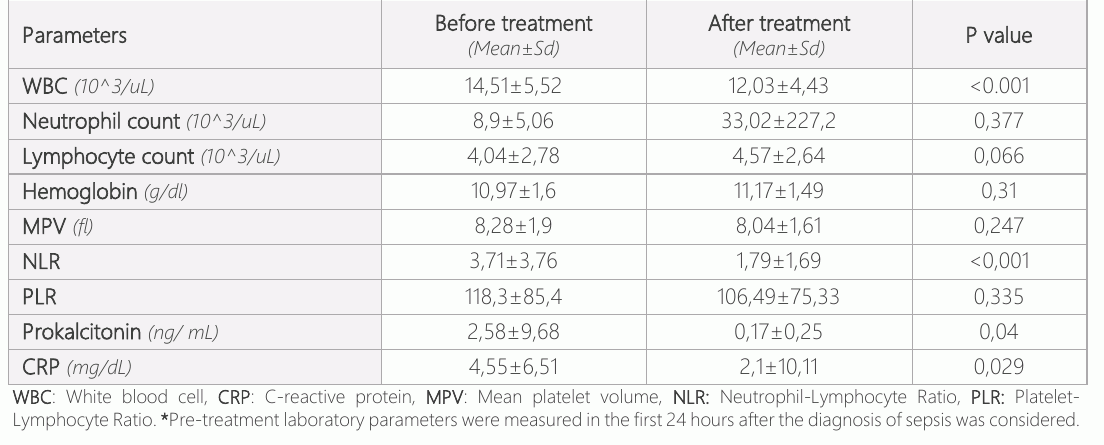
A comparison of the laboratory parameters measured in the first 24 hours after the diagnosis of sepsis and at the end of the treatment showed significant differences between WBC, NLR, CRP, and Procalcitonin levels (p<0.05, Table 3, Figure 2). There was no significant difference regarding the other parameters. A correlation analysis between NLR and the other parameters showed a positive correlation between NLR, CRP and PCT (p=0.036, p=0.012/ r=0.251, r:0.299, respectively).
Discussion
Pediatric intensive care units are services that serve diverse groups of patients requiring a multidisciplinary approach. Mortality risk can be high in patients managed in intensive care units. Sepsis is one of the leading risk factors for increased mortality (14, 15).
Pediatric intensive care units admit patients aged 1 month to 18 years for treatment. According to our literature review, Xiao et al.(16) reported that 80.8% of patients admitted to PICU for treatment of sepsis were children aged 1 month to 5 years; Navin et al. reported that the same age group comprised 68% of patients. Our study findings showed similarity with literature data, with 70% of our patients being 1 month to 5 years old. Sepsis can be observed in any age group, and care should be exercised to detect sepsis during intensive care unit follow-up of children under the age of 5 and to remember that early diagnosis may be life-saving in those patients. A review of the previous studies investigating the cause of intensive care unit admission showed that respiratory infections were the most common cause, affecting 76% of patients in the study of Xiao et al. (16), 34.6% of patients in the study of Workman et al. (17), 21% of patients in the study of Rey et al. (10), and 36.8% of patients in the study of Aygün (13). Similarly, our study found that respiratory infections were the most common etiological cause, being responsible for 70% of PICU admissions. A separate analysis of the former studies regarding the focus of the infection-causing sepsis showed that respiratory and urinary infections were the most common foci (10, 16, 17). In our study, the most common foci of infection-causing sepsis, in decreasing order of frequency, were respiratory and urinary infections. It is of vital importance to determine the foci causing sepsis and to start empirical antibiotic therapy as fast as possible at an early stage. It should be kept in mind that the respiratory and urinary systems are typically responsible for sepsis, and empirical therapy against these foci should be initiated until culture results become available.
Variable results have been reported in the literature regarding the detection of micro organisms in culture samples of children with sepsis. Katu et al. (18) found culture positivity in 66.7% of patients, Demirdağ et al. (19) in 77%, and Workman et al. (17) in 67%. We detected culture positivity in 74.2% of our patients. The most commonly isolated bacteria in our patients, in decreasing order of frequency, were Proteus mirabilis, Acinetobacter baumanii, Pseudomonas aeruginosa, Carbapenem-resistant enterococci and Methicillin-resistant staphylococcus aureus (MRSA). Using blood samples to diagnose sepsis is of great importance for preventing mortality in critically ill patients with a life-threatening infection. However, it is not always a simple task to obtain an adequate blood volume for cultures in children and infants. Furthermore, possible contamination of blood culture samples especially by skin saprophytes despite efforts to reduce contamination continues to be an important clinical problem. Despite several challenges posed by blood cultures, they continue to guide clinicians for the treatment of sepsis for the sake of efficacy and, if necessary, modification of the treatment.
Sepsis is a pathophysiological process rather than a specific syndrome and is too complex to be defined by a single parameter. A perfect marker is yet to be found to diagnose sepsis. Since the clinical signs and symptoms of sepsis are not specific and may frequently vary, there is a potential for some biomarkers to be used for determining the severity of sepsis, clinical monitoring, and assessing treatment response. There are a plethora of studies in this field (8–12, 19).
Among such biomarkers, PCT and CRP are the most widely used ones. Procalcitonin is a protein containing 116 amino acids, which is a precursor protein of the calcitonin hormone released by the medullary neuroendocrine C cells of the thyroid gland, typically in response to a high calcium level in blood. Procalcitonin level is thought to increase 2 hours after the onset of an infection, to become detectable by 4 hours, to peak at 6 hours, and to maintain its level for 8-24 hours. Thanks to such favorable kinetics, PCT is an ideal candidate for use as a biomarker (20). C-reactive protein (CRP) is a pentameric acute-phase protein of hepatic origin circulating in the blood. Serum CRP level rises dramatically in the case of inflammation, tissue injury, and cytokine-mediated reaction against an infection. Quantification of acute-phase reaction may provide clinical information about the presence of tissue injury or inflammation as well as the treatment response (21). Serum CRP level is elevated by all inflammatory processes including infections and sepsis. Despite being a classical and sensitive biomarker of inflammation, CRP cannot distinguish bacterial and other types of inflammatory infections (20). Using PCT for the diagnosis of sepsis may offer an advantage since it may have better specificity for bacterial infections, unlike many other biomarkers that may be elevated in conditions other than bacterial infection (22).
Bustos et al. (8) reported that, unlike CRP and lactate, PCT was a good predictor of mortality and septic shock, being capable of categorizing patients by the severity of sepsis at the time of pediatric intensive care unit admission. In a study in adults, Luzzani et al. (22) found that PCT was a better marker of sepsis and showed a better correlation to the severity of infection and organ dysfunction than CRP. Rey et al. (10) found that PCT was a better diagnostic marker than CRP among critically ill children with sepsis. Nargis et al. (23). studied the specificities of PCT and CRP in patients treated for sepsis and reported a specificity of 72.2% for PCT and 33.3% for CRP.
Our comparison of the laboratory parameters measured in the first 24 hours after the diagnosis of sepsis and at the end of the treatment detected significant differences in WBC, CRP, PCT, and NLR levels. While 20 (28.5%) of our patients had a normal CRP level and 50 (71.5%) of them had an elevated CRP level in the first 24 hours of sepsis, all of our patients had an elevated PCT level. These results support the literature data indicating that PCT rapidly rises and reaches a detectable level in blood at an earlier stage than CRP; they also suggest that PCT may be a better parameter than CRP for assessing bacterial sepsis. In addition, we believe that CRP and PCT should be effectively used to monitor patients with sepsis and to assess their treatment response.
Neutrophils are the first-line cellular defense cells of innate immunity against infectious agents. Lymphocytes play an important role in adaptive immunity. The immune response against various triggers causes an increase in neutrophil count and a decrease in lymphocyte count. Neutrophil lymphocyte ratio (NLR) is a readily available, rapidly obtained, and inexpensive parameter. Several researchers have studied NLR with CRP and PCT as an important marker for an early diagnosis of bacteremia and assessment of the outcomes of sepsis (24, 25, 26). Several studies have shown that, in comparison to standard diagnostic biomarkers, NLR made meaningful contribution to the early prediction of sepsis (25-27). Furthermore, several studies in infants and newborns have indicated that NLR was an accurate predictor of sepsis along with CRP (28,29). We detected a significant difference between the NLR levels at the onset of sepsis and at the end of the treatment; we also demonstrated that NLR had a positive correlation with CRP and PCT. We believe that the neutrophil-lymphocyte ratio can be used for disease monitoring similarly to CRP and PCT.
Conclusion
Respiratory and urinary infections are the most common foci of infection among patients treated for sepsis at pediatric intensive care units. The proliferation of microorganisms in culture samples is time-consuming and sometimes even not possible; we, therefore, believe that empirical antibiotic treatment should be commenced as soon as possible after determining the most probable focus and microorganism.
We believe that PCT, CRP, and NLR, the effectiveness of which has been recently shown, may be used as biomarkers to support the diagnosis of sepsis and to assess treatment efficacy. In the light of our results, we may suggest that PCT is a more effective marker than CRP in the first 24 hours among patients suspected to have sepsis. In conclusion, markers such as PCT, CRP, and NLR should not be used alone as the definitive tests but should be used in conjunction with physical examination, clinical features, and microbiological culture results to diagnose sepsis and to assess treatment response.
Declarations
- Author contributions (CRediT)
- All authors contributed equally to all 14 CRediT roles (Conceptualization, Data curation, Formal analysis, Funding acquisition, Investigation, Methodology, Project administration, Resources, Software, Supervision, Validation, Visualization, Writing – original draft, Writing – review & editing).
- Funding
-
The authors report no involvement in the research by the sponsor that could have influenced the outcome of this work.
- Conflicts of interest
- The authors declared no conflict of interest for the present article.
- Ethics approval
- The study was conducted in compliance with the criteria of the Helsinki Declaration. All the participants (family members) accepted the informed/written consent. It was approved by the Dicle University Faculty of Medicine Institutional Ethics Committee (Date: 23.05.2019 No: 200).
- Data availability
- A data availability statement was not reported in the published version of this article.
References (25)
21 of 25 references are linked to their DOI.
- Bone RC, Balk RA, Cerra FB, Dellinger RP, Fein AM, Knaus WA, Schein RM, Sibbald WJ. Definitions for sepsis and organ failure and guidelines for the use of innovative therapies in sepsis. The ACCP/SCCM Consensus Conference Committee. American College of Chest Physicians/Society of Critical Care Medicine. Chest. 1992 Jun;101(6):1644-55 DOI Crossref PubMed Google Scholar
- Levy MM, Fink MP, Marshall JC, Abraham E, Angus D, Cook D, Cohen J, Opal SM, Vincent JL, Ramsay G; International Sepsis Definitions Conference. 2001 SCCM/ESICM/ACCP/ATS/SIS International Sepsis Definitions Conference. Intensive Care Med. 2003 Apr;29(4):530-8 DOI Crossref PubMed Google Scholar
- Gotts JE, Matthay MA. Sepsis: pathophysiology and clinical management. BMJ. 2016 May 23;353:i1585. DOI Crossref PubMed Google Scholar
- Uhle F, Lichtenstern C, Brenner T, Weigand MA. Sepsis und Multiorganversagen - Pathophysiologie der Sepsis. Anasthesiol Intensivmed Notfallmed Schmerzther. 2015 Feb;50(2):114-22 DOI Crossref PubMed Google Scholar
- Font MD, Thyagarajan B, Khanna AK. Sepsis and Septic Shock - Basics of diagnosis, pathophysiology and clinical decision making. Med Clin North Am. 2020 Jul;104(4):573-585 DOI Crossref PubMed Google Scholar
- Hotchkiss RS, Karl IE. The pathophysiology and treatment of sepsis. N Engl J Med. 2003 Jan 9;348(2):138-50 DOI Crossref PubMed Google Scholar
- Goldstein B, Giroir B, Randolph A. International Consensus Conference on Pediatric Sepsis. International pediatric sepsis consensus: definitions for sepsis and organ dysfunction in pediatrics. Pediatr Crit Care Med. 2005 Jan;6(1):2-8 DOI Crossref PubMed Google Scholar
- Bustos B R, Padilla P O. Valor predictivo de la procalcitonina en niños con sospecha de sepsis [Predictive value of procalcitonin in children with suspected sepsis]. Rev Chil Pediatr. 2015 Sep-Oct;86(5):331-6 DOI Crossref PubMed Google Scholar
- Luzzani A, Polati E, Dorizzi R, Rungatscher A, Pavan R, Merlini A. Comparison of procalcitonin and C-reactive protein as markers of sepsis. Crit Care Med. 2003 Jun;31(6):1737-41 DOI Crossref PubMed Google Scholar
- Rey C, Los Arcos M, Concha A, Medina A, Prieto S, Martinez P, Prieto B. Procalcitonin and C-reactive protein as markers of systemic inflammatory response syndrome severity in critically ill children. Intensive Care Med. 2007 Mar;33(3):477-84 DOI Crossref PubMed Google Scholar
- Standage SW, Wong HR. Biomarkers for pediatric sepsis and septic shock. Expert Rev Anti Infect Ther. 2011 Jan;9(1):71-9 DOI Crossref PubMed Google Scholar
- Lanziotti VS, Póvoa P, Prata -Barbosa A, et al. Patterns of C-reactive protein ratio response to antibiotics in pediatric sepsis: A prospective cohort study. J Crit Care. 2018;44:217-222 DOI Crossref PubMed Google Scholar
- Aygun F. Procalcitonin Value Is an Early Prognostic Factor Related to Mortality in Admission to Pediatric Intensive Care Unit. Crit Care Res Pract. 2018;2018:9238947 DOI Crossref PubMed Google Scholar
- Lindell RB, Nishisaki A, Weiss SL, Traynor DM, Fitzgerald JC. Risk of Mortality in Immunocompromised Children With Severe Sepsis and Septic Shock. Crit Care Med. 2020;48(7):1026 -1033 DOI Crossref PubMed Google Scholar
- Boeddha NP, Schlapbach LJ, Driessen GJ, et al. Mortality and morbidity in community-acquired sepsis in European pediatric intensive care units: a prospective cohort study from the European Childhood Life-threatening Infectious Disease Study (EUCLIDS). Crit Care. 2018;22(1):143 DOI Crossref PubMed Google Scholar
- Xiao C, Wang S, Fang F, et al. Epidemiology of Pediatric Severe Sepsis in Main PICU Centers in Southwest China. Pediatr Crit Care Med. 2019;20(12):1118-1125 DOI Crossref PubMed Google Scholar
- Workman JK, Ames SG, Reeder RW, et al. Treatment of Pediatric Septic Shock With the Surviving Sepsis Campaign Guidelines and PICU Patient Outcomes. Pediatr Crit Care Med. 2016;17(10):e451-e458 DOI Crossref PubMed Google Scholar
- Katu S, Suwarto S, Pohan H, Abdullah M. Factors that influence the success of empirical antibiotic therapy in patients with severe sepsis and septic shock in the inpatient ward of the Hospital Cipto Mangunkusumo. Indones Int Med J. 2015;2:96- PubMed Google Scholar
- Demirdag K, Özden M, Gödekmerdan A, Cihangiroğlu M, Kalkan A. Sepsis olgularında prokalsitonin, TNF-α ve C-reaktif protein düzeylerinin değerlendirilmesi. Klimik dergisi. 2003;16(1): 21-4 PubMed Google Scholar
- Cleland DA, Eranki AP. Procalcitonin. In: StatPearls. Treasure Island (FL): StatPearls Publishing; September 3, 2020 PubMed Google Scholar
- Faraj M, Salem N. C-reactive protein. Intech Journal, 2012, 89-100 PubMed Google Scholar
- Luzzani A, Polati E, Dorizzi R, Rungatscher A, Pavan R, & Merlini A. Comparison of procalcitonin and C-reactive protein as markers of sepsis. Critical care medicine. 2003;31(6),1737–41 DOI Crossref PubMed Google Scholar
- Nargis W, Ibrahim, M, Ahamed BU. Procalcitonin versus C - reactive protein: Usefulness as biomarker of sepsis in ICU patient. International journal of critical illness and injury science. 2014; 4(3), 195–199 DOI Crossref PubMed Google Scholar
- Djordjevic D, Rondovic G, Surbatovic M, et al. Neutrophil-to-Lymphocyte Ratio, Monocyte-to-Lymphocyte Ratio, Platelet-to-Lymphocyte Ratio, and Mean Platelet Volume-to-Platelet Count Ratio as Biomarkers in Critically Ill and Injured Patients: Which Ratio to Choose to Predict Outcome and Nature of Bacteremia?. Mediators Inflamm. 2018;2018:3758068 DOI Crossref PubMed Google Scholar
- de Jager CP, van Wijk PT, Mathoera RB, de Jongh - Leuvenink J, van der Poll T, Wever PC. Lymphocytopenia and neutrophil-lymphocyte count ratio predict bacteremia better than conventional infection markers in an emergency care unit. Crit Care. 2010;14(5):R192 26Ljungström L, Pernestig AK, Jacobsson G, Andersson R, Usener B, Tilevik D. Diagnostic accuracy of procalcitonin, neutrophil-lymphocyte count ratio, C-reactive protein, and lactate in patients with suspected bacterial sepsis. PLoS One. 2017;12(7):e0181704 85 77 Sepsis Diagnosis in Pediatric Intensive Care Unit Cengiz et al. 27. Loonen AJ, de Jager CP, Tosserams J, et al. Biomarkers and molecular analysis to improve bloodstream infection diagnostics in an emergency care unit. PLoS One. 2014;9(1): e87315 28. Alkan Ozdemir S, Arun Ozer E, Ilhan O, Sutcuoglu S. Can neutrophil to lymphocyte ratio predict late-onset sepsis in preterm infants?. J Clin Lab Anal. 2018;32(4):e22338 29. Can E, Hamilcikan Ş, Can C. The Value of Neutrophil to DOI Crossref PubMed Google Scholar
Read the full text
Open the PDF here
Loading the PDF…
If the PDF does not show here, use the download button above.
How to cite this article
Cengiz H, Yilmaz K, Pirinccioglu AG, Kan A. Evaluation of Biomarkers in Patients with Sepsis Diagnosis in Pediatric Intensive Care Unit. Ulutas Med J. 2021;7(2):77-85. doi:10.5455/umj.20210504075319
Cengiz, H., Yilmaz, K., Pirinccioglu, A. G., & Kan, A. (2021). Evaluation of Biomarkers in Patients with Sepsis Diagnosis in Pediatric Intensive Care Unit. The Ulutas Medical Journal, 7(2), 77–85. https://doi.org/10.5455/umj.20210504075319
Cengiz, H. et al. (2021) 'Evaluation of Biomarkers in Patients with Sepsis Diagnosis in Pediatric Intensive Care Unit', The Ulutas Medical Journal, 7(2), pp. 77–85. doi: 10.5455/umj.20210504075319.