The Ulutas Medical Journal · ISSN 2149-0430 (print), 2149-388X (online) · https://www.ulutasmedicaljournal.com/?mno=80412
Original Article · Open access
Dosimetric Comparison of Scalp Protection in Whole Brain Radiotherapy Due to Brain Metastasis
Also available in: العربية · Türkçe · 中文
- Department of Radiation Oncology, Inonu University, Faculty of Medicine, Turkey ROR
- Department of Radiation Oncology, Ankara Yıldırım Beyazıt University, Faculty of Medicine, Turkey ROR
Abstract
- Introduction
- The objective is to demonstrate dosimetrically preserving scalp in whole-brain irradiation in the treatment of brain metastases and making dosimetry at 20 points determined on the rando phantom while comparing between Linac and Tomotherapy devices.
- Materials and Methods
- 10 randomized patients, who had previously undergone radiotherapy for whole-brain metastasis cancer, were determined prospectively. In Helical Tomotherapy (HT) and Linac devices, Intensity-modulated radiation therapy (IMRT) and volumetric modulated arc therapy (VMAT) were planned to be 30 Gray(Gy) 10 Fractions for the whole brain region.
- Results
- The average target volume (PTV), Homogeneity Index (HI), conformity index (CI), and integral dose (ID) for IMRT and VMAT were 0.075, 0.77, 0.94, 0.97 and 29.67, 23.57, respectively. Mean median doses for scalp IMRT and VMAT were 19.71Gy and 18.01 Gy (p<0.005). Lenses and body doses were significant for IMRT and VMAT. The mean median plan doses for Rando phantom scalp were 19.43Gy and 19.55Gy in IMRT and VMAT, respectively. The mean median film doses for Rando phantom scalp were 17.03Gy and 20.64Gy in IMRT and VMAT, respectively (p<0.005).
- Conclusion
- By using both VMAT and Helical Tomotherapy techniques, it is possible to dry the lens and scalp without low PTV doses in whole-brain irradiation.
Keywords
Full text
Introduction
Brain metastasis is known as the spread of a tumor occurring in tissues and organs other than the brain through the blood circulation or lymphatic system and forming a tumor on the brain. These types of tumors are the most common cases in the brain. With the treatment of visible and hidden lesions, reduced symptoms, and rapid application, whole-brain radiotherapy is a common treatment feature (1). Although the most common clinical findings in brain metastase include headache, vomiting, convulsion, and different neurological findings, diagnosis can be made without any clinical symptoms, and approximately 20% of lung, breast and colorectal cancers are cancer types that cause brain metastasis (2–4).
Upon diagnosis, radiotherapy, chemotherapy, surgery, and supportive treatments are often applied to brain metastases. Radiotherapy is an important and effective treatment method in the treatment of brain metastases(3, 5). Since brain metastases spread through the blood and lymph system, one of the precautions to be taken against this situation is to consider the whole brain as a target and apply whole-brain radiotherapy.
There are many significant parameters in determining the treatment option for brain metastases. The most significant of these are listed the age, general condition, quality of life and performance of the patients, and the toxicities that may occur (4, 8). Temporary or permanent hair loss is observed in whole-brain radiotherapy (WBRT). Together with hair loss (alopecia) is an important psychological problem in terms of the quality of life of patients, it has a deeply negative impact on their social life (6, 7).
When the dose and fraction values prescribed in whole-brain radiotherapy treatments were compared; the best dosage regimen in terms of mean survival, local control, and neurological function is the administration of a 3 Gy dose in 10 fractions with a total of 30 Gy. This dose fractionation regimen is used as a standard in WBRT (9, 10).
In this study, we aimed to minimize such negativities, 10 patients randomly selected with Intensity-modulated radiation therapy (IMRT) and volumetric modulated arc therapy (VMAT) techniques in scalp-protected Helical Tomo therapy and Linac devices in whole-brain radiotherapy, and dosimetric comparison of scalp doses in different techniques, and the same techniques on the rando phantom by planning the parietal and occipital bones at 20 points.
Material and Method
Patient Selection
Ten randomized patients, who had undergone radiotherapy for whole-brain metastasis cancer previously were determined prospectively. Our study is approved by the ethics committee following the Helsinki Declaration.
Simulation and Contouring of Targets and OARs Computed tomography (CT) images of 3 mm thickness were obtained by thermoplastic head mask to provide head & neck immobilization in the supine position of all patients.
From the computerized tomography (CT) images recorded in the planning computer of 10 randomly selected patients, the whole brain was contoured prospectively and the planning target volume (PTV) was determined by giving the whole brain a 3-millimeter margin. The organs at risk (OARS) (lens, eye, optic nerve) were contoured (11).
Between skull and skin tissue was determined as scalp by giving a margin of 3 millimeters to the planning target volume and drawing 2 millimeters from the external (skin tissue) contour. Based on these drawn contours, IMRT and VMAT planning was made in Helical Tomotherapy (HT) and Linac devices with 30 Gray 10 Fractions to the whole brain region. Similarly in the Rando phantom, scalp contouring with PTV was made with a 30Gy 10 fraction planning, and 20 points were determined on the rando phantom, 10 points were determined on the frontal-upper parietal and occipital bones, 5 points were determined in the right parietal and 5 were determined with 2 cm intervals in the left parietal, and a CT image was taken. In addition, planning was made in both devices, and irradiation was performed in tomotherapy and linac devices by placing gafchromic EBT3-1417 film on these points. Homogeneity and conformity indices and integral dose calculations were made for plan evaluation.
Treatment Planning
The planning target volume (PTV) was obtained by giving a 3-millimeter margin to the whole brain. By giving a margin of 3 millimeters to the planning target volume and drawing 2 millimeters from the external contour, between the skull and skin tissue was determined as scalp. Prescription dose 30 Gy was determined as 10 fractions. The dose was prescribed to cover 95% of PTV.
A 0.03cc volume in any PTV was planned not to receive>110% of the prescribed dose. Compliance with critical organ doses determined by RTOG protocol was demanded. In the Helical Tomotherapy device, planning was made with Intensity-modulated radiation therapy (IMRT) to be 30Gray 10 Fractions to the whole brain region. During the treatment, the gantry rotates 360 degrees continuously and at a constant speed and applies RT. During helical treatment, the MLC layout changes in each projection by dividing 360 degrees into 51 projection angles while Linac rotates continuously.
Tomotherapy planning system (Hi-Art Tomo therapy, version 5.1.2, Accuray, Madison, WI, USA) was used in planning HT. MRT and VMAT plans were contoured at the Velocity contouring station. For Helical Tomotherapy device, field size shaping field width (jaw) field width 2.5cm, opening and closing time of MLCs modulation factor (MF) 2.8, table movement speed pitch factor 0.215, and fine dose calculation grid were used.
In the Linac device, planning was made with volumetric modulated arc therapy (VMAT) to be 30Gray 10 Fractions for the whole brain region. Varian Eclipse planning system (version 13.7-Varian Medical Systems, Palo Alto, USA) was used in VMAT planning. The structures contoured in the Velocity contouring station were transferred to the Eclipse planning system in DICOM (Digital Imaging and Communication in Medicine) format.
Two full arcs were used in the VMAT plans at 181°-179° clockwise and 179° 181° counter clockwise. A total of 177 firing points were used at 2 degrees for each full arc. PO 13.6 algorithm was used for fiber positions, dose rate, gantry speed. The maximum dose rate was determined as 600 MU/min. Dose calculation matrix resolution 2.5mm and final dose calculation Anisotropic Analytical Algorithm (AAA) photon dose calculation algorithm was used for all plans. To reduce the tongue and groove effect of the fibers, 30˚ and 330˚ collimator angles were used in arc treatment. 120-fiber (central 20-cm of field uses, 0.5-cm-wide leaves, the outer field uses 1-cm-wide leaves) dynamic multileaf collimator (MLC) was used in VMAT plans. The maximum MLC speed was determined to be 2.5 cm/s.
According to RTOG protocol, 50% of the doses (D50)-V5(volume of received 5 grays), V10(volume of received 10 grays), V15 (volume of received 5 grays), V20(volume of received 20 grays) doses of critical organs and scalp dose were measured with maximum, minimum and average doses (24). 6MV energy was used in all VMAT and HT plans.
Homogeneity and conformity indices and integral dose calculations were made for plan evaluation.
Evaluation Tools
Plan evaluation was made by examining all slides one by one and looking at dose-volume histograms (DVHs). Homogeneity index (HI) was calculated as HI=D2-D98/Dp. D2 is the minimum dose for 2% of the target volume, D98 is the minimum dose for 98% of the target volume, and Dp is the anticipated dose. This is the commonly used formula in the literature. The ideal value of this formula, which evaluates the degree of dose distribution homogeneity of in the target volume, is equal to 0 (12).
Conformity index (CI) was calculated as RTOG CI=VRI/TV. VRI is the volume of the reference dose (cm3), TV is the target volume. CI is defined as the ratio based on the exact volume of the prescribed dose to match the target volume. Its ideal value is equal to 1 (13). Integral dose (ID) calculated= Mean dose (Gy) * volume (L); Calculated as dose-volume histogram received by normal tissue (14).
Statistics
Analyzes were performed using the Statistical Package for Social Science version 22.0, software (SPSS, Chicago, IL, USA). Data were summarized as mean±standard deviation (SD). To evaluate the normality of distribution, Unpaired T-test, data with normal distribution were analyzed with repeated measures analysis of variance, and Bonferroni post-hoc method was used. Non-normally distributed data were analyzed by the Independent Samples test and a Bonferroni adjusted pairwise comparison was used. The significance level was admitted as 0.05 in all analyzes.
Results
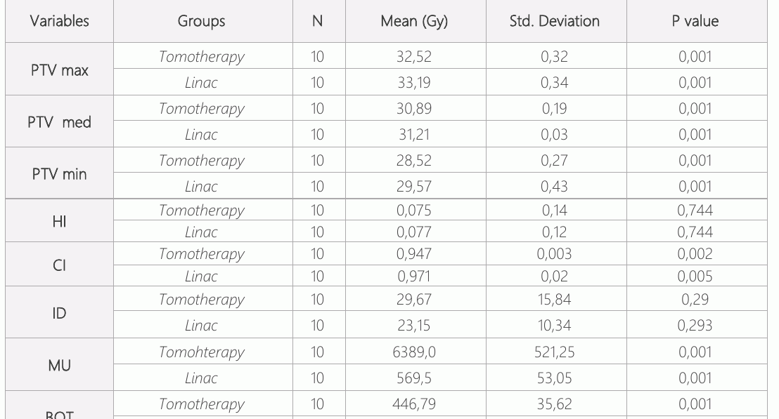
Significant differences were observed in tomotherapy and linac device in the maximum, median, and minimum values for the planned target volume (p<0.005). In the plans made with Tomotherapy IMRT, the maximum (max) dose value of PTV is 108%, the median (med) value is 102%, the minimum (min) values are 95%, while this value is 110% maximum, 103% median and 98% minimum in Linac VMAT. For the target volumes with calculated dosimetric data, for HI, CI, ID, monitor unit (MU), and beam-on time (BOT) values; Although there is no significant difference in HI, ID dose calculations (p>0.05), CI, MU, and BOT in Linac VMAT plans were found to be higher than HT IMRT (p<0.005). Comparison values are shown in table 1.
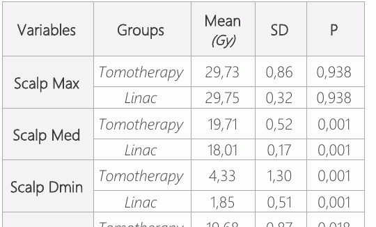
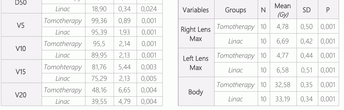
Although the maximum values in scalp doses according to both plan criteria are not close to each other (p>0.05), the median and minimum values and the VMAT plan technique are significantly superior with the V5(volume of received 5 grays), V10, V15 and V20 and 50% of the doses (D50) (p=0.005). The comparison values are shown in table 2.
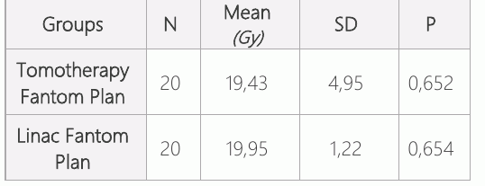
Although there is no significant change in IMRT and VMAT plans in the dose values taken at 20 points in the plan made on Rando phantom, the values are close to each other and the dose values are shown in Table 3.
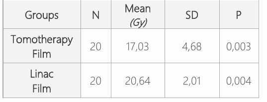
There was a significant difference (p<0.005) in the irradiation made with gapchromic film to 20 points in the plan made on the Rando phantom, and the HT IMRT plan was superior to the VMAT plan. Comparison values are shown in table 4. While lens doses and body doses are lower in HT IMRT, these values are higher in the Linac VMAT plans (p<0.05). Comparison values are shown in table 5.
Discussion
In this study, we searched for an answer to the question of whether scalp protection is possible in patients undergoing WBRT with two different modern techniques and is there any difference between these techniques. In previous studies, it has been shown that scalp doses with the IMRT technique are considerably lower than conventional WBRT treatments (15, 16). In the study conducted by Kao et al., conventional WBRT and IMRT-WBRT were compared. While the mean scalp dose was 26.2 Gy in conventional treatment, it was 16.4 Gy with IMRT (26.2Gy vs. 16.4Gy, p<0.001). PTV 30 was lower in the IMRT arm (38.4 Gy vs. 32 Gy, p<001). Hair protection was provided 50% in 4 of 15 patients, and 25-50% in 6 patients (17). In the study of Witek et al, it was shown that IMRT and WBRT can protect not only the hippocampus but also the scalp. Mean scalp V10 and V20 were found to be 46% and 35%, respectively (18). In our study, scalp Dmean is found 19.7 Gy with HT and 18.01 for VMAT; V10 is 95.5% for HT and 89.95% for VMAT; the V20 value is 48.16% for HT and 39.55% for VMAT. Volumetric modulated arc therapy (VMAT) is a new radiation technique that can provide highly compatible dose distributions with better target volume range and preservation of normal tissues compared to conventional radiotherapy techniques (19). In a prospective study conducted in Australia, 9 patients who underwent VMAT-WBRT were given scalp protection with hippocampus protection. PTV was prescripted at 30Gy. The scalp was divided into 2 as superior and inferior 4 cm above the most superior pinna. Superior scalp Dmax was found as 20.5Gy, Dmean: 10.4Gy, and for inferior scalp Dmax: 23.1 Gy and Dmean: 12.2 Gy (20).
Helical Tomotherapy is a novel method and arc-based application of IMRT. During the treatment, the gantry rotates 360° at a constant and fixed speed to apply RT. In a study conducted by Hu et al., scalp doses could be reduced up to 52% of the PTV prescribe dose in the application of radiotherapy to the brain region (21). There are many studies comparing Tomotherapy and VMAT techniques for various tumor types. Similar studies have been conducted in whole-brain radiotherapy, as well. In the study of Doğan et al. in which they applied whole-brain radiotherapy for prophylactic cranial radiotherapy (PCI) with hippocampus protection; the minimum, maximum and mean values of PTV brain doses were higher in the VMAT arm(p=0,0001). When HI and CI values were compared, PTV was significantly superior in VMAT CI (p=0.033), however, there was no significant difference in HI values. When the lens doses were analyzed, it was observed that the mean and maximum dose values of the right and left lenses were much lower in helical tomotherapy (22).
In the study of Rong et al., HI was also found to be 0.15 and better than other techniques, IMRT and VMAT (23). In our study, on the other hand, PTV max, PTV median, and PTV min were found lower in HT (p<0.001). While there was no difference in terms of HI; CI was superior in VMAT. MU and Beam on time values were found to be much lower with the VMAT technique. In our study, the measurements made on TPS were measured and verified with Gafcromic films at 20 different points on the phantom. The measurement results were found to be similar.
The limitations of the present study are that Gafrocrmic film measurements are made on the phantom and not on the real patient.
Conclusion
By using both VMAT and Helical Tomotherapy techniques, it is possible to dry the lens and scalp without low PTV doses in whole-brain irradiation.
Declarations
- Author contributions (CRediT)
- All authors contributed equally to all 14 CRediT roles (Conceptualization, Data curation, Formal analysis, Funding acquisition, Investigation, Methodology, Project administration, Resources, Software, Supervision, Validation, Visualization, Writing – original draft, Writing – review & editing).
- Funding
-
Funding information was not reported in the published version of this article.
- Conflicts of interest
- A conflict of interest statement was not reported in the published version of this article.
- Ethics approval
- The present study was approved by the Inonu University Clinical Research Ethics Committee (Approval number: 2019/218).
- Data availability
- A data availability statement was not reported in the published version of this article.
References (24)
21 of 24 references are linked to their DOI.
- Achrol AS, Rennert RC, Anders C, Soffietti R, Ahluwalia MS, Nayak L, Peters S, Arvold ND, Harsh GR, Steeg PS, Chang SD. Brain metastases. Nat Rev Dis Primers. 2019 Jan 17;5(1):5 PubMed Google Scholar
- Zhi-zhen W, Zhi-yong Y, Wen- cheng Z, et al. Brain metastasis treated with Cyberknife, Chin Med J 2009; 122 (16): 1847-1850 PubMed Google Scholar
- Murai T, Ogino H, Manabe Y, et al. Fractionated stereotactic radiotherapy using Cyberknife for the treatment of large brain metastases: a dose-escalation study, ClinicalOncology 26 (2014) 151-158 DOI Crossref PubMed Google Scholar
- Lippitz B, Lindquist C, Paddick I, et al. Stereotactic radiosurgery in the treatment of brain metastases: The current evidence, Cancer Treatment Reviews 40 (2014) 48-59 DOI Crossref PubMed Google Scholar
- Sio TT, Jang S, Lee SW, et al. Comparing Gamma KnifeandCyberknife in patients with brain metastases, Journal of Applied Clinical Medical Physics, Vol. 15, No. 1, 2014 DOI Crossref PubMed Google Scholar
- S. Scoccianti, G. Simontacchi, C. Talamonti, L.Marrazzo, G.A. Carta, L. Visani, M. Baki, L. Poggesi, D. Greto, B. Detti, P. Bonomo, M. Loi, S. Pallotta, L.Livi. Scalp-sparing radiotherapy to minimize alopecia in patients with primary brain cancer DOI Crossref PubMed Google Scholar
- Ahmad I, Sardana K, Chufal KS, Bhatt CP (2018) Radiation - Induced Alopecia: An Under-Appreciated Side Effect of Whole Brain Radiotherapy and Strategies to Ameliorate it J Nucl Med Radiat Ther S9: 002 DOI Crossref PubMed Google Scholar
- Mehta MP, Tsao MN, Whelan TJ, et al. The American society for therapeutic radiology and oncology (ASTRO) evidence - based review of the role of radiosurgery for brain metastases, Int. J. Radiation Oncology Biol. Phys., Vol. 63, No. 1, pp. 37-46, 2005 DOI Crossref PubMed Google Scholar
- Gaspar LE, Mehta MP, Patchell RA, et al. The role of whole-brain radiation therapy in the management of newly diagnosed brain metastases: a systematic review and evidence-based clinical practice guideline, J Neurooncol (2010) 96: 17-32 DOI Crossref PubMed Google Scholar
- Hunter GK, Suh JH, Reuther AM, et al. Treatment of five or more brain metastases with radiosurgery, Int. J. RadiationOncologyBiol. Phys.,Vol. 83, No. 5, pp. 1394 -1398, DOI Crossref PubMed Google Scholar
- Webpages:https://www.qarc.org/cog/ACNS1721_Contouring Guidelines_20190306.pdf PubMed Google Scholar
- Kataria T, Sharma K, Subramani V, Karrthick KP, Bisht SS. Homogeneity Index: An objective tool for assessment of conformal radiation treatments. Journal of medical physics / Association of MedicalPhysicists of India. 2012;37(4):207 -213. DOI Crossref PubMed Google Scholar
- Feuvret Liocet al., Conformity Index: a review, Int J RadiatOncolBiolPhys, 64 (2), 2006, 333-342 DOI Crossref PubMed Google Scholar
- Aoyama H, Westerly DC, Mackie TR, Olivera GH, Bentzen SM, Patel RR,. Integral radiation dose to normal structures with conformal external beam radiation. Int J Radiat Oncol Biol Phys. 2006 Mar 1;64(3):962-7 DOI Crossref PubMed Google Scholar
- Roberge, D., Parker, W., Niazi, T. M., Olivares, M. Treating the contents and not the container: dosimetric study of hair-sparing whole brain intensity modulated radiation therapy. Technol Cancer Res Treat 4, 567-570 (2005) DOI Crossref PubMed Google Scholar
- Lawenda, B. D., Gagne, H. M., Gierga, D. P., Niemierko, A., Wong, W. M., Tarbell, N. J., Chen, G. T., Hochberg, F. H., Loeffler, J. S. Permanent alopecia after cranial irradiation: dose-response relationship. Int J Radiat Oncol Biol Phys 60, 879-887 (2004) DOI Crossref PubMed Google Scholar
- Kao J, Darakchiev B, Conboy L, et al. Tumor Directed, Scalp Sparing Intensity Modulated Whole Brain Radiotherapy for Brain Metastases. Technology in Cancer Research & Treatment. October 2015:547-555 DOI Crossref PubMed Google Scholar
- Witek M, Vahknenko Y, Siglin J, Harrison A, Xiao Y, Lui H, Andrews D, Shi W. Dose Reduction to the Scalp with Hippocampal Sparing Is Achievable with Intensity Modulated Radiotherapy. Int J Med Phys Clin Eng Radiat Oncol. 2014 Aug;3(3):176-182 DOI Crossref PubMed Google Scholar
- Teoh M, Clark C.H, Wood K, Whitaker S, Nisbet A. Volumetric modulated arc therapy: a review of current literature and clinical use in practice. The British Journal of Radiology, 84 (2011), 967–996 DOI Crossref PubMed Google Scholar
- Williams N, Boyle F, Hong A, et al. Whole brain radiotherapy with partial hair-sparing technique - a feasibility study. Int J Radiol Radiat Ther. 2020;7(4):104-110 DOI Crossref PubMed Google Scholar
- Hu, YA, et al. How Well Do We Spare Scalp with Image-Guided Rotational IMRT. International Journal of Radiation Oncology, Biology, Physics, 2011; Volume 81, Issue 2, S840 DOI Crossref PubMed Google Scholar
- Yetmen Dogan O, Cini N, Nasır Can SS. Dosimetric comparison of volumetric modulated arc therapy (VMAT) and Helical tomotherapy for hippocampalavoidance prophylactic whole brain radiotherapy (HA-PWBRT) in small cell lung cancer: Treatment plans based study. Med J Bakirkoy 2020;16(3):263-71 DOI Crossref PubMed Google Scholar
- Rong Y, Evans J, Xu-Welliver M, et al. Dosimetric evaluation of intensity-modulated radiotherapy, volumetric modulated arc therapy, and helical tomotherapy for hippocampal - avoidance whole brain radiotherapy. PLoS One. 2015;10(4):e0126222 Scalp Protection in Whole Brain Radiotherapy Demirtaş et al. DOI Crossref PubMed Google Scholar
- Krayenbuehl J, Di Martino M, Guckenberger M, Andratschke N. Improved plan quality with automated radiotherapy planning for whole brain with hippocampus sparing: a comparison to the RTOG 0933 trial. Radiat Oncol. 2017 Oct 2;12(1):161 DOI Crossref PubMed Google Scholar
Respond to this article
Comments on a published article are welcome as a Letter to the Editor.
Get an e-mail when new articles in your fields are published
Questions to the authors
Questions are moderated: an editor approves each question, the authors answer, and an editor approves the answer before both appear here.
No answered questions yet.
Ask the authors a question
Discussion
Comments are moderated: they appear after the writer confirms the e-mail address and an editor approves them. Each comment carries a conflict-of-interest statement.
No comments yet.
Write a comment
Read the full text
Open the PDF here
Loading the PDF…
If the PDF does not show here, use the download button above.
How to cite this article
Demirtas M, Arslan SA. Dosimetric Comparison of Scalp Protection in Whole Brain Radiotherapy Due to Brain Metastasis. Ulutas Med J. 2021;7(2):69-76. doi:10.5455/umj.20210510081414.online
Demirtas, M., & Arslan, S. A. (2021). Dosimetric Comparison of Scalp Protection in Whole Brain Radiotherapy Due to Brain Metastasis. The Ulutas Medical Journal, 7(2), 69–76. https://doi.org/10.5455/umj.20210510081414.online
Demirtas, M. and Arslan, S.A. (2021) 'Dosimetric Comparison of Scalp Protection in Whole Brain Radiotherapy Due to Brain Metastasis', The Ulutas Medical Journal, 7(2), pp. 69–76. doi: 10.5455/umj.20210510081414.online.
@article{umj80412,
  title = {Dosimetric Comparison of Scalp Protection in Whole Brain Radiotherapy Due to Brain Metastasis},
  author = {Demirtas, Mehmet and Arslan, Suheyla Aytac},
  journal = {The Ulutas Medical Journal},
  year = {2021},
  volume = {7},
  number = {2},
  pages = {69--76},
  doi = {10.5455/umj.20210510081414.online},
  url = {https://www.ulutasmedicaljournal.com/?mno=80412},
  issn = {2149-388X}
}