The Ulutas Medical Journal · ISSN 2149-0430 (print), 2149-388X (online) · https://www.ulutasmedicaljournal.com/?mno=282922
Original Research · Open access
The Regenerative Effects of Hemostatic Plant Extract in Second Degree Burn Wound
- Elazig Training and Research Hospital, Department of Plastic and Reconstructive Surgery ROR
- Elazig Training and Research Hospital, Department of Cardivascular Surgery ROR
- Adnan Menderes University Medical Faculty, Department of Pediatrics ROR
- Fırat University Medical Faculty Department of Histology and Embriology ROR
Abstract
- Introduction
- The effects of Ankaferd Blood Stopper (ABS), a hemostatic herbal agent, on wound healing of experimental second degree thermal burn model were analyzed in this study. Material and Methods: 24 wistar albino rats were used. All rat subjects were divided into 4 equal groups as control, burn control, Ankaferd Blood Stopper and serum physiologic. In all rats except for those in negative control group, second degree thermal burn was formed with metal plates heated on their backs. Biopsies were taken on 0th,7th,14th and 21st days. In tissue samples; they were evaluated in terms of fibroblast increase histopathologically, congestion, inflammatory cell increase and epithelization findings and they were also scored. The values obtained were compared statistically.
- Results
- No significant difference was observed between groups with regard to fibroblast increase (p>0.05). On the 7th day, congestion values in burn control group was determined higher than ABS group (p<0.005). On 7th day again, inflammatory cell increase in ABS group was identified quite low compared to that of control groups (p<0.005). While a distinct increase was determined in epithelization in ABS group on 7th day, this increase was observed as at medium level in serum physiologic group. There was no increase in epithelization in burn control group. When epithelization findings on 7th day of burn formation were exained, the values in ABS group were determined significantly high compared to other groups (p<005).
- Conclusion
- Our findings were of supportive quality that ABS has more positive effect on second degree thermal burn healing.
Keywords
Full text
Introduction
Burn is a trauma continuing to be a cause of morbidity and mortality which is frequently encountered owing to thermal, electricity, radiation and chemical with a developing industry today (1). The damage amount and degree that burn forms in the skin vary with our treatment options. While first degree burns restricted with epidermis recover with a simple medical treatment, there is no treatment apart from surgical option in third degree burns which are full-thickness skin and subcutan tissues. In the second degree burn treatment (burn with a partial thickness) that affects epidermis together with a part of dermis, no full consensus could be reached in literature (2–5). Nevertheless, the treatment way generally accepted is to clean the dead tissue firstly, prevent barrier gap formed due to skin loss, protect body against infection and fluid loss and potential secondary pathologies. A vast quantity of chemical and herbal drug was used to ensure burn wound healing that leaves minimum scars in the fastest way, however, no ideal consequence could be reached in these treatment options (6–9).
Ankaferd-5 used as a medical, hemostatic agent is composed of standardised extracts obtained from the so-called plant’s dried root and leaves. It contains thymus vulgaris (thymus), glycrrhiza glabra (licorice), vitis vinifera (vitis), alpinia officinarum (galgant) ve Urticadioica (nettle) (10–12). This mixture is utilized in the control of minor and major bleedings (11, 13–18). It has been reported that ankaferdin, proved its efficiency in bleeding control with many studies (19), has a positive and accelerator impact on tissue regeneration and wound healing (8, 12,19-22,) and also antimicrobial (20), antifungal (23), antiinflammatory (24) and anti-carcinogenic (25), antioxidant and antimutagenic features (26). However, when we look at literature, there are no studies examining the effects on thermal wound healing of ABS.
In this study, the effects of herbal Ankaferd Blood Stopper (ABS) have been analysed experimentally in rat model on second degree thermal burn wound.
Material and Methods
Fırat University Animal Experiments Local Ethics Board approval was attained with 2014/2 and 25 numbered decision.
Ankaferd Blood Stopper (ABS)
ABS is a patented product (Turkish Patent No. 2007-0-114485) comprising a standardized mixture of plants G.glabra (90 μg/mL), V. Vinifera (80 μg/mL), U.Dioica (60 μg/mL), T. Vulgaris (50 μg/mL), and A. Officinarum (70 μg/mL) (27). A 2-mL vial of ABS was obtained from the manufacturer (Trend Teknoloji Ilac AS, Turkey).
The Preparation of Subjects
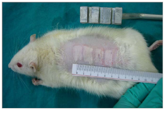
24 Wistar Albino male rats were used in this study. The subjects were randomly divided into 4 groups as control, burn control, Ankaferd and Serum Physiologic. On the first day of the experiment, full-thickness skin biopsies were obtained from the shaved backs of the subjects of control group. After the biopsies attained on the 0th day, the subjects in the control group were sacrificed with carbondioxide gas. In the other groups, however, second degree thermal burn was formed by pressing 2x1 cm 4 metal plates after they had been kept in boiling water for 30 seconds in the way that their body surfaces did not exceed 10% of them under anesthesia (Figure-1).
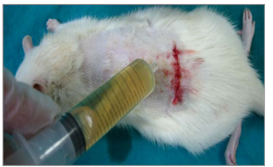
Following the procedure, the wounds of all 21 subjects were dressed specific to their groups. Closed injury dressing was carried out with only sterile gauze dressing without using any substance in burn control group. The subjects in Ankaferd group, however, ankaferd was dripped to burn wounds and closed injury dressing was performed with sterile gauze dressing (Figure-2). Burn wounds of subjects in serum physiologic group were made wet with serum physiologic and closed injury dressing was conducted with sterile gauze dressing. Full-thickness skin biopsies were obtained from burn areas on 0th, 7th, 14 and 21 days of the study for histopathologic evaluation. After the biopsies attained on the 21st day of study subjects were sacrificed with carbondioxide.
The Evaluation of Skin Samples
The obtained tissue samples were fixed in 10% neutral formaldehyde and waited in 5% formic acid. After the routine histopathologic preparations, the materials taken into paraffin blocks were divided into vertical sections as 5μm with help of Leica Rotary microtome. The attained sections were analysed under the light microscope after they had been stained with Masson Trikrom.
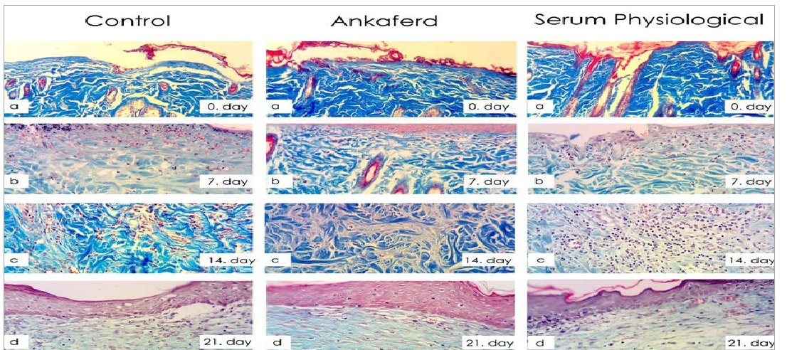
Taking the control groups as a reference, the changes (fibroblast increase, congestion, inflammatory cell increase, epithelization) were evaluated as none (0), low (1), medium (2), extreme (3), severe (4) according to their histopathologic situations (Figure 3, 4, 5). Scoring was carried out for every rat and average values were determined for every group.
Statistical Analysis
Statistical evaluation of the collected data via subject follow-up forms was performed by usingSPSSv21.5 package programme. Statistical significance was accepted as p<0.05. Since sample size was composed of 24 subjects, nonparametric tests were utilized in statistical analyses as variations did not fit to normal distribution. Kruskal-Wallis test was used in the comparison of congestion, fibroblast increase, inflammatory cell increase and epithe-lization values of control, burn, isotonic and Ankaferd groups and then Mann Whitney U test was utilized in Post Hoc group comparisons.
Results
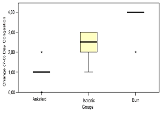
A significant difference was not determined in biopsies obtained on 0th, 7 , 14 and 21 days when fibroblast increase was evaluated (p>0.05). In the congestion evaluation; a significant difference was established between Ankaferd and burn control groups when change amounts between histopathologic data on 0th and 7th days were examined. While less increase was seen in congestion in Ankaferd group on 7th day, a severe increase was observed in congestion in burn control group (Figure-6). The difference between them was statistically significant (p<0.05).
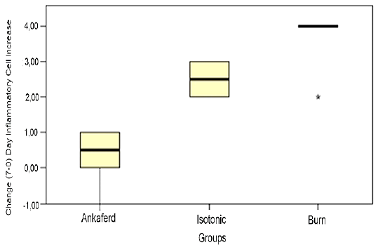
In the inflammatory cell increase evaluation; a significant difference was identified between Ankaferd and burn control group when change amounts between histopathologic data were analysed on 0th-7th days and 0 -21 days. While less increase was seen in inflammatory cells in Ankaferd group on the 7th day, a severe increase was observed in inflammatory cells in the burn control group (Figure-3b,4b,7). The difference between them was statistically significant (p<0.05).
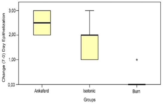
In the epithelization evaluation; a significant difference was established between the burn control group and serum physiologic and the Ankaferd groups when change amounts were observed between histopathologic data on 0th- 7thand 0th-21st days. While there was an extreme increase seen in Ankaferd epithelization on the 7th day, an average increase was seen in the epithelization of serum physiologic group. Yet, there was no increase in the epithelization of burn control group on the 7th day (Figure-3b, 4b,5b,8). The difference between them was statistically significant (p<0.005).
Discussion
As long as skin protects its completeness, it can fulfillits duties,on the other hand, a complex recovery period commences that many cells play part including keratinocyte, fibroblast, endothelium cells, macrophage and thrombocytes in case of deterioration of this completeness. New tissue formation initiates with migration, infiltration and proliferation of these cells and wound closes (14, 28, 29). To accelerate the recovery of burn wound, the aim in the treatment methods used was to affect the factors playing a role in wound healing and shorten the periods regarding these phases and ensure an ideal scar formation. Therefore, many topical and systemic agents were used and delay and irregularities were tried to be inhibited in the wound healing. None of these treatment options have a feature that is alternative to others (8). On the other hand, advanced fibroblastic activity and increased inflammatory period cause irregularities in many wound healings includingburn wounds and lead to hypertrophic scar or keloid development (15). Therefore, the control of these periods in wound healing and additional agents to be used while recovering are of importance (16).
In line with our results, Arslan et al. applied ABS to a soft tissue laceration developed based on a trauma and with no repair chance with primary closure on a 80-year-old female patient. In the control performed 24 hours later, it was determined that wound opening started to close and was of the opinion that ABS is effective in superficial wound healing (8).
In a study in which the efficiency of Celox and Ankaferd® was analysed as styptic in experiment animals given varfarin by Aktop et al., fibrosis values were found significantly low in Celox® group compared to other two groups. This situation was described to be late in Celox® group but a better improvement. As a result, both agents were effective in bleeding control, it was determined that ABS causes a rapid improvement while Celox®forms a late recovery period (21). In our study, ABS that affects fibrosis in a way equivalent to serum physiologic supports this perspective that it forms a better epithelization.
In a study that full thickness skin defect was formed on the backs of rats by Akalın et al., they evaluated the effects of ABS on a secondary wound healing as histologically and photographically. Similar to our study; fibroblast proliferation, inflammatory cell increase, collagen storage, wound contraction and congestion increase were examined on the 3rd, 7th, 10th and 14th days of skin defects formed in full thickness. Results were obtained in parallel with findings of our study. According to the outcomes of this study, in the experiment group that ABS was applied to wound surface fibroblast proliferation degree, vascularization degree, wound contraction rate and type-I/type-III collagen rate were statistically and significantly found high compared to those of control group (22).
In a study that Isler etal.created a bone defect in tibias of rats, they studied the effects of ABS on early bone recovery. In the histopathological evaluation of tissues on 7th day, less inflammation and necrosis were seen in the group that ABS was applied compared to control group and it was reported that there was more new bone formation in the course of early bone amelioration (12). This study complying with our findings showed that ABS which is determined to increase epithelization in an isolated way without forming more activities than serum physiologic in fibroblastic activity affected the other ways of cellular healing.
In the partial nephrectomies performed on 24 rats by Huri et al., they stated that in a group ABS was applied, it reduced partial nephrectomy and hot ischemia and ensured hemostasis. They analysed the effect of the Ankaferd by evaluating glomerular necrosis, acute inflammation, calcification, fibrosis, fibroblast activation and microvascular proliferation histopathologically. They informed that inflammation, fibrosis, fibroblast activation and microvascular proliferation were not significantly affected. It was also supported in this study in a similar way that ABS displayed a regenerative effect beyond its fibroblastic activity. In addition, no symptoms seen such as glomerular necrosis and calcification was evalauted as a positive effect on renal tissue (19).
When the samples were compared in terms of congestion, inflammation, fibroblast proliferation and epithelization in our study, the serum physiologic and ABS groups provided better results compared to burn control group. However, ABS was determined to have superiority over serum physiologic with regard to inflammation control and fibroblast increase (p>0.005). Nevertheless, providing an apparent effect of ABS compared to serum physiologic in increasing epithelization (p<0.005) was interpreted as Ankaferd has an effect on wound healing and epithelization apart from inflammatory periods.
As a result, Ankaferd seems to contribute to effective wound healing by accelerating epithe-lization similar to literature data in our study. However, ensuring efficiency without increasing inflammatory period and fibroblast activity makes us think that it can have an effect to cellular regeneration. Moreover, if uncontrolled scar tissue is thought to develop in burn wounds where inflammation and fibroblastic effectivity are high, ABS can be thought to yield better results in burn isolated wound healing. Nonetheless, controlled studies performed in people are required to support this prediction.
Declarations
- Author contributions (CRediT)
- All authors contributed equally to all 14 CRediT roles (Conceptualization, Data curation, Formal analysis, Funding acquisition, Investigation, Methodology, Project administration, Resources, Software, Supervision, Validation, Visualization, Writing – original draft, Writing – review & editing).
- Funding
-
No financial support was used for the study.
- Conflicts of interest
- A conflict of interest statement was not reported in the published version of this article.
- Ethics approval
- Fırat University Animal Experiments Local Ethics Board approval was attained with 2014/2 and 25 numbered decision.
- Data availability
- A data availability statement was not reported in the published version of this article.
References (29)
17 of 29 references are linked to their DOI.
- Stephen Basil AP, Steven EW, Arthur DM. Epidemiology, demographic, and outcome characteristics of burn injury. In: Herndon DN, ed. Total Burn Care. 4thed. Galveston, TX, USA: Saunders Elsevier; 2012. p.16-45. PubMed Google Scholar
- Griffs RW. Management of multiple casualties with burns. Br Med J (Clin Res Ed). 1985;5:917-8. PubMed Google Scholar
- Oryan A, Alemzadeh E, Moshiri A. Burn wound healing: present concepts, treatment strategies and future directions. J wound care 2017;2;26:5-19. DOI Crossref PubMed Google Scholar
- Kahn SA, Patel JH, Lentz CW. Fire fighter Burn injures: Predictable patterns influenced by Turn out Gear. J Burn Care Res 2012;33(1):152-6. PubMed Google Scholar
- Rezaei E, Beiraghi-Toosi A, Ahmadabadi A, Tavousi SH, Alipour Tabrizi A, Fotuhi K, Jabbari Nooghabi M, Manafi A, Ahmadi Moghadam S. Can Skin Allograft Occasionally Act as a Permanent Coverage in Deep Burns? A Pilot Study. World J Plast Surg 2017;6(1):94-99. PubMed Google Scholar
- Bayram Y, Parlak M, Aypak C, Bayram I. Three-year review of bacteriological profile and antibiogram of burn wound isolates in Van, Turkey. Int J Med Sci 2013;10(1):19-23. DOI Crossref PubMed Google Scholar
- Rosenberg L, Krieger Y, Bogdanov-Berezovski A, Silberstein E, Shoham Y, Singer AJ. A novel rapid and selective enzymatic debridement agent for burn wound management: a multi-center RCT. Burns 2014;40(3):466-74. DOI Crossref PubMed Google Scholar
- Arslan S, Yeşil Y, Ülger Z, Cankurtaran M, Göker H, Haznedaroğlu IC. Yaşlı Bir Hastada Yumuşak Doku Travmasına Bağlı Gelişen Yara İyileşmesinde Ankaferd. Akademik Geriartri Dergisi 2010;2(1):58-60. Regenerative Effects of ABS in Burn Wound Ozcan, et al. PubMed Google Scholar
- Bahramsoltani R, Farzaei MH, Rahimi R. Medicinal plants and their natural components as future drugs for the treatment of burn wounds: an integrative review. Arch Dermatol Res 2014;306(7):601-17. DOI Crossref PubMed Google Scholar
- Uygun M, Yavuz OY, Uraş İ, Duman İG, Haltaş H, Kömürcü M. Effect of Ankaferd Blood Stopper on muscle healing. Clin Invest Med 2016;39(6):27526. DOI Crossref PubMed Google Scholar
- Goker H, Haznedaroglu IC, Ercetin S, Kirazli S, Akman U. Haemostatic actions of the folkloric medicinal plant extract Ankaferd Blood Stopper. J Int Med Res 2008;36:163‐ 1170. DOI Crossref PubMed Google Scholar
- İsler SC, Demircan S, Cakarer S, Cebi Z, Keskin M, Keskin C, Soluk M, Yüzbaşioğlu E. Effects of folk medicinal plant extract Ankaferd Blood Stopper on early bone healing. J Appl Oral Sci 2010;18:409-414. DOI Crossref PubMed Google Scholar
- Kosar A, Cipil HS, Kaya A, Uz B, Haznedaroglu IC, Goker H. The efficacy of Ankaferd Blood Stopper in antithrombotic drug induced primary and secondary hemostatic abnormalities of a rat bleeding model. Blood Coagul Fibrinolysis 2009:20;185‐ 190. DOI Crossref PubMed Google Scholar
- Theoret CL. Update on wound repair. Clin Tech Equine Pract 2004;3:110-122. DOI Crossref PubMed Google Scholar
- Gauglitz GG, Korting HC, Pavicic T, Ruzicka T, Jeschke MG. Hypertrophic scarring and keloids: pathomechanisms and current and emerging treatment strategies. Mol Med 2011;17(1-2):113-25. DOI Crossref PubMed Google Scholar
- Penn JW, Grobbelaar AO, Rolfe KJ. The role of the TGF-β family in wound healing, burns and scarring: a review. Int J Burns Trauma 2012;2(1):18-28. PubMed Google Scholar
- Jimenez PA, Jimenez SE. Tissue and cellula rapproaches to wound repair. Am J Surg 2004;187:56-64. PubMed Google Scholar
- Cipil HS, Kosar A, Kaya A. Invivo hemostatic effect of the medicinal plant extract Ankaferd Blood Stopper in rats pretreated with warfarin. Clin Appl Thromb Hem 2009;15:270-276. DOI Crossref PubMed Google Scholar
- Huri E, Akgul T, Ayyıldız A, Ustun H, Germiyanoglu C. Hemostatik role of a folkloric medicinal plant extract in a rat partial nephrectomy model: controlled experimental trial. J Urol 2009;181:2349-54. DOI Crossref PubMed Google Scholar
- Akkoc N, Akcelik M, Haznedaroglu IC. Invitro anti-bacterial activities of Ankaferd Medicinal Plant Extract. Int J Lab Hematol 2008;30:95. PubMed Google Scholar
- Aktop S, Emekli- Alturfan E, Ozer C, Gonul O, Garip H, Yarat A, Goker K. Effects of Ankaferd Blood Stopper and Celox on the tissue factor activities of warfarin-treated rats. Clin Appl Thromb Hemost 2014;20(1):16-21. DOI Crossref PubMed Google Scholar
- Akalin C, Kuru S, Barlas AM, Kismet K, Kaptanoglu B, Demir A, Astarci HM, Ustun H, Ertas E. Beneficial effects of Ankaferd Blood Stopper on dermal wound healing: an experimental study. Int Wound J 2014;11:64–68. PubMed Google Scholar
- Akkoç N, Akçelik M, Haznedaroğlu İC. Göker H, Turgut M, Aksu S, Kirazlı Ş, Fırat HC. Definition of in vitro anti-fungal effects of Ankaferd medical plant extract. In: The 34th National Congress of Haematology (Proceedings Book); 8 – 11 October 2008; Çeşme, İzmir, Turkey: abstract no: S017 (in Turkish). PubMed Google Scholar
- Koçak E, Akbal E, Taş A, Köklü S, Karaca G, Can M, Kösem B, Üstün H. Anti-inflammatory efficiency of Ankaferd blood stopper in experimental distal colitis model. Saudi J Gastroenterol 2013;19:126–130. DOI Crossref PubMed Google Scholar
- Göker H, Kılıç E, Uçar Çetinkaya D, Büyükaşık Y, Aksu S, Turgut M, Haznedaroğlu İC. The in vitro anticancer activity of Ankaferd on human colon cancer (CaCo-2) cells. The 10th National Congress of Internal Medicine Congress Book; 15 – 19 October 2008; Antalya, Turkey: p044 (in Turkish). PubMed Google Scholar
- Uğur A, Saraç N, Çankal DA, Özle M. The antioxidant and antimutagenic activities of Ankaferd blood stopper,a natural hemostatic agent used in dentistry. Turk J Med Sci 2016;19;46(3):657-63. DOI Crossref PubMed Google Scholar
- Beyazit Y, Kurt M, Kekilli M, Goker H, Haznedaroglu IC. Evaluation of hemostatic effects of Ankaferd as an alternative medicine. Altern Med Rev 2010;15:329-336. PubMed Google Scholar
- Werner S, Grose R. Regulation of wound healing by growth factors and cytokines. Physiol Rev 2003;83:835-870. DOI Crossref PubMed Google Scholar
- Li J, Chen J, Kirsner R. Pathophysiology of acute wound healing. Clin Dermatol 2007; 25: 9-18. DOI Crossref PubMed Google Scholar
Respond to this article
Comments on a published article are welcome as a Letter to the Editor.
Get an e-mail when new articles in your fields are published
Questions to the authors
Questions are moderated: an editor approves each question, the authors answer, and an editor approves the answer before both appear here.
No answered questions yet.
Ask the authors a question
Discussion
Comments are moderated: they appear after the writer confirms the e-mail address and an editor approves them. Each comment carries a conflict-of-interest statement.
No comments yet.
Write a comment
Read the full text
Open the PDF here
Loading the PDF…
If the PDF does not show here, use the download button above.
How to cite this article
Ozcan EC, Senol S, Tokgoz Y, Kuloglu T. The Regenerative Effects of Hemostatic Plant Extract in Second Degree Burn Wound. Ulutas Med J. 2017;3(4):78-84. doi:10.5455/umj.20171108031208
Ozcan, E. C., Senol, S., Tokgoz, Y., & Kuloglu, T. (2017). The Regenerative Effects of Hemostatic Plant Extract in Second Degree Burn Wound. The Ulutas Medical Journal, 3(4), 78–84. https://doi.org/10.5455/umj.20171108031208
Ozcan, E.C. et al. (2017) 'The Regenerative Effects of Hemostatic Plant Extract in Second Degree Burn Wound', The Ulutas Medical Journal, 3(4), pp. 78–84. doi: 10.5455/umj.20171108031208.