The Ulutas Medical Journal · ISSN 2149-0430 (print), 2149-388X (online) · https://www.ulutasmedicaljournal.com/?mno=182557
Case Report · Open access
Gingival Depigmentation: Esthetic Enhancement Made Simple and Efficient
Also available in: العربية · Türkçe · 中文
- Department of Paedodontics and Preventive Dentistry Subharti Dental College, Swami Vivekanand Subharti University, Meerut, India ROR
Abstract
Background: Dental esthetic problems in adolescence have significant impact on the psychologic health and well being during this important period of development. These dental problems not only hinder the normal oral functioning of these children, but also have a considerable psychological impact. Dental problems in anterior region may be related to abnormality in shape, size, color and structure of the tooth, pigmentation of the gingiva, traumatic injuries, orthodontic problems. One of the frequently encountered dental problems is gingival pigmentation, which is seen as a purplish staining or as brown or light brown patches. Case: The aim of the report is to describe the management of gingival pigmentation by means of electrocautery as a feasible esthetic alternative in an adolescent girl.
Keywords
Full text
Introduction
Dental esthetic problems in adolescence have illnesses (e.g. Addison’s disease) to malignant neoplasm significant impact on the psychologic health and well being during this critical period of development. These dental problems not only hinder the normal oral functioning of these children but also have a significant psychological impact. It also affects the interaction of these children with their peers. Dental problems in anterior region may be related to abnormality in shape, size, color and structure of the tooth, pigmentation of the gingiva, traumatic injuries, orthodontic problems. One of the common dental problems reported is the pigmentation of the gingiva which is seen as a diffuse purplish discoloration or as irregularly shaped brown or light brown patches (1). It is found in all races and at any age without any gender predilection (2, 3). This discoloration of the gingiva may occur due to a variety of conditions. These may range from physiologic reasons (e.g. racial pigmentation) to manifestations of systemic (e.g. melanoma and Kaposi sarcoma)(4). The physiological gingival melanin hyperpigmentation usually does not present as a medical problem, but the patient may complain that their ‘black gums’ are unaesthetic. This problem is aggravated in the patients with a ‘gummy smile’ or an excessive gingival display while they smile or talk. For depigmentation of gingiva different treatment modalities are reported which includes bur abrasion, scraping, partial thickness flap, cryotherapy, electro-cauterization and laser-aided depigmentation. The aim of this case report is to describe the management of gingival pigmentation using electro cautery as a viable esthetic option in an adolescent girl. The aim of the report is to describe the management of gingival pigmentation by means of electrocautery as a feasible esthetic alternative in an adolescent girl.
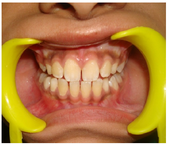
A 15 year old adolescent girl reported with her parents, to the department of Pedodontics and Preventive Dentistry with the chief complaint of brown-black pigmentation on her gingiva and resultant embarrassment due to this gingival discoloration. The patient's history revealed that the blackish discoloration of gingiva was present since birth suggestive of physiologic melanin pigmentation (Fig. 1). Medical history of the case was noncontributory.
By intraoral examination, a melanin pigmentation was seen on the facial gingiva extending from maxillary right first premolar to left premolar. A treatment plan was drawn for the patient involving surgical depigmentation of anterior maxillary gingiva under local anesthesia.
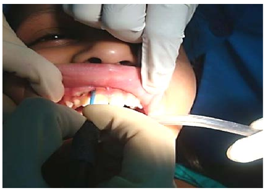
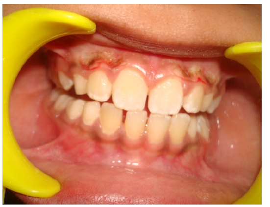
Informed written consent was obtained from the patient’s parents prior to the procedure. Oral prophylaxis was carried out and local anesthetic administered at the beginning of the depigmentation procedure. The pigmented gingival epithelium from the distal surface of right maxillary first premolar to the distal surface of the left first premolar was excised using an electrocautery loop electrode. Any remnants of the pigmented areas that were left were removed (Fig. 2 and 3).
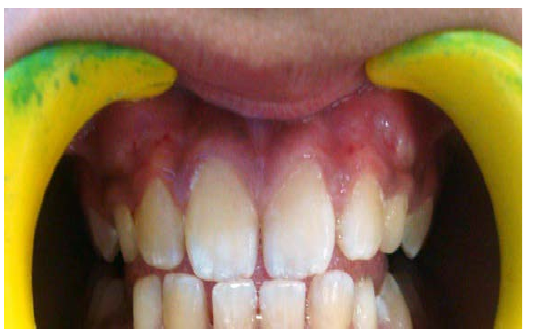
Postoperative instructions were applied. Analgesic medication was prescribed for the management of postoperative pain and the patient was instructed to use a chlorhexidine mouth and continue regular oral hygiene practices thereafter. After 1 week the surgical area was examined. The healing was uneventful without any post-surgical complications. The gingiva appeared pink, healthy, and firm giving a normal appearance with improved esthetics (Fig. 4).
Discussion
There are wide variations in gingival color in normal healthy persons. The degree of vascularization, the thickness of the keratinized layer, and the amount of the pigment containing cells will determine the color of the gingiva (5). There are few studies published to date regarding clinical methods of treatment of pigmented gingiva. The techniques that were tried in the past to treat gingival pigmentation include chemical cauterization, gingivectomy, scalpel scraping procedure, and abrasion of gingiva. The recent techniques of gingival depigmentation in practice are cryotherapy free gingival autograft and laser therapy and all these methods have achieved satisfactory results (6).
Oringer (1975) mentioned electrocautery as a superior method of depigmentation over scalpel on the basis of the ‘exploding cell theory’ which states that molecular disintegration of melanin pigments occur in the basal and suprabasal layer of the operated and surrounding area due to electoral energy generated during the electrocautery procedure (7). Although healing is uneventful it has a disadvantage of undesired tissue destruction on repeated and continuous application.
Conclusion
The depigmentation technique induces minimal trauma with reduced surgical intervention is an efficient treatment option to improve the facial gingival esthetics of an adolescent at a sensitive period of life.
Declarations
- Author contributions (CRediT)
- All authors contributed equally to all 14 CRediT roles (Conceptualization, Data curation, Formal analysis, Funding acquisition, Investigation, Methodology, Project administration, Resources, Software, Supervision, Validation, Visualization, Writing – original draft, Writing – review & editing).
- Funding
-
Funding information was not reported in the published version of this article.
- Conflicts of interest
- There is no conflict of interest or not any sources of support.
- Ethics approval
- Ethics approval details were not reported in the published version of this article.
- Data availability
- A data availability statement was not reported in the published version of this article.
References (7)
3 of 7 references are linked to their DOI.
- NamdeoraojiBahadure R, Singh P, Jain E, Khurana H, Badole G. Management of pigmented gingiva in child patient: a new era to the pediatric dentistry. Int J Clin Pediatr Dent. 2013;6(3):197-200. PubMed Google Scholar
- Dummet CO. Oral pigmentation: First symposium on oral pigmentation. J Periodontol. 1960;31(7):356-360. PubMed Google Scholar
- Page LR, Corio RL, Crawford BE, Giansanti JS, Weathers DR. The oral melanotic macule. Oral Sung Oral Med Oral Pathol. 1977;44(2):219-226. DOI Crossref PubMed Google Scholar
- Ship Jonathan A, Greenberg Martin S, Glick Michael., editors. Burket’s oral medicine: Pigmented lesions of the oral mucosa. 10th. Hamilton Ontario: BC Decker Inc; 2003 PubMed Google Scholar
- Begamaschi O, Kon S, Doine AI, Ruben MP. Melanin repigmentation after gingivectomy: A five year clinical and transmission electron microscopic study in humans. Int J Periodontics Restorative Dent.1993;13:85-92. PubMed Google Scholar
- Ko HJ, Park JW, Suh JY, Lee JM. Esthetic treatment of gingival melanin hyperpigmentation with a Nd:YAG laser and high speed rotary instrument: comparative case report. J Periodontal Implant Sci. 2010 Aug;40(4):201-5. DOI Crossref PubMed Google Scholar
- Kathariya R, Pradeep AR. Split mouth de-epithelization techniques for gingival depigmentation: A case series and review of literature. J Indian SocPeriodontol. 2011 Apr;15(2):161- DOI Crossref PubMed Google Scholar
Respond to this article
Comments on a published article are welcome as a Letter to the Editor.
Get an e-mail when new articles in your fields are published
Questions to the authors
Questions are moderated: an editor approves each question, the authors answer, and an editor approves the answer before both appear here.
No answered questions yet.
Ask the authors a question
Discussion
Comments are moderated: they appear after the writer confirms the e-mail address and an editor approves them. Each comment carries a conflict-of-interest statement.
No comments yet.
Write a comment
Tell me when this article is cited
Leave your e-mail address and confirm it. These notices are not being sent yet; your confirmed address is kept for them and used for nothing else.
Read the full text
Open the PDF here
Loading the PDF…
If the PDF does not show here, use the download button above.
How to cite this article
Adlakha VK, Srivastava N, Chandna P, Grover J. Gingival Depigmentation: Esthetic Enhancement Made Simple and Efficient. Ulutas Med J. 2015;1(1):7-9. doi:10.5455/umj.2015.1.2
Adlakha, V. K., Srivastava, N., Chandna, P., & Grover, J. (2015). Gingival Depigmentation: Esthetic Enhancement Made Simple and Efficient. The Ulutas Medical Journal, 1(1), 7–9. https://doi.org/10.5455/umj.2015.1.2
Adlakha, V.K. et al. (2015) 'Gingival Depigmentation: Esthetic Enhancement Made Simple and Efficient', The Ulutas Medical Journal, 1(1), pp. 7–9. doi: 10.5455/umj.2015.1.2.
@article{umj182557,
  title = {Gingival Depigmentation: Esthetic Enhancement Made Simple and Efficient},
  author = {Adlakha, Vivek K. and Srivastava, Nikhil and Chandna, Preetika and Grover, Jyotika},
  journal = {The Ulutas Medical Journal},
  year = {2015},
  volume = {1},
  number = {1},
  pages = {7--9},
  doi = {10.5455/umj.2015.1.2},
  url = {https://www.ulutasmedicaljournal.com/?mno=182557},
  issn = {2149-388X}
}