The Ulutas Medical Journal · ISSN 2149-0430 (印刷版), 2149-388X (在线版) · https://www.ulutasmedicaljournal.com/?mno=37498
原创论著 · 开放获取
儿童体内的Varicella Zoster病毒会影响临时动脉结构还是血液流动?
标题、摘要和全文为机器翻译。 英文原文
- Department of Physical Medicine and Rehabilitation, Giresun University, Faculty of Medicine, Giresun, TURKEY ROR
- Department of Pediatrics, Giresun University, Faculty of Medicine, Giresun, TURKEY ROR
- Department of Radiology, Giresun University, Faculty of Medicine, Giresun, TURKEY ROR
- 开放获取
- CC BY-NC 4.0
- ORCID
- 资助
- 伦理批准
- 数据可用性
摘要
导言:Varicella-Zoster Virus(VZV)是病毒的Herpesviridae家族的一员,并造成两种不同的临床条件. 我们的研究主要目的不是诊断GCA,而是想评估在童年时期VZV之后是否有结构变化或流量变化,使用美国的时间动脉,这是GCA在晚年时最常受到影响的动脉,并审查患者是否表现出与GCA相关的临床症状. 材料和方法: 该研究被设计为一种预期的单盲研究,包括30名被同名儿科医生诊断患有16岁以下阴道炎的患者,并显示出感染特征. 结果:研究中包括的病人中,没有任何人表现出光环迹象。 此外,没有任何患者显示压缩迹象、隔离或速成结果。 在常见的表面时间动脉中,Intima-media复合厚度的平均值被测量为0.25毫米(0.18-0.27毫米)。 结论:鸡瘟患者在时间动脉上没有表现出任何超音速变化,也没有与GCA有关的临床症状,然而我们认为VZV感染与GCA发育之间的关系值得进一步研究,研究范围更大,后续期更长.
关键词
全文
全文为机器翻译;以英文原文为准。 英文原文
导言
Varicella-Zoster病毒(VZV)是病毒的Herpesviridae家族的一员,引起两种不同的临床条件. 瓦里切拉(Varicella),或俗称"鸡痘",是一种相对温和的临床病症,其特征是广泛出现异端性皮疹,在没有病毒免疫力的人体内接触病毒引起. Shin(Shigles),或称pe疹(herpes zoster)是VZV在一次感染后在机体内休眠的反复感染. (1)鸡毒杆菌很容易从感染者通过空气、水滴或直接接触传播 (2)尽管大多数阴道感染在7-10天之内就已经明显,但据了解,并发症率随着年龄的增长而增加,在罕见的病例中会导致严重发病甚至死亡。
巨型细胞动脉炎(GCA)是一种涉及中到大口径动脉的系统型血管炎,通常见于晚年. 头痛是最常见的症状,可以伴有其他的抱怨,如下巴的夸张、视力问题和头皮敏感性。 诊断除实验室参数和时间动脉活检外,还采用超音速(美国)和其他成像方法 (3)事实证明,与活检相比,美国GCA诊断的敏感性和特殊性要好得多,而且事实证明,在文献中,它比活检更具成本效益。 (4)。 。 。
VZV并发症包括肝后神经病、脑膜炎、肌炎和心肌炎。 (5). 血管病通常会影响大脑动脉,并在某些情况下会造成瞬变性异体攻击甚至中风等情况. (6)此外,其他研究以GCA的形式报告VZV或VZV疫苗在外侧动脉、眼科和视网膜动脉中的并发症及其症状和结果 (7, 8)另一方面,一些其他研究没有报告在时间动脉中看到VZV,而VZV在GCA病理学中也没有发挥作用. (9). 之前没有研究评估时间动脉是否在文献中使用美国进行阴道感染后受到影响. 这些与文献中的观点相矛盾的观点造成了一个难题,即VZV在GCA病理学中是否有作用. 我们的研究主要目的不是诊断GCA,而是想评估在童年时期VZV之后是否有结构变化或流量变化,使用美国的时间动脉,这是GCA在晚年时最常受到影响的动脉,并审查患者是否表现出与GCA相关的临床症状.
材料和方法
该研究被设计为大学医学院物理医学和康复系,儿科和放射学系的远景单盲研究. 该研究获得了大学道德操守委员会的道德操守批准。 所有参与者和/或其父母在参加本研究之前都签署了知情同意表。
该研究包括30名被同一名儿科医生诊断患有16岁以下瓦氏菌的患者,并显示出感染特征. Varicella特征的定义是,损伤最初是红毛球,然后转变为具有明显的内液体的冰囊,在冰囊周围形成光环状的红毛球,且损伤显示出多态性(同一区域不同阶段的损伤)(4)参与者的社会人口财产(年龄、性别、教育)、阴道免疫史、杂症、药物、与时间动脉有关的症状(阴道、头痛、视力问题、耳痛、寺庙的敏感性),他们是否接受过任何对鸡毒的症状治疗,在后续治疗期间是否出现并发症(皮肤感染、CNS参与、肺炎、鼠疫解剖、肝炎、心肌炎、肾炎、出血性糖尿病)。 排除了可能影响到血管结构(血管炎、高血压、糖尿病)、严重感染、发烧和疾病、合作有限或智力迟钝、恶性肿瘤和手术现场开口的患者。
儿科医生利用上述标准诊断了鸡瘟,评估了这些患者的临床症状,如发烧,新开始头痛,以及其局部化,时间动脉敏感度和脉搏,下巴发光的存在,视力丧失,脉冲变化. 使用灰度和色度的多普勒(Doppler US)来评估初步诊断期间的时动脉,以及一名有10年经验的放射科医生(T.B.)在为期6个月的后续访问时对病人的初步诊断及其评估原因视而不见。 (10). 美国使用东芝Applio 500美国设备(日本东京东芝医疗系统公司)进行了评估,采用了支持多普勒模式的高频线性探测器(PLT-1005 BT [4-14 MVZV])。 多普勒频率被设定为10MVZM,PRF 2-3 kVZV,焦点设定为5mm,深度设定为1-2cm. 在纵向和轴线飞机上对常见的表面动脉及其前部和腹部分支进行了双边审查。 GCA特征标志,如光环标志、压缩标志、闭塞和快感存在得到评估 (4)此外,据了解,静脉炎患者体内数值增加的Intima-media综合测量是使用Schäfer VS等人的研究作为参考进行的。 (11)“抑制信号”的定义是,在美国探测器施压后没有压缩动脉,“隔离”的定义是,尽管PRF不断减少,颜色增益的设定和无法获得多普勒信号,但时间动脉却充满了低血压材料,最后,“激素化”的定义是,多普勒美国存在动荡的彩色图案和持续的二聚体填充,同时,在压力波多普勒美国,压力波多普勒美国中最大静脉流动增加,这些动脉被记录为“阳性”或负性”。 (11)。 。 。
统计分析
用于统计分析的是社会科学统计包第22.0版(美国芝加哥)。 描述性数据作为±标准偏差和中位数(最小最大)列报。
结果
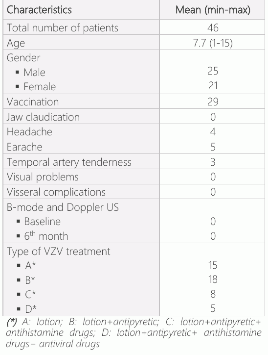
表1显示了患者的人口统计值。 研究中包括的30名患者中,没有任何人表现出光环痕迹. 此外,没有任何患者显示压缩迹象、隔离或速成结果。 在常见的表面时间动脉中,Intima-media复合厚度的平均值被测量为0.25毫米(0.18-0.27毫米)。
讨论情况
文献中对VZV感染与GCA发展的关系存在矛盾的研究. 在我们的研究中,被诊断出有鸡毒杆菌的病人中,没有一个显示出任何短期或长期可归因于GCA的临床或美国结果,而且童年的阴道感染和晚年GCA发育之间也没有临床或美国的联系.
GCA病原学在文献中还不清楚. 然而,有人认为,船壁与免疫系统之间的关系可以在疾病的发病过程中发挥作用。 (12)活性血管腺细胞收集CD4+ T细胞和巨噬细胞向着船壁 然后这些细胞从冒险地层穿透到Intima层 由Vasa Vasorum 导致颗粒瘤形成 (14)由于细胞内病原体也经常引起类似的颗粒性炎症,因此一些患者认为感染是触发因素。 (15)至于VZV,VZV被证明有能力引起幻觉,受影响船只的病理学样本显示多核巨细胞存在,类似于GCA. (16)其他一些研究报告说,在对有阳性和阴性活检结果的GCA患者的时间动脉和主动脉标本进行详细审查时,发现了VZV抗体和DNA (17–21)相反,一些病人报告说,在GCA病人的时间动脉标本中没有发现VZV (22–28)另一项案例研究报告VZV和GCA之间没有关系 (29),另一个案例控制研究报告称,在VZV感染后,GCA的风险增加。 (30) 在文献中看到这些自相矛盾的结果后,它让我们好奇我们是否能够发现与受VZV感染儿童的GCA患者有关的任何临床或美国发现,以及VZV感染引起的超声波学变化,这种感染一般是童年时期的一种儿童疾病,尽管GCA是老年病人所看到的疾病。 从这个意义上说,这项研究是第一次,值得对这一问题感兴趣。 我们使用美国评估而不是时间动脉活检,这是今天GCA诊断中最常用的方法,原因是我们的主要目标不是诊断GCA,而是检测儿童VZV感染后时间动脉是否发生超声波变化,以及时间动脉活检是一种具有并发症的侵入性和高风险程序,它只评估了某部分血管结构,只有一个容器,最后证明美国成像是一种成本效益高得多的非侵入方法,对GCA诊断具有更高的敏感性和特殊性. (4). 使用美国可以检测到GCA的4个特征病理学症状. 这些是船体壁(卤素标志)加厚,无法压缩动脉(压缩标志),激素化和船体隔离。 细胞渗透和水肿可以收集Intima,然后扩散到GCA的介质和冒险层. 舰壁上的厚度显示舰体周围的低温视野,被命名为光环标志 (30)尽管在美国,有严肃的研究显示光环标志敏感度为55-100%,特异性为78-100%,但这种成像方法在GCA诊断中仍不能取代时间动脉活检. (6) TABUL研究对时间动脉活检和多普勒美国进行预测性比较,报告敏感性为39%至54%,特异性为100%至81%。 (10)超声波评估的关键要点之一是需要一位有经验的放射学家来进行评估。 在文献中,大多数执行美国手术的临床医生 被证明在时间动脉评估上无能 (10)为了在研究中避免这一障碍,我们雇用了一位有经验的放射学家,他在美国时间动脉评估方面有经验。
由于上述原因,我们计划利用美国评估VZV病人的时动脉,但这也对我们的研究造成了一些限制。 其中一个局限性是,尽管是由经验丰富的放射学家完成的,但超声波学评估方法仍然被广泛研究,用于明确的GCA诊断,尽管它被证明是一种可靠的方法,今天仍在讨论. 另一个局限性是,我们无法在任何一个病人身上进行时间动脉活检,这是诊断的关键所在. 然而,我们并不认为进行时间动脉生物检查,只是为了在没有GCA临床症状或实验室发现的患者中诊断或检测相关组织上的VZV是合乎道德的. 最后,我们最后的限制是短时间的后续期,即六个月。 研究中包括的病人今后有可能发现GCA的临床/超声波迹象,而且我们也许能够在常规后续期间发现这些变化。 因此,我们认为有必要以上述这些局限性进一步研究这一问题。
结论
鸡瘟病人在时间动脉上没有表现出任何超音速变化,也没有与GCA有关的临床症状,然而我们认为VZV感染和GCA发育之间的关系值得进一步研究,研究范围更大,后续期更长.
声明
- 作者贡献(CRediT)
- 所有作者对全部 14 项 CRediT 角色贡献均等(Conceptualization, Data curation, Formal analysis, Funding acquisition, Investigation, Methodology, Project administration, Resources, Software, Supervision, Validation, Visualization, Writing – original draft, Writing – review & editing)。
- 资助
-
Funding information was not reported in the published version of this article.
- 利益冲突
- The authors declared no conflict of interest for the present article.
- 伦理批准
- The study was conducted according to the declaration of Helsinki. The study was approved by the Ethic Committee of Giresun University (Date: 07.11.2018; No :5/1/29) and the patients gave informed consent prior to participation.
- 数据可用性
- A data availability statement was not reported in the published version of this article.
参考文献 (30)
30 条参考文献中有 27 条已链接 DOI。
- Azap A, Kurt H, Varisella Zoster Virüs İnfeksiyonları. Ankara Üniversitesi Tıp Fakültesi Mecmuası, 2001 54.04 PubMed Google Scholar
- Kanra G, Kara A. Varisella zoster virus enfeksiyonları. Çocuk Sağlığı ve Hastalıkları Dergisi 2002;45:260-74 PubMed Google Scholar
- Ulusoy, Hasan, Nebahat Sarıca, and Şule Arslan. "Polymyalgia Rheumatica and Giant Cell Arteritis: A Case Report and Review of Literature." Turkish Journal of Physical Medicine and Rehabilitation 55.1 (2009) PubMed Google Scholar
- Schmidt, Wolfgang A. "Ultrasound in the diagnosis and management of giant cell arteritis." Rheumatology 57.suppl_2 (2018): ii22- ii31 DOI Crossref PubMed Google Scholar
- Gilden D, Cohrs RJ, Mahalingam R, Nagel MA. Varicella zoster virus infection of the nervous system. In: Johnson MV, Adams HP, Fatemi SA, eds. Neurobiology of disease. 2nd ed. New York: Oxford University Press, 2015. In press DOI Crossref PubMed Google Scholar
- Nagel MA, Gilden D. Update on varicella zoster virus vasculopathy. Curr Infect Dis Rep 2014; 407:1–8 DOI Crossref PubMed Google Scholar
- Nagel MA, Khmeleva N, Boyer PJ, Choe A, Bert R, Gilden D. Varicella zoster virus in the temporal artery of a patient with giant cell arteritis. J Neurol Sci 2013; 335:229 –30 DOI Crossref PubMed Google Scholar
- Lotan, I., & Steiner, I. (2017). Giant cell arteritis following varicella zoster vaccination. Journal of the neurological sciences, 375, 158-159 DOI Crossref PubMed Google Scholar
- Muratore, Francesco, et al. "No detection of varicella-zoster virus in temporal arteries of patients with giant cell arteritis." Seminars in arthritis and rheumatism. Vol. 47. No. 2. WB Saunders, 2017 DOI Crossref PubMed Google Scholar
- Luqmani, R. et al. The Role of Ultrasound Compared to Biopsy of Temporal Arteries in the Diagnosis and Treatment of Giant Cell Arteritis (TABUL): a diagnostic accuracy and cost-effectiveness study. Health Technol. Assess. (Rockv). 20, 1–238 (2016) DOI Crossref PubMed Google Scholar
- Schäfer VS, Juche A, Ramiro S, Krause A, Schmidt WA. Ultrasound cut-off values for intima-media thickness of temporal, facial and axillary arteries in giant cell arteritis. Rheumatology 2017;56:1479– 83 DOI Crossref PubMed Google Scholar
- England, B. R., Mikuls, T. R., Xie, F., Yang, S., Chen, L., & Curtis, J. R. (2017). Herpes zoster as a risk factor for incident giant cell arteritis. Arthritis & Rheumatology, 69(12), 2351-2358 DOI Crossref PubMed Google Scholar
- Weyand CM, Goronzy JJ. Immune mechanisms in medium and large-vessel vasculitis. Nat Rev Rheumatol 2013;9:731–40 DOI Crossref PubMed Google Scholar
- Russo MG, Waxman J, Abdoh AA, Serebro LH. Correlation between infection and the onset of the giant cell (temporal) Varicella Zoster Virus in Children Kulaklı et al. arteritis syndrome: a trigger mechanism? Arthritis Rheum 1995;38:374–80 DOI Crossref PubMed Google Scholar
- Nagel MA, Cohrs RJ, Mahalingam R, Wellish MC, Forghani B, Schiller A, et al. The varicella zoster virus vasculopathies: clinical, CSF, imaging, and virologic features. Neurology 2008;70:853–60 DOI Crossref PubMed Google Scholar
- Mitchell BM, Font RL. Detection of varicella zoster virus DNA in some patients with giant cell arteritis. Invest Ophthalmol Vis Sci 2001;42:2572– 7 DOI Crossref PubMed Google Scholar
- Nagel MA, White T, Khmeleva N, Rempel A, Boyer PJ, Bennett JL, et al. Analysis of varicella-zoster virus in temporal arteries biopsy positive and negative for giant cell arteritis. JAMA Neurol 2015;72:1281–7 DOI Crossref PubMed Google Scholar
- Gilden D, White T, Khmeleva N, Katz BJ, Nagel MA. Blinded search for varicella zoster virus in giant cell arteritis (GCA)- positive and GCA-negative temporal arteries. J Neurol Sci 2016;364:141–3 DOI Crossref PubMed Google Scholar
- Gilden D, White T, Khmeleva N, Heintzman A, Choe A, Boyer PJ, et al. Prevalence and distribution of VZV in temporal arteries of patients with giant cell arteritis. Neurology 2015;84:1948– 55 DOI Crossref PubMed Google Scholar
- Gilden D, White T, Boyer PJ, Galetta KM, Hedley -Whyte ET, Frank M, et al. Varicella zoster virus infection in granulomatous arteritis of the aorta. J Infect Dis 2016;213:1866–71 DOI Crossref PubMed Google Scholar
- Kennedy PG, Grinfeld E, Esiri MM. Absence of detection of varicella-zoster virus DNA in temporal artery biopsies obtained from patients with giant cell arteritis. J Neurol Sci 2003;215:27–9 DOI Crossref PubMed Google Scholar
- Nordborg C, Nordborg E, Petursdottir V, LaGuardia J, Mahalingam R, Wellish M, et al. Search for varicella zoster virus in giant cell arteritis. Ann Neurol 1998;44:413–4 DOI Crossref PubMed Google Scholar
- Alvarez-Lafuente R, Fernandez-Gutierrez B, Jover JA, Judez E, Loza E, Clemente D, et al. Human parvovirus B19, varicella zoster virus, and human herpes virus 6 in temporal artery biopsy specimens of patients with giant cell arteritis: analysis with quantitative real time polymerase chain reaction. Ann Rheum Dis 2005;64:780–2 DOI Crossref PubMed Google Scholar
- Helweg-Larsen J, Tarp B, Obel N, Baslund B. No evidence of parvovirus B19, Chlamydia pneumoniae or human herpes virüs infection in temporal artery biopsies in patients with giant cell arteritis. Rheumatology (Oxford) 2002;41:445– 9 DOI Crossref PubMed Google Scholar
- Rodriguez-Pla A, Bosch-Gil JA, Echevarria-Mayo JE, Rossello-Urgell J, Solans-Laque R, Huguet-Redecilla P, et al. No detection of parvovirus B19 or herpesvirus DNA in giant cell arteritis. J Clin Virol 2004;31:11–5 DOI Crossref PubMed Google Scholar
- Solomon, I. H., Docken, W. P., & Padera, R. F. (2019). Giant cell arteritis is not associated with varicella zoster virus in temporal artery biopsies or ascending aortic resections. The Journal of Rheumatology, jrheum-180912 DOI Crossref PubMed Google Scholar
- BuckinghamEM, FoleyMA, Grose C, et al. Identification of herpes zoster-associated temporal arteritis among cases of giant cell arteritis. Am J Ophthalmol. 2018;187:51–60. doi:10.1016/j.ajo.2017.12.017 DOI Crossref PubMed Google Scholar
- Schafer VS, Kermani TA, Crowson CS, Hunder GG, Gabriel SE, Ytterberg SR, et al. Incidence of herpes zoster in patients with giant cell arteritis: a population-based cohort study. Rheumatology (Oxford) 2010;49:2104–8 DOI Crossref PubMed Google Scholar
- Rhee RL, Grayson PC, Merkel PA, Tomasson G. Infections and the risk of incident giant cell arteritis: a population-based, casecontrol study. Ann Rheum Dis 2017;76:1031–5 DOI Crossref PubMed Google Scholar
- Schmidt WA, Kraft HE, Volker L, Vorpahl K, Gromnica-Ihle EJ. Colour Doppler sonography to diagnose temporal arteritis. Lancet 1995;345:866 DOI Crossref PubMed Google Scholar
阅读全文
在此打开 PDF
正在加载 PDF…
若 PDF 未在此显示,请使用上方的下载按钮。
如何引用本文
Kulakli F, Bulut M, Bekci T. Does Varicella Zoster Virus in Children Affect Temporal Artery Structure or Blood Flow?. Ulutas Med J. 2021;7(1):54-59. doi:10.5455/umj.20201229071421
Kulakli, F., Bulut, M., & Bekci, T. (2021). Does Varicella Zoster Virus in Children Affect Temporal Artery Structure or Blood Flow?. The Ulutas Medical Journal, 7(1), 54–59. https://doi.org/10.5455/umj.20201229071421
Kulakli, F., Bulut, M. and Bekci, T. (2021) 'Does Varicella Zoster Virus in Children Affect Temporal Artery Structure or Blood Flow?', The Ulutas Medical Journal, 7(1), pp. 54–59. doi: 10.5455/umj.20201229071421.