The Ulutas Medical Journal · ISSN 2149-0430 (印刷版), 2149-388X (在线版) · https://www.ulutasmedicaljournal.com/?mno=283352
病例报告 · 开放获取
埃尔博周围的肿瘤:案例报告
标题、摘要和全文为机器翻译。 英文原文
- Department of Radiology, Malatya State Hospital, Malatya, Turkey ROR
- Department of Radiology, Inonu University School of Medicine, Malatya, Turkey ROR
- 开放获取
- CC BY-NC 4.0
- ORCID
- 资助
- 伦理批准
- 数据可用性
摘要
肿瘤性钙化是一种在大关节周围看到的罕见的经理钙化. 大关节见于臀部,肩部和肘部,脚踝中较少见. 不清楚为什么前20年的生活是由家庭倾向性阿法基亚引起的,以及更频繁的出现. 我们提出了磁共振成像 患者的诊断结果 肿瘤钙化肘痛诊所。 当在大关节附近看到带有软组织腹腔钙化的质量时,在差别诊断中应铭记肿瘤钙化.
关键词
全文
全文为机器翻译;以英文原文为准。 英文原文
导言
肿瘤性钙化(TC)是一种在大关节周围观察到的罕见的环状钙化. (1)在臀部、肩部和肘部可以看到大关节,在脚和脚踝中也不太常见 (2)不清楚为什么头20年的生活是由家庭倾向性阿法基亚引起的,以及人们更经常地看到 (3)我们介绍了接受TC肘痛诊疗的病人的磁共振成像结果。
案例介绍
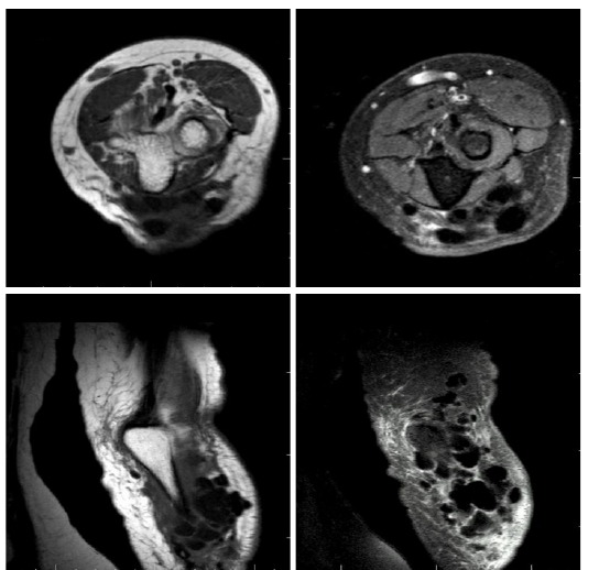
T1AG 星座图 T2AG 星座图 星座图 PDW 冕座图 PDW SPIR 在抱怨肘部疼痛的患者体内获得了轴序,并向放射科申请提取MR.
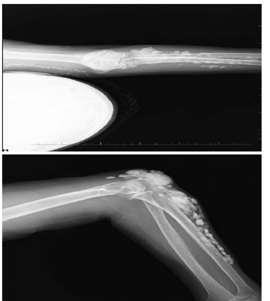
在多个不同体积中观察到了计算法中心(图1、2、3和4),T1AG轴和冠状、PDW冠状和PDWSPIR冠状序列聚集在众多低强度腹膜组件中。 直接的放射图显示这些损伤是钙化中心(图5和图6)。
讨论情况
TC显示周边局部化,特别是在压缩区,其形式为质量与邻接联合空间没有直接关系. 组织中有白沙、瘀青颜色和纹理或钙化 (4)在TC中,这种钙化病原体是由于创伤-营养障碍或创伤症造成的。 (5, 6)在TC中可以看到疼痛和皮肤侵蚀等临床症状. (7)。 。 。
TC的异病形式是家庭. 另一种形式的TC是由于慢性肾衰竭中观察到的高钙磷比. (8). 关于CT的诊断可以通过辐射学发现和病人的医学故事来进行. 无线电图示;TC和Idiopathic肾脏不足都是次于钙化地点,造成多个定义明确的铺路石. 核磁共振在所有序列中都被视为低强度损伤。 此外,由于病理钙化周围的炎症引起的水肿在T2AG和Inversion Recovery(IR)序列中被视为超强信号. (8, 9). 异位骨化、钙化肝脏瘤或淋巴瘤、钙化软组织钙化、伞骨质骨质瘤和骨质疏松症。 在骨骼骨骼疏松的躯体中,可能是钙化的,但是它们的位置是内在的。 病情的临床史和病情的发生地点对差别诊断非常重要 (7, 10). 当大型关节附近看到具有软组织近缘钙化的质量时,在不同的诊断中应铭记TC。
声明
- 作者贡献(CRediT)
- 所有作者对全部 14 项 CRediT 角色贡献均等(Conceptualization, Data curation, Formal analysis, Funding acquisition, Investigation, Methodology, Project administration, Resources, Software, Supervision, Validation, Visualization, Writing – original draft, Writing – review & editing)。
- 资助
-
Funding information was not reported in the published version of this article.
- 利益冲突
- A conflict of interest statement was not reported in the published version of this article.
- 伦理批准
- Ethics approval details were not reported in the published version of this article.
- 数据可用性
- A data availability statement was not reported in the published version of this article.
参考文献 (10)
10 条参考文献中有 4 条已链接 DOI。
- Oeggerli P, Maurer R, Kistler H. Tumoral calcinosis with superinfection and sepsis Schweiz Med Wochenschr 1991; 121 (38): 1387-139 PubMed Google Scholar
- Prahinski JR, Schaefer RA. Tumoral calcinosis of the foot. Foot Ankle Int 2001; 22: 911-913 DOI Crossref PubMed Google Scholar
- Sledz K., et al., Tumoral Calcinosis of the Temporomandibular Joint: CT and MR Findings, AJNR 16: 782-785, Apr 1995 0195-6108 / 95 / 1604-0782 PubMed Google Scholar
- Yurdoğlu C., Özbaydar MU., Adaş M. et al. Familial tumoral calcinosis in three patients in the same family, Acta Orthop Traumatol Turc 2007; 41 (3): 244-248. PubMed Google Scholar
- Slavin RE, Wen J, Kumar D, Evans EB. Familial tumoral calcinosis. A clinical, histopathologic, and ultrastructural study with an analysis of its calcifying process and pathogenesis. Am J Surg Pathol 1993; 17: 788-802 DOI Crossref PubMed Google Scholar
- Boskey AL, Vigorita VJ, Sencer O, Stuchin SA, Lane JM Chemical, Microscopic, and ultrastructural characterization of the mineral deposits in tumoral calcinosis. Clin Orthop Relat Res 1983; (178): 258-69. DOI Crossref PubMed Google Scholar
- Kumara, Singh P, Dobhal H. Case of Recurrent Tumoral Calsinosis in a Young Female, World Journal of Endocrine Surgery, January-April 2013; 5 (1): 18-20 PubMed Google Scholar
- Steinbach LS et al., Tumoral calcinosis: radiologic-pathologic correlation, Skeletal Radiol (1995) 24: 573-578 DOI Crossref PubMed Google Scholar
- Farzan M, Farhood AR., Tumoral Calcinosis: What is the treathment? Report of two cases of different types and review of the literature, Am J Orthop. 2011; 40 (9): E170-E176. PubMed Google Scholar
- Sledz K, Ortiz O, Wax M, Bouguot J, Tumoral Calcinosis of the Temporomandibular Joint: CT and MR Findings, AJNR: 16, April 1995 PubMed Google Scholar
阅读全文
在此打开 PDF
正在加载 PDF…
若 PDF 未在此显示,请使用上方的下载按钮。
如何引用本文
Unlu S, Sanli B, Yildirim IO, Oner S, Dogan M. Tumoral Calcinosis Around the Elbow: Case Report. Ulutas Med J. 2017;3(4):85-87. doi:10.5455/umj.20171114024323
Unlu, S., Sanli, B., Yildirim, I. O., Oner, S., & Dogan, M. (2017). Tumoral Calcinosis Around the Elbow: Case Report. The Ulutas Medical Journal, 3(4), 85–87. https://doi.org/10.5455/umj.20171114024323
Unlu, S. et al. (2017) 'Tumoral Calcinosis Around the Elbow: Case Report', The Ulutas Medical Journal, 3(4), pp. 85–87. doi: 10.5455/umj.20171114024323.