The Ulutas Medical Journal · ISSN 2149-0430 (print), 2149-388X (online) · https://www.ulutasmedicaljournal.com/?mno=82600
Original Article · Open access
The Relationship of Non-dipper Blood Pressure Pattern with the Fragmented QRS Morphology on ECG and the Role of Inflammatory Biomarkers in Polycystic Ovary Syndrome
Abstract
- Introduction
- This study aimed to investigate the relationship between the fragmented QRS (fQRS) morphology and hematological markers and non-dipper blood pressure patterns in patients with polycystic ovary syndrome (PCOS).
- Materials and Methods
- Sixty-five patients with PCOS between the ages of and 18-40 years were included in the study. Control and patients were compared in terms of laboratory findings, electrocardiograms, and holter results.
- Results
- In the PCOS, non-dipper patterns in systolic and diastolic blood pressure were more common, but only the non-dipper pattern showed significance in systolic blood pressure (p=0.007). In both groups, the non-dipper blood pressure was more frequent in those with fragmented QRS. Neutrophil (p=0.01), lymphocyte (p=0.002), neutrophil-lymphocyte ratio(p<0.0001), monocyte-lymphocyte ratio(p=0.014), platelet lymphocyte ratios (p=0.001), erythrocyte distribution width (p<0.0001), and platelet distribution width (p=0.001) were higher in the PCOS than in the control.
- Conclusion
- The levels of inflammatory markers and the frequency of non-dipper blood pressure patterns are increased in PCOS. In these patients, the fQRS morphology may be used to distinguish those with non-dipping blood pressure patterns.
Keywords
Full text
Introduction
Polycystic ovary syndrome (PCOS) is a syndrome characterized by amenorrhea, oligomenorrhea, irregular bleeding, hirsutism, and/or obesity in 5%–10% of women in their reproductive period (1). The risk of cardiovascular disease increases in addition to Type-2 DM, endometrial cancer, and spontaneous abortion in PCOS patients (2). The risk of death and myocardial infarction increased 3.3fold in these individuals, followed by cardiovascular reasons (3).
Many studies have been conducted in recent years on the relationship between PCOS and an enhanced risk of cardiovascular disease. A contributing factor to the elevated cardio vascular risk status in these patients may be the high frequency of non-dipper blood pressure patterns. The non-dipping blood pressure pattern was a prominent prognostic indicator for cardiovascular morbidity and mortality in hypertensive and non-hypertensive individuals (4, 5). Increased inflammation in these patients may also be another mechanism to explain the increased cardiovascular risk.
It has been reported that fragmented QRS (fQRS) morphology can be used to predict non-dipper blood pressure patterns on electrocardiography (ECG). To the best of our knowledge, no study has investigated the connection between the non-dipper blood pressure pattern and fQRS in patients with PCOS. In this study, we aimed to examine the relationship between fQRS morphology and hematological inflammation markers, which are potential threat indicators of cardiovascular diseases in patients with PCOS, and the non-dipper blood pressure pattern.
Materials and Methods
Study Design
The present study analyzed the relationship between fQRS morphology and hematological inflammation markers, which are potential threat indicators of cardiovascular diseases in patients with PCOS, and the non-dipper blood pressure pattern. Ninety patients who applied to the Obstetrics and Gynecology outpatient clinic were included in the present study. The patients were divided into two groups as PCOS and controls.
Ethical Statement
The study was administered after consent was obtained from the ethics committee. Approval from the local ethics committee was obtained (date: 24/06/2020, Session: 2020/12, Decision Number:09). Following the Helsinki Declaration, voluntary consent forms of patients included in the study were obtained (06/03/2017-No:02).
Inclusion/Exclusion Criteria
Patients with two positive findings of hirsutism, polycystic ovarian appearance, and menstrual irregularity were included in the PCOS (n:65), and patients without two positive findings were included in the control group (n:35). ECG without sinusoidal rhythm, left ventricular systolic dysfunction, heart valve disease, left ventricular hypertrophy, atrial fibrillation, morbid obesity (BMI>40 kg/m2), smoking and alcohol use, history of systemic disease (hypertension, diabetes mellitus, renal failure, connective tissue disease, thyroid dysfunction, etc.), patients with fluid-electrolyte imbalance were excluded from the study. Demographic and laboratory data of all patients included in the study were recorded.
Cardiological Assessment
In the study and control groups, 12-lead ECGs were derived at 10 mm/mV amplitude and 25 mm/s. The fQRS component was evaluated for all ECG leads. Fragmented QRS was defined as notching in two adjacent R or S waves, where QRS lasted <120 ms without any bundle branch block in the areas of blood supply to the major coronary artery (6). 24-h Holter blood pressure was measured to investigate the diurnal rhythm of the patients' daily blood pressure. For the Holter recording to be considered valid, the conditions of using it for at least 24 hours, measuring blood pressure every 15 minutes during the day, every 30 minutes at night, and an erroneous measurement rate of less than 20% was stipulated.From Holter blood pressure records, daytime, nighttime, and 24-h systolic blood pressure and diastolic blood pressure, as well as the mean daytime-nighttime systolic and diastolic blood pressure differences, were calculated. Individuals with a blood pressure decrease of more than 10% at night were accepted as dipper and those with less than 10% of the non-dipper blood pressure pattern (7). 12-lead surface ECG recordings at rest were stored to evaluate the presence of fQRS morphology and its association with the non-dipper blood pressure pattern. Patients in both groups were asked to visit on the second or third day of the study period.
Demographics and Lanoratory Analysis
Patient age, height, weight, body mass index (BMI), fasting glucose (FBG), total cholesterol, triglyceride, LDL, HDL, BUN, creatinine, AST, ALT, FSH, LH, sodium, potassium, prolactin, estradiol, TSH, and hemogram were ordered. The sub-parameters of neutrophil, lymphocyte, monocyte, platelet, erythrocyte distribution width (RDW-SD), platelet distribution width (PDW), and red blood cells (RBC) in the hemogram were evaluated. Neutrophil lymphocyte ratio (NLR), monocyte lymphocyte ratio (MLR), and platelet lymphocyte ratio (PLR) were calculated. The relationship between hematological markers, 24-hour ambulatory blood pressure results, non-dipper blood pressure pattern, and the presence of fQRS morphology on ECG were investigated.
Statistical Data
All analysis were given with details in the tables. The frequency and percentage of categorical data and median (clearance) for continuous data are presented as descriptive values. For comparisons between groups, the "Mann- Whitney U-test was used for the two groups, while the " chi-square test was used to compare categorical variables. Results were considered statistically significant when the p-value was less than 0.05 (p<0.05). The collected patient data were analyzed using IBM Statistical Package for the Social Sciences (SPSS) for Windows 23.0.
Results
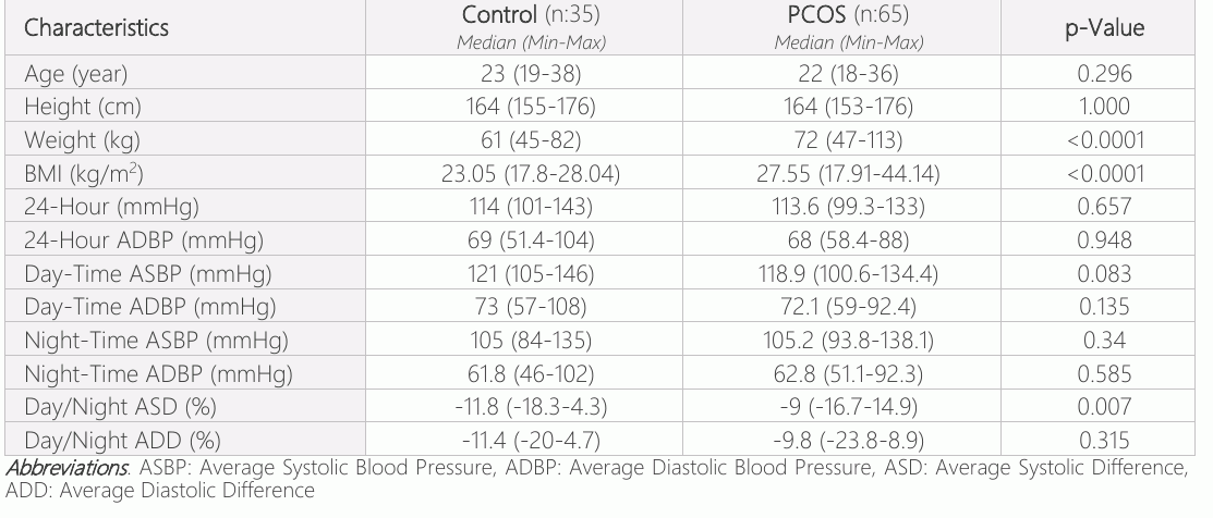
In demographic assessmnets, there were no differences in age and height between the two groups. A significant difference was found in weight and BMI in the PCOS (p<0.0001 and p<0.0001,respectively). No significant difference was observed between the two groups for 24- hour mean systolic, diastolic, mean daytime systolic, diastolic, and mean nighttime systolic and diastolic blood pressures. A significant difference was found in the PCOS group in terms of the mean daytime/nighttime systolic blood pressure difference. However, when both groups were compared in terms of daytime/nighttime diastolic blood pressure difference, although the change in the PCOS group was less, it was not considered significant.
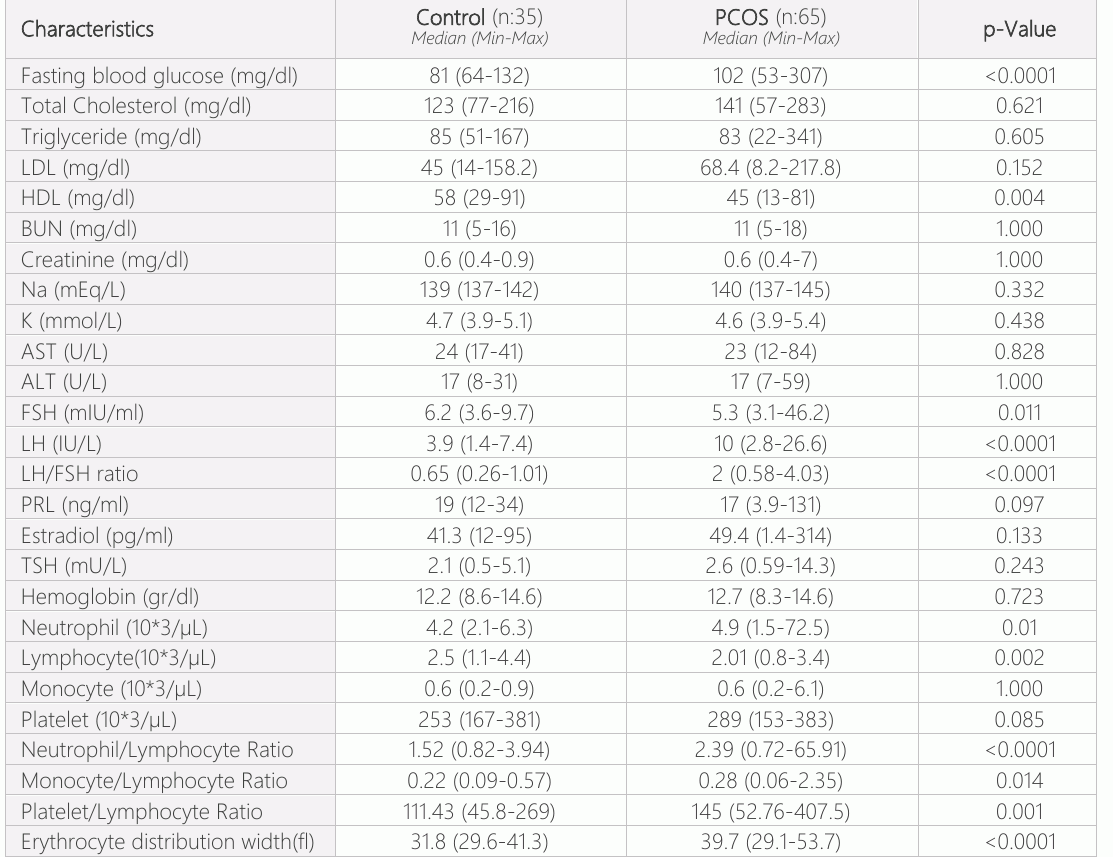
Although significant differences were observed in FBG and HDL in both groups, no difference was found in terms of total cholesterol, triglyceride, LDL, BUN, creatinine, potassium, sodium, AST, and ALT values. In terms of LH/FSH, this ratio was observed to be increased in the PCOS group and was found to be significant. There were no differences in PRL, estradiol, TSH, and hemoglobin values.
In terms of hemogram, monocyte and platelet counts were not significantly different in both groups, while neutrophil and lymphocyte counts showed an increase in favor of PCOS, and this difference was considered significant. In terms of NLR, MLR, PLR, RDW-SD, and PDW values, these values were found to be increased in the PCOS group, and this was considered significant. No difference was found in either group in terms of RBC values (Tables 1 and 2). When patients with ECG fragmentation were evaluated in terms of the daytime/nighttime mean systolic blood pressure difference, although the difference in the PCOS group was less, this was not significant (p=0.765). When both groups without ECG fragmentation were compared, no significant difference was found in terms of daytime/nighttime systolic pressure difference (p=0,289). ECG fragmentation had a non-dipper pressure pattern, and patients without fragmentation had a dipper blood pressure pattern in both groups. In addition, the change in the daytime/nighttime systolic blood pressure difference between both groups with ECG fragmentation was greater than the change between both groups without ECG fragmentation. When we evaluated the patients with and without ECG fragmentation in terms of daytime/nighttime systolic blood pressure difference in the control group and the PCOS group, it was observed that the change in the daytime/nighttime systolic blood pressure difference was less in patients with ECG fragmentation. When patients with and without ECG fragmentation were compared, in both groups, a difference was seen (p<0.0001 and p<0.0001, respectively) (Table 3).
When patients with ECG fragmentation were evaluated in terms of the daytime/nighttime mean diastolic blood pressure difference, although the difference in the control was less, this was not significant (p=0.222). When both groups without ECG fragmentation were compared, although the difference in daytime /nighttime systolic blood pressure was lower in the PCOS, the difference was not significant (p=0.888). Patients with ECG fragmentation had a non-dipper blood pressure pattern, and patients without fragmentation had a dipper blood pressure pattern in both groups. When the difference in daytime/nighttime diastolic blood pressure was compared between those with and without ECG fragmentation, a significant difference was found in both groups (p<0.0001 and p<0.0001, respectively) (Table 4).
Discussion
In clinical studies of ambulatory blood pressure, a circadian rhythm was observed, and it has been shown that blood pressure drops at night and is higher during the daytime. This condition, which is part of the normal circadian rhythm, is defined as the nocturnal dipper pattern. The absence of observation of this physiological process is called the non-dipper blood pressure pattern, a condition closely connected to the dangers of a cardiovascular event and end-organ damage (8). It has been reported that nocturnal sympathetic nervous system discharge plays a role in the formation of this pattern (9). Deterioration of autonomic nervous system functions, which is the main cause of non-dipper blood pressure patterns, may cause adverse events by increasing peripheral vascular resistance. It has been reported that this condition emerges more frequently in obese or overweight people than in the healthy population (10). The prevalence of cardiovascular diseases in those with non-dipper patterns is twice as high as that in healthy people of similar age (11). It is also a condition associated with a worse prognosis of the non-dipper blood pressure pattern than the dipper blood pressure pattern (12).


In PCOS, the three key inflammatory markers are highly and significantly correlated with BMI, according to last reports. In addition, there were correlations with other cardiovascular risk markers, including HDL, triglycerides, diastolic pressure, as well as with insulin resistance, quantified by HOMA-IR. In the previous reports, untreated PCOS demonstrated significantly increased serum concentrations of hsCRP, IL-6, and increased numbers of leukocytes, granulocytes, and lymphocytes compared to a sample of healthy women which were, however, not matched by BMI. In a study, Kargılı et al. reported that the non-dipping blood pressure pattern was higher in PCOS patients than in the control group. In addition, it has been emphasized that the non-dipper pattern may be the sole reason for creating a basis for cardiovascular diseases (13). In line with the literature, we observed the non-dipper pattern in the PCOS group, mainly in terms of the mean daytime/nighttime systolic and diastolic blood pressure differences between the groups in our study. However, we found that the frequency of increased non-dipper pattern in patients with PCOS was statistically significant, with only a difference in systolic blood pressure. It was considered that this situation might be due to the small study volume.
Distinguishing those with non-dipper blood pressure patterns in patients with PCOS may be important in reducing cardiovascular risk. Therefore, these patients need to have markers that indicate a non-dipper blood pressure pattern. In a recent study, Eyüboğlu & Akdeniz showed that fQRS may be predictive of non-dipper pressure patterns in prehypertensive patients. (14). fQRS is a finding that occurs during many cardiovascular diseases. It has been shown that fQRS is also associated with increased blood pressure in patients with a non-dipper cardiac pattern, and a narrow fQRS complex seen on ECG indicates delayed ventricular conduction disturbance as well as myocardial damage and fibrosis (15, 16). Therefore, determining the predictive role of the non-dipper blood pressure pattern of fQRS in PCOS patients may be important for the early detection of patients with high cardiovascular risk. In this study, we found that fQRS data correlated with non-dipper blood pressure in both groups. We also found that the non-dipper blood pressure pattern was more frequent in the PCOS group than in the control group. These results show that fQRS can be used in ECG to predict non-dipper blood pressure in patients with PCOS.
In studies in the literature, many different inflammatory markers have been observed to increase in patients with PCOS. Due to its relationship with obesity, insulin resistance has been identified in patients with PCOS, and increased levels of free fatty acids, TNF-α, and CRP have been detected (10). In a study, Patel et al. observed increases in all inflammatory markers and intima-media thickness in PCOS patients (17). Samy et al. demonstrated that inflammatory markers were investigated in 108 PCOS patients and 75 healthy volunteers, and it has been shown that CRP, IL-6, and TNF-α significantly increased and correlated with BMI and HOMA-IR when measured against the control group (18). In this study, while statistically significantly high FBG and low HDL were observed in both PCOS groups, no statistically significant difference was found for total cholesterol, triglyceride, LDL, BUN, creatinine, sodium, potassium, AST, and ALT levels. In terms of LH/FSH, this ratio was observed to be increased in the PCOS group and was found to be statistically significant. There were no statistical differences in PRL, estradiol, TSH, and hemoglobin values. In terms of hemogram sub-parameters, neutrophil and lymphocyte counts showed an increase in favor of PCOS, and this difference was considered statistically significant. Neutrophil, lymphocyte, NLR, MLR, PLR, RDW, and PDW values, which are hematological inflammation markers, were found to be significantly higher in the PCOS group. These high levels of hematological inflammation markers may explain the rise in cardiovascular threats in patients with PCOS (19). In our study, we showed that the fQRS pattern is closely associated with the non-dipping blood pressure pattern. We uniquely demonstrated in the literature that the fQRS pattern may play a predictive role in distinguishing those with non-dipping blood pressure patterns in PCOS patients. In addition, consistent with the literature, the presence of increased inflammation in PCOS patients was shown through the hematological inflammation markers used in the study.
As a result, the dominance of the non-dipper blood pressure pattern may be a mechanism contributing to the high cardiovascular risk in this group. Fragmented QRS morphology on ECG may be a parameter in the differentiation of PCOS patients with increased cardiovascular risk due to non-dipper blood pressure.
Declarations
- Author contributions (CRediT)
- All authors contributed equally to all 14 CRediT roles (Conceptualization, Data curation, Formal analysis, Funding acquisition, Investigation, Methodology, Project administration, Resources, Software, Supervision, Validation, Visualization, Writing – original draft, Writing – review & editing).
- Funding
-
The author(s) received no financial support for the research, authorship, and/or publication of this article.
- Conflicts of interest
- The authors declare no conflict of interest.
- Ethics approval
- The study was administered after consent was obtained from the ethics committee. Approval from the local ethics committee was obtained (date: 24/06/2020, Session: 2020/12, Decision Number:09). Following the Helsinki Declaration, voluntary consent forms of patients included in the study were obtained (06/03/2017-No:02).
- Data availability
- A data availability statement was not reported in the published version of this article.
References (19)
19 of 19 references are linked to their DOI.
- Sherwood A, Bower JK, Routledge FS, Blumenthal JA, McFetridge-Durdle JA, Newby LK, et al. Nighttime blood pressure dipping in postmenopausal women with coronary heart disease. Am J Hypertens. 2012 Oct;25(10):1077-82 DOI Crossref PubMed Google Scholar
- Cai A, Zhong Q, Liu C, Zhou D, Li X, Zhang Y, et al. Associations of systolic and diastolic blood pressure night-to-day ratios with atherosclerotic cardiovascular diseases. Hypertens Res. 2016 Dec;39(12):874-8 DOI Crossref PubMed Google Scholar
- Routledge FS, Hinderliter AL, McFetridge-Durdle J, Blumenthal JA, Paine NJ, Sherwood A. Endothelial function in postmenopausal women with nighttime systolic hypertension. Menopause. 2015 Aug;22(8):857-63 DOI Crossref PubMed Google Scholar
- Hansen TW, Li Y, Boggia J, Thijs L, Richart T, Staessen JA. Predictive role of the nighttime blood pressure. Hypertension. 2011;57(1):3-10 DOI Crossref PubMed Google Scholar
- de la Sierra A, Gorostidi M, Banegas JR, Segura J, de la Cruz JJ, Ruilope LM. Nocturnal hypertension or nondipping: which is better associated with the cardiovascular risk profile?. Am J Hypertens. 2014;27(5):680-7 DOI Crossref PubMed Google Scholar
- Stewart JM. Mechanisms of sympathetic regulation in orthostatic intolerance. J Appl Physiol (1985). 2012;113(10): 1659-68 DOI Crossref PubMed Google Scholar
- Cuspidi C, Giudici V, Negri F, Sala C. Nocturnal nondipping and left ventricular hypertrophy in hypertension: an updated review. Expert Rev Cardiovasc Ther. 2010;8(6):781-92 164 77 Polycystic Ovary Syndrome Erkayiran et al. DOI Crossref PubMed Google Scholar
- Passino C, Magagna A, Conforti F, Buralli S, Kozáková M, Palombo C, et al. Ventricular repolarization is prolonged in nondipper hypertensive patients: role of left ventricular hypertrophy and autonomic dysfunction. J Hypertens. 2003 Feb;21(2):445-51 DOI Crossref PubMed Google Scholar
- Salles GF, Reboldi G, Fagard RH, Cardoso CR, Pierdomenico SD, Verdecchia P, et al. ABC-H Investigators. Prognostic Effect of the Nocturnal Blood Pressure Fall in Hypertensive Patients: The Ambulatory Blood Pressure Collaboration in Patients With Hypertension (ABC-H) Meta-Analysis. Hyper tension. 2016 Apr;67(4):693-700 DOI Crossref PubMed Google Scholar
- Nakano S, Kitazawa M, Tsuda S, Himeno M, Makiishi H, Nakagawa A, et al. Insulin resistance is associated with reduced nocturnal falls of blood pressure in normotensive, nonobese type 2 diabetic subjects. Clin Exp Hypertens. 2002 Jan-Feb;24(1-2):65-73 DOI Crossref PubMed Google Scholar
- Sherwood A, Steffen PR, Blumenthal JA, Kuhn C, Hinderliter AL. Nighttime blood pressure dipping: the role of the sympathetic nervous system. Am J Hypertens. 2002;15(21): 111-8 DOI Crossref PubMed Google Scholar
- Das MK, Saha C, El Masry H, Peng J, Dandamudi G, Mahenthiran J, et al. Fragmented QRS on a 12 -lead ECG: a predictor of mortality and cardiac events in patients with coronary artery disease. Heart Rhythm. 2007;4(11):1385-92 DOI Crossref PubMed Google Scholar
- Kargili A, Karakurt F, Kasapoglu B, Derbent A, Koca C, Selcoki Y. Association of polycystic ovary syndrome and a non-dipping blood pressure pattern in young women. Clinics (Sao Paulo). 2010;65(5):475-9 DOI Crossref PubMed Google Scholar
- Eyuboglu M, Akdeniz B. Association Between Non-Dipping and Fragmented QRS Complexes in Prehypertensive Patients. Arq Bras Cardiol. 2019;112(1):59-64 DOI Crossref PubMed Google Scholar
- Romero-Corral A, Caples SM, Lopez-Jimenez F, Somers VK. Interactions between obesity and obstructive sleep apnea: implications for treatment. Chest. 2010;137(3):711-9 DOI Crossref PubMed Google Scholar
- Jain R, Singh R, Yamini S, Das MK. Fragmented ECG as a risk marker in cardiovascular diseases. Curr Cardiol Rev. 2014;10 (3):277-86 DOI Crossref PubMed Google Scholar
- Patel SS, Truong U, King M, Ferland A, Moreau KL, Dorosz J, et al. Obese adolescents with polycystic ovarian syndrome have elevated cardiovascular disease risk markers. Vasc Med. 2017 Apr;22(2):85-95 DOI Crossref PubMed Google Scholar
- Samy N, Hashim M, Sayed M, Said M. Clinical significance of inflammatory markers in polycystic ovary syndrome: their relationship to insulin resistance and body mass index. Dis Markers. 2009;26(4):163-70 DOI Crossref PubMed Google Scholar
- Polónia J, Amaral C, Bertoquini S, Martins L. Attenuation of heart rate recovery after exercise in hypertensive patients with blunting of the nighttime blood pressure fall. Int J Cardiol. 2006;106(2):238-43 DOI Crossref PubMed Google Scholar
Read the full text
Open the PDF here
Loading the PDF…
If the PDF does not show here, use the download button above.
How to cite this article
Erkayiran U, Aksu E, Arslanca T. The Relationship of Non-dipper Blood Pressure Pattern with the Fragmented QRS Morphology on ECG and the Role of Inflammatory Biomarkers in Polycystic Ovary Syndrome. Ulutas Med J. 2021;7(3):157-164. doi:10.5455/umj.20210520114119
Erkayiran, U., Aksu, E., & Arslanca, T. (2021). The Relationship of Non-dipper Blood Pressure Pattern with the Fragmented QRS Morphology on ECG and the Role of Inflammatory Biomarkers in Polycystic Ovary Syndrome. The Ulutas Medical Journal, 7(3), 157–164. https://doi.org/10.5455/umj.20210520114119
Erkayiran, U., Aksu, E. and Arslanca, T. (2021) 'The Relationship of Non-dipper Blood Pressure Pattern with the Fragmented QRS Morphology on ECG and the Role of Inflammatory Biomarkers in Polycystic Ovary Syndrome', The Ulutas Medical Journal, 7(3), pp. 157–164. doi: 10.5455/umj.20210520114119.