The Ulutas Medical Journal · ISSN 2149-0430 (印刷版), 2149-388X (在线版) · https://www.ulutasmedicaljournal.com/?mno=221058
病例报告 · 开放获取
昆德拉图斯隆博鲁姆肌肉内的初级Hydatid疾病
标题、摘要和全文为机器翻译。 英文原文
- Istanbul Training and Research Hospital, Department of General Surgery ROR
- Haseki Training and Research Hospital, Department of General Surgery ROR
- 开放获取
- CC BY-NC 4.0
- ORCID
- 资助
- 伦理批准
- 数据可用性
摘要
引言:Hydatid病是一种寄生虫病,由胶虫(genus Echinococus)引起. 它可能位于人体的任何地方. 最常见的器官是肝脏(52-72%)和肺部(10-40%)。 Hydatid疾病很少在肌肉结构中发展,占所有病例的1-5%. 案例介绍 : 在这项研究中,我们报告了一名53岁的男性病人,他左侧腰部区域出现肿胀,并被诊断出患有四重脑膜炎。 结论: 如果生活在土耳其流行国家的任何病人在软组织中出现肿胀,如果他或她应当接受活体检查或囊肿渴望等调查,那么就必须进行谨慎的身体检查和准确的成像研究,同时牢记孝德病是一种可能的诊断.
关键词
全文
全文为机器翻译;以英文原文为准。 英文原文
导言
海达氏病(学名:Echinococcosis),又名海达氏病(学名:Echinococcusis)或海达氏病(学名:Echinococcus),是盘虫科海达氏病(学名:Echinococcus)的一种寄生虫. Echinococus granulosus(英語:E. granulosus)是该病最常见的发病者,其最终宿主是肉食动物如狗,狼和狐狸. 食草动物如羊,山羊,骆驼和马作为中间宿主. 在人类中,通过口摄入后,带虫的幼虫会穿透小肠的壁,然后通过门户血管到达肝脏. 从这里,它可以传入系统循环. Hydatid囊肿可能在体内任何地方发育,其中肝脏是主要器官(52-72%),其次是肺(10-40%) (1)Hydatid囊囊很少位于肌肉中,占所有病例的1-5%。 (2)。 。 。
案例介绍
一位53岁的男性病人向我们的门诊诊所展示了他左下腰肿胀的1年历史. 他的个人医学史和家族史是无足轻重的. 物理检查显示,在隆巴地区,一个不运动、无痛和清晰可见的质量约为15x10厘米。 E. granulosus的间接异庚丁酸试验(IHA)在1/512乳化时呈阳性. 完整的血数和生化测试是正常的。 Ultrasonography(美国),计算成型(CT)和磁共振-ce成像(MRI)显示,位于四极体韧带肌肉内的多层、外围的增强对比性囊肿体大小为14x13毫米,表明有hy症(图1)。 根据这些发现,该患者在一般麻醉下进行了手术。 囊肿质通过在左侧腰椎区域进行截面切除而进入。
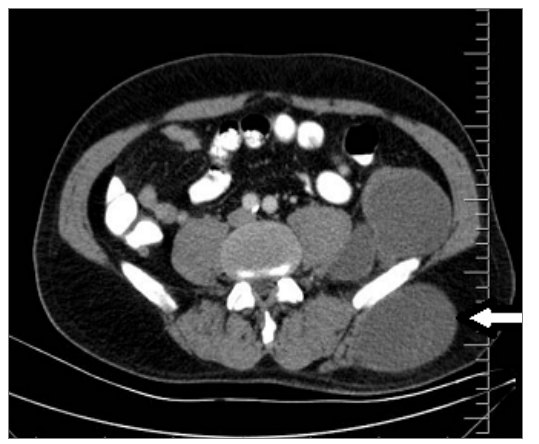
在将盐水湿润的膝垫放置在其周围并获得一个润滑液后,囊泡被吸入。 囊壁无屋顶. 人们看到女儿的睾丸,并诊断出感染的Hydatid囊肿。 为培养和敏感度采集了一批液体样本。 囊肿的含量完全空出后,空间被灌溉,上面有超音速盐碱溶液和10%的povidoneiodine. 增加了部分切除囊肿壁,手术完成. 术后课程是没有创意的。 文化结果为阴性,病理检查与hydatid囊肿一致. Albendazol治疗始于术后早期.
讨论情况
虽然Hydatid病多分布在肝脏和肺部,但是在肌肉结构中也很少出现,特别是在来自地方病区的病人中. (3)涉及肌肉的Hydatid疾病有3-5%的先发性 (4, 5) 通常由身体遥远部位的hy囊自发或创伤后破裂所致,并可能次于 囊手术 (6)。 。 。
在诊断中,美国应该是第一选择。 另一方面,在出现晚期疾病的情况下,可以确定损伤与周围组织的关系和适当的分解幅度,并且可以通过核磁共振进行差别诊断。 (7)阳性时,可以考虑血清测试的结果,但据报告,在半个主要的肌肉Hydatid囊肿病例中,这些结果为虚假阴性,发现IHA敏感度为67%。 (8)我们的病人呈现出一个明显的质量 被美国确定为囊肿, 之后被核磁共振发现为Hydatid疾病。 此外,核磁共振和核磁共振在1/512乳位上呈阳性。 应避免诊断性活检或渴望,因为疾病可能破裂和传播 (9). 外科手术目前为首选治疗 (10)我们的病例也是通过手术治疗的 在手术中,感染的囊肿被部分切除 并完全排水。
总之,在像土耳其这样的地方病流行国家,必须记住,任何软组织出现肿胀的病人,都应该通过身体检查和对hydatid疾病的成像研究,谨慎地加以评价。 此外,除非排除hydatid疾病,否则不应为进行活检或愿望而付出任何努力。
声明
- 作者贡献(CRediT)
- 所有作者对全部 14 项 CRediT 角色贡献均等(Conceptualization, Data curation, Formal analysis, Funding acquisition, Investigation, Methodology, Project administration, Resources, Software, Supervision, Validation, Visualization, Writing – original draft, Writing – review & editing)。
- 资助
-
Funding information was not reported in the published version of this article.
- 利益冲突
- A conflict of interest statement was not reported in the published version of this article.
- 伦理批准
- Ethics approval details were not reported in the published version of this article.
- 数据可用性
- A data availability statement was not reported in the published version of this article.
参考文献 (10)
10 条参考文献中有 7 条已链接 DOI。
- Sayek I, Tirnaksiz M. Cystic hydatid disease: Current trends in diagnosis and management. Surg today 2004; 34(8):987-96. DOI Crossref PubMed Google Scholar
- Digesu G, Picone A, La Bianca A, Massaro M, Vetri G. Muscular and subcutanuous hydatidosis. Minerva Med 1987;78(7):835-40. PubMed Google Scholar
- Eckert J, Deplazes P. Biological, epidemiological, and clinical aspects of echinococcosis, a zoonosis of increasing concern. Clin Microbiol Rev 2004;17:107–35. DOI Crossref PubMed Google Scholar
- Gougoulias NE, Varitimidis SE, Bargiotas KA, Dovas TN, Karydakis G, Dailiana ZH. Skeletal muscle hydatid cysts presenting as soft tissue masses. Hippokratia 2010;14:126– 30. PubMed Google Scholar
- García-Alvarez F, Torcal J, Salinas JC, Navarro A, García-Alvarez I, Navarro-Zorraquino M, et al. Musculoskeletal hydatid disease: A report of 13 cases. Acta Orthop Scand 2002;73:227– 31. DOI Crossref PubMed Google Scholar
- Guidelines for treatment of cystic and alveolar echinococcosis in humans. WHO Informal Working Group on Echinococcosis. Bull World Health Organ1996;74:231– 42. PubMed Google Scholar
- Memis A, Arkun R, Bilgen I, Ustun EE. Primary soft tissue hydatid disease: report of two cases with MRI characteristics. Eur Radiol 1999;9:1101– 3. DOI Crossref PubMed Google Scholar
- Durakbasa CU, Tireli GA, Sehiralti V, Sander S, Tosyali AN, Mutus M. An audit on pediatric hydatid disease of uncommon localization: incidence, diagnosis, surgical approach, and outcome. J Pediatr Surg 2006;41:1457–63. DOI Crossref PubMed Google Scholar
- Tatari H, Baran O, Sanlidag T, Gore O, Ak D, Manisalı M, et al. Primary intramuscular hydatidosis of supraspinatus muscle. Arch Orthop Trauma Surg 2001;121:93-4. DOI Crossref PubMed Google Scholar
- Yorganci K, Sayek I. Surgical treatment of hydatid cysts of the liver in the era of percutaneous treatment. Am J Surg 2002;184:63–9. DOI Crossref PubMed Google Scholar
阅读全文
在此打开 PDF
正在加载 PDF…
若 PDF 未在此显示,请使用上方的下载按钮。
如何引用本文
Ari A, Buyukasik K, Akce B, Tatar C, Vardar YM. Primary Hydatid Disease within the Quadratus Lumborum Muscle. Ulutas Med J. 2016;2(1):63-65. doi:10.5455/umj.20160317093539
Ari, A., Buyukasik, K., Akce, B., Tatar, C., & Vardar, Y. M. (2016). Primary Hydatid Disease within the Quadratus Lumborum Muscle. The Ulutas Medical Journal, 2(1), 63–65. https://doi.org/10.5455/umj.20160317093539
Ari, A. et al. (2016) 'Primary Hydatid Disease within the Quadratus Lumborum Muscle', The Ulutas Medical Journal, 2(1), pp. 63–65. doi: 10.5455/umj.20160317093539.