The Ulutas Medical Journal · ISSN 2149-0430 (print), 2149-388X (online) · https://www.ulutasmedicaljournal.com/?mno=18828
Original Article · Open access
The Evaluation of Risk Factors and Results in Pregnant Women with Intrauterine Fetal Hydrocephalus: A Single Center Experience
- Department of Obstetrics and Gynecology, Demiroglu Bilim University, Istanbul, Turkey
- Department of Neurosurgery, Baskent University-Istanbul Hospital, Istanbul, Turkey ROR
- Department of Obstetrics and Gynecology, Kanuni Sultan Süleyman Research Center, Istanbul, Turkey ROR
- Department of Obstetrics and Gynecology, Atakoy Towers Private Clinic, Istanbul, Turkey
- Department of Obstetrics and Gynecology, Sarıyer Hamidiye Etfal Training & Research Hospital, Istanbul, Turkey ROR
Abstract
Introduction Fetal hydrocephalus (FHC) occurs as a result of excessive production of cerebrospinal fluid (CSF) or CSF absorption or circulation disorders.We aim to evaluate risk factors, and to analyze the management and results in pregnant women diagnosed with FHC. Material and Methods Pregnant women diagnosed with prenatal FHC in the perinatology center between January 2015-December 2020 were included in the study, retrospectively. Demographic datas and anomalies accompanying FHC were analyzed. The frequency of fetal magnetic resonance imaging(MRI), amniocentesis and/or karyotype analysis rates, and termination frequency were examined. Results A total of 117 patients with intrauterine FHC were included. The median age and gestational week were 25.5 years and 21+4 weeks. Isolated FHC was observed in 32.5%. The frequency of fetal USG use is 97.4%; MRI was performed only in 3 (2.6%) patients. The amniocentesis and karyotype analysis were performed in 17.1% and 15.4%. While the presence of isolated FHC or accompanying anomalies has no effect on performing amniocentesis and karyotype analysis; it was determined that karyotype analysis was applied significantly less in case of NTD diagnosis(p=0.001). While the termination rates was 47.9; live birth occurred in 51.3%. The gestational week was significantly lower in patients who were terminated than in those whose delivery was decided (median 20+3 vs 23+2) (p=0.015). Termination rates were found to be significantly lower than those with anomaly accompanying FHC(p=0.018). Conclusion Accuracy rates of fetal USG in the diagnosis of prenatal FHC are quite high. Prenatal diagnosis and timing are important factors affecting termination and live birth rates.
Keywords
Full text
Introduction
Fetal hydrocephalus (FHC) occurs as a result of excessive production of cerebrospinal fluid (CSF)/CSFabsorption disorderor CSF circulation disorders (1). Fetal ventriculomegaly is defined as a lateral ventricular atrial diameter of ≥10 mm (normal range 6.1±1.3 mm) (1, 2). Increasing the CSF concentration in the ventricular system causes ventricular dilatation and increased intracranial pressure (1). In the literature, the incidence of FHC is reported to be between 0.3-2/1000 live births (3, 4). The most common finding of abnormal brain development in the second and third trimesters of pregnancy is FHC (1, 2).
Chromosomal anomalies, genetic defects and intrauterine infections are generally reported for the etiology of FHC (5, 6). In addition to the fact that its etiology is quite complicated, neurodevelopmental outcomes of the fetus also differ after birth. This situation causes many difficulties for clinical management, prenatal follow-up, delivery and postnatal management (1–3). After the diagnosis of FHC is made in pregnant women, scans are made in terms of accompanying anomalies [chromosomal anomaly and central nervous system anomaly (Chiari-II, Dandy-Walker malformation and corpus callosum agenesis, etc.)] and a decision is made with the parents in terms of the progression or termination of pregnancy. The easy use of ultrasonography (USG) makes it the most commonly used method in the diagnosis of FHC; depending on fetal position, cranial ossification, and technical reasons, diagnosis may be delayed or missed (1, 3, 5). In the literature, the rate of misdiagnosis of the first USG was reported to be approximately 12.8% (7). Magnetic resonance imaging (MRI), which is not affected by maternal and fetal factors, can be used to diagnose FHC and detect other abnormalities (8). Determining whether demographic characteristics have an effect on the development of FHC in pregnants can prevent FHC before and during pregnancy or take necessary precautions in early period. Our primary is to evaluate the demographic datas and risk factors of pregnants who diagnosed with FHC in the perinatology. The investigations of management, amniocentesis rates, genetic etiologies and termination rates were second aim of the study.
Materials and Methods
Patients Selection
All pregnants diagnosed with FHC between January 2015 and December 2020 in the tertiary gynecology & obstetrics clinic and perinatology center were included in the present study. After giving written or verbal information about the study, written consent was obtained from the patients who agreed to participate in the study. Ethics committee approval was given by the faculty scientific research ethics committee. Data Collection and Assessment of Patients Demographic datas (gender, age, gestational week), gravida, parity, abortion and curettage numbers, and anomalies accompanying FHC were analyzed. Tge results of the pregnants in the perinatology council, the frequency of fetal MRI use, amniocentesis and/or karyotype analysis rates, and termination frequency were examined. In addition, the factors affecting the amniocentesis and termination/follow-up decision in pregnants who diagnosed with FHC in the prenatal period (gestational age, week, accompanying anomalies) were investigated.
Statistical Analysis
All data were analysed using the SPSS 25 (IBM, Armonk, NY: IBM Corp.) program Continuous variables were expressed as mean±standard deviation, median (interquartile range, IQR), and categorical variables as numbers (n) and percentages (%). When the parametric test assumptions were met, t-test was used to compare differences between independent groups. When parametric test assumptions were not met, Mann-Whitney U test was used to compare differences between independent groups. The Fisher's exact probability or Chi-squared tests used to compare demographics. p<0.05 was considered statistically significant.
Results
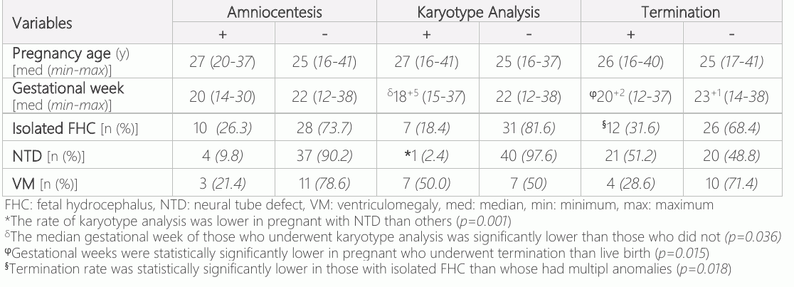
The medical records of 129 pregnants who diagnosed with intrauterine FHC were analyzed. Twelve pregnants were excluded from the study due to missing data. 117 pregnants who diagnosed with FHC were included in the study. The median age at the time of diagnosis was 25.5 (min: 16 – max: 41, IQR 22 - 30) years, while the median gestational week was 21+4 (min: 12+3- max: 38, IQR 18 - 26) weeks. In addition,gravida, parity, abortion and curettage are shown in Table-1. One third of pregnants [34.2% (n=40)] were primiparous. While 32.5% (n=38) of the pregnants had "isolated FHC" and 6.0% (n=7) had "triventricular hydrocephalus"; “ventriculomegaly (VM)” in 12% (n=14), “neural tube defects (NTD)” in 35% (n=41), “cerebellar hypoplasia” in 6.8% (n=8), and % 4.3 (n=5) had accompanying “Dandy-Walker Anomaly”. NTD diagnosis (p=0.001) (Table-3). Live birth occurred in 75% (n=15) of the pregnants who underwent amniocentesis, termination was performed in 5 pregnants. Multipl anomalies with FHC were observed in 3 pregnants and isolated FHC was observed in 2 pregnants who underwent amniocentesis and were terminated. No any chromosomal anomalies were in any of the fetuses whose karyotype analysis was done. The termination rate was 47.9% (n=56) and live birth occurred in 51.3% (n=60) (Table-2). The gestational week was significantly lower in the pregnants who were terminated than in those who decided to live birth (median 20+3 vs 23+2) (p=0.015) (Table-3). In addition, termination rates in pregnants with isolated FHC were lower than those with anomaly accompanying FHC (p=0.018). On the other hand, it was observed that the accompanying NTD or VM had no affect on the termination decision (p=0.593 and p=0.865, respectively)(Table-3).
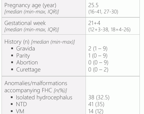

Gestational age and week and number of abortion and/or curettage did not have any effect on isolated FHC/accompanying anomaly /malformation (p>0.05). In our study, fetal USG was performed in all pregnants who diagnosed with FHC; fetal MRI was performed only in 3 (2.6%). In addition, while amniocentesis was performed in 17.1% (n=20), karyotype analysis was performed in 15.4% (n=18) (Table-2). No complications were observed in any of the pregnants who underwent amniocentesis.It was observed that the age and gestational week of the pregnants had no effect on the decision to perform amniocentesis (Table-3). While the presence of isolated FHC or accompanying anomalies has no effect on performing amnio centesis and karyotype analysis; karyotype analysis was applied significantly less in case of
Discussion
Our study includes the characteristics, risk factors and management of pregnants who diagnosed with prenatal FHC, and anomalies accompanying the fetus with FHC. Although the incidence of FHC is reported as 0.3-2/1000 live births, the actual incidence is probably higher since many fetal death cases are missed (3, 4, 10, 11). However, although it has been reported that FHC is seen mostly in primiparous women, this rate is controversial because the frequency of curettage is unknown (9–12). In our study, 34.2% of the pregnants who diagnosed with FHC were found to be primiparous. In addition, it was found that 13.7% of the pregnants had a curettage history at least once. Contrary to the literature, the number of primiparous was low in our study; however, this rate might actually be higher because the numbers of fetuses who underwent curettage were not knew.
Although prenatally detected FHC is seen as isolated, it is seen accompanied by triventricular (communicating HC, aqueductus stenosis, etc.), Dandy-Walker Anomaly and other anomalies (cerebellar, ventriculomegaly, corpus callosum agenesis). Approximately 60% of non-isolated FHC cases are accompanied by Dandy-Walker anomalies, NTDs and anomalies of the corpus callosum (13–15). In our study, similar to the literature, approximately one third of FHC cases were isolated, while NTD accompanied the other third. The primarily option for the diagnosis of FHC is fetal USG. Although it is easy to apply, fetal MRI is needed due to its nature, the inability to visualize some anomalies, and limiting factors such as technical reasons (16, 17). While Wang et al. found the accuracy of USG to be 92.9% in the diagnosis of FHC in pregnants, Parazzini C et al. reported that USG could not diagnose in 27.4% of patients compared to MRI in 179 fetal ventriculomegaly cases(1, 18). In our study, 97.4% of the pregnants with FHC were diagnosed with USG, while fetal MRI was performed in only 3 patients. Similar with literature, fetal MRI usage was lower in our study. The high experience of USG and technological developments of physicians in the perinatology center may explain this result. In case of malformations, advanced maternal age, anomalies, genetic syndromes in fetal USG, or uncertain USG findings, further evaluation with genetic counseling, amniocentesis, and karyotype analysis methods is recommended (19, 20). Although amniocentesis is a reliable diagnostic method, complications may develop after the procedure. These complications are mostly inversely proportional to the experience of the person performing the procedure (21). In our study, the frequency of amniocentesis was found to be 17.1%, while our genetic analysis rate was 15.4%. The absence of complications in any pregnant after amniocentesis can be explained by the experienced physicians in the perinatology center. In addition, it has been reported that the incidence of chromosomal anomaly in genetic examinations performed after USG anomaly is seen is between 4-27% (22–24). In our study, chromosomal anomaly was not detected in any of the samples. An insufficient number of patients who underwent amniocentesis. Therefore the genetic analysis performed in fewer patients may explain this. Inclusion of who were diagnosed with FHC (although some accompanying anomalies) and the absence of pregnants with suspected fetal chromosomal anomaly explains the result that anomaly was not observed in any fetus, contrary to the literature. In addition, it was observed that the age and gestational week of the pregnants did not have any effect on the decision to perform amniocentesis in our study. Also, the rate of amniocentesis/genetic analysis in pregnants who were diagnosed with NTD on fetal USG or MRI was lower than isolated FHC or other concomitant anomalies. The fact that the probability of chromosomal anomaly in pregnants diagnosed with fetal NTD is higher than the isolated FHC or other accompanying anomalies may explain this result.
In a study conducted by Garne E et al with the data of 4 countries in Europe, it was observed that the live birth rate was 47% in pregnants diagnosed with FHS, while the termination rate was 48% and the stillbirth rate was 5% (25). On the other hand, the same study reported that, in pregnants diagnosed with FHC in the prenatal period, live birth was reported as 35.7%, termination was 60% and stillbirth rate was 4.3% (25). In line with these results, it was suggested that prenatal diagnosis and timing are the main criteria that are effective in making the termination decision. The termination rate varies considerably due to the socioeconomic status of the countries, the role of women in social life, education levels, cultural factors and religious differences (26, 27). The termination is not allowed except in life- threating situations for women and/or in 120 days of pregnancy in Muslim countries. For this reason, termination cannot be applied in pregnants who have fetal anomalies after 120 days and babies with anomalies are born (28). In our country, education levels, socio-economic conditions, ethnic and religious differences also affect the termination rates (29). In our study, while the live birth rate was 51.3%, the termination frequency was 47.9%. Although all pregnants were diagnosed prenatally, the termination frequency was lower; it should be kept in mind that socio-economic conditions and religious differences in our country may also affect this decision. Similar to the literature, in our study, it was observed that the gestational week of the pregnants who underwent termination was lower than those who decided to live birth. This result supports that early prenatal diagnosis increases the frequency of termination.
Limitations of the study; (1) the single-center nature of our study constitutes an important limitation in the generalization; (2) Since it is retrospective, the follow-up of pregnants and the postpartum life expectancy are unknown.
Conclusion
FHC is one of the most common central nervous system anomalies in pregnants. The gestational week was significantly lower in the pregnants with FHC who were terminated than in those who decided to live birth. Termination rates in pregnants with isolated FHC were lower than those with anomaly accompanying FHC. Amniocentesis / genetic analysis is performed less frequently in case of NTD diagnosis.
Declarations
- Author contributions (CRediT)
- All authors contributed equally to all 14 CRediT roles (Conceptualization, Data curation, Formal analysis, Funding acquisition, Investigation, Methodology, Project administration, Resources, Software, Supervision, Validation, Visualization, Writing – original draft, Writing – review & editing).
- Funding
-
Funding information was not reported in the published version of this article.
- Conflicts of interest
- All authors declare no conflict of interest.
- Ethics approval
- Ethics committee approval was given by the faculty scientific research ethics committee.
- Data availability
- A data availability statement was not reported in the published version of this article.
References (29)
23 of 29 references are linked to their DOI.
- Wang R, Ding Y, Yang M, Lai W. Diagnosis, management, and neurodevelopmental outcomes of fetal hydrocephalus: an observational prospective study. Childs Nerv Syst. 2021 DOI Crossref PubMed Google Scholar
- Pilu G, Falco P, Gabrielli S, Perolo A, Sandri F, Bovicelli L. The clinical significance of fetal isolated cerebral borderline ventriculomegaly: report of 31 cases and review of the literature. Ultrasound Obstet Gynecol. 1999;14(5):320-6 DOI Crossref PubMed Google Scholar
- Pisapia JM, Sinha S, Zarnow DM, Johnson MP, Heuer GG. Fetal ventriculomegaly: Diagnosis, treatment, and future directions. Childs Nerv Syst. 2017;33(7):1113-1123 DOI Crossref PubMed Google Scholar
- Peiro JL, Fabbro MD. Fetal therapy for congenital hydro cephalus-where we came from and where we are going. Childs Nerv Syst. 2020;36(8):1697-1712 DOI Crossref PubMed Google Scholar
- Cavalheiro S, Moron AF, Almodin CG, Suriano IC, Hisaba V, Dastoli P, Barbosa MM. Fetal hydrocephalus. Childs Nerv Syst. 2011;27(10):1575-83 63 77 Intrauterine Fetal Hydrocephalus Korkmaz et al. PubMed Google Scholar
- Karimy JK, Reeves BC, Damisah E, Duy PQ, Antwi P, David W, Wang K, Schiff SJ, Limbrick DD Jr, Alper SL, Warf BC, Nedergaard M, Simard JM, Kahle KT. Inflammation in acquired hydrocephalus: pathogenic mechanisms and therapeutic targets. Nat Rev Neurol. 2020;16(5):285-296 DOI Crossref PubMed Google Scholar
- Tugcu AU, Gulumser C, Ecevit A, Abbasoglu A, Uysal NS, Kupana ES, Yanik FF, Tarcan A. Prenatal evaluation and postnatal early outcomes of fetal ventriculomegaly. Eur J Paediatr Neurol. 2014;18(6):736-40 DOI Crossref PubMed Google Scholar
- Di Mascio D, Sileo FG, Khalil A, Rizzo G, Persico N, Brunelli R, Giancotti A, Panici PB, Acharya G, D'Antonio F. Role of magnetic resonance imaging in fetuses with mild or moderate ventriculomegaly in the era of fetal neurosonography: systematic review and meta-analysis. Ultrasound Obstet Gynecol. 2019;54(2):164-171 DOI Crossref PubMed Google Scholar
- Nomura ML, Barini R, De Andrade KC, Milanez H, Simoni RZ, Peralta CF, Machado IN, Zambelli H, Maio KT. Congenital hydrocephalus: gestational and neonatal outcomes. Arch Gynecol Obstet. 2010;282(6):607-11 DOI Crossref PubMed Google Scholar
- Moutard ML, Kieffer V, Feingold J, Kieffer F, Lewin F, Adamsbaum C, Gélot A, Campistol I Plana J, van Bogaert P, André M, Ponsot G. Agenesis of corpus callosum: prenatal diagnosis and prognosis. Childs Nerv Syst. 2003;19(7-8):471-6 DOI Crossref PubMed Google Scholar
- Cavalheiro S, Sparapani FV, Moron AF, da Silva MC, Stávale JN. Fetal meningeal hemangiopericytoma. Case report. J Neurosurg. 2002;97(5):1217-20 DOI Crossref PubMed Google Scholar
- Stein SC, Feldman JG, Apfel S, Kohl SG, Casey G. The epidemiology of congenital hydrocephalus. A study in Brooklyn. Childs Brain. 1981;8(4):253-62 DOI Crossref PubMed Google Scholar
- Baffero GM, Crovetto F, Fabietti I, Boito S, Fogliani R, Fumagalli M, Triulzi F, Mosca F, Fedele L, Persico N. Prenatal ultrasound predictors of postnatal major cerebral abnormalities in fetuses with apparently isolated mild ventriculomegaly. Prenat Diagn. 2015;35(8):783-8 DOI Crossref PubMed Google Scholar
- Özkavukcu E, Haliloğlu N. Fetal Kraniospinal ve Fasiyal Anomaliler. Trd Sem 2017;5:211-33 PubMed Google Scholar
- Koyuncu Arslan M, Akar M, Pala HG, Taner CE, Oncel MY. Fetal Santral Sinir Sistemi Anomalilerinin Değerlendirilmesi; Referans Merkez Perinatoloji Konsey Verileri. Forbes J Med. 2020;1(3):75-78 PubMed Google Scholar
- Levine D, Barnes PD, Robertson RR, Wong G, Mehta TS. Fast MR imaging of fetal central nervous system abnormalities. Radiology. 2003;229(1):51-61 DOI Crossref PubMed Google Scholar
- Glenn OA, Barkovich AJ. Magnetic resonance imaging of the fetal brain and spine: an increasingly important tool in prenatal diagnosis, part 1. AJNR Am J Neuroradiol. 2006;27(8): 1604-11 PubMed Google Scholar
- Parazzini C, Righini A, Doneda C, Arrigoni F, Rustico M, Lanna M, Triulzi F. Is fetal magnetic resonance imaging indicated when ultrasound isolated mild ventriculomegaly is present in pregnancy with no risk factor? Prenat Diagn 2012;32(8):752-7 DOI Crossref PubMed Google Scholar
- Flick A, Krakow D, Martirosian A, Silverman N, Platt LD. Routine measurement of amniotic fluid alpha-fetoprotein and acetylcholinesterase: the need for a reevaluation. Am J Obstet Gynecol. 2014;211(2):139.e1-6 DOI Crossref PubMed Google Scholar
- Yayla M, Bayhan G, Yalınkaya A, Alp N, Fidanboy M, Görmüş H.Yüksek riskli gebeliklerde 2. trimester genetik amniyosentez: 165 olgunun klinik değerlendirilmesi. Perinatoloji Dergisi. 1999; 7(1):40-46 PubMed Google Scholar
- Leschot NJ, Verjaal M, Treffers PE. Risks of midtrimester amniocentesis; assessment in 3000 pregnancies. Br J Obstet Gynaecol. 1985;92(8):804-7 DOI Crossref PubMed Google Scholar
- Stoll C, Dott B, Alembik Y, Roth MP. Evaluation of routine prenatal ultrasound examination in detecting chromosomal abnormalities in a low risk population. Hum Genet. 1993;91(1): 37-41 DOI Crossref PubMed Google Scholar
- Lockwood DH, Neu RL. Cytogenetic analysis of 1375 amniotic fluid specimens from pregnancies with gestational age less than 14 weeks. Prenat Diagn. 1993;13(9):801-5 DOI Crossref PubMed Google Scholar
- Eddleman KA, Malone FD, Sullivan L, Dukes K, Berkowitz RL, Kharbutli Y, Porter TF, Luthy DA, Comstock CH, et al. Pregnancy loss rates after midtrimester amniocentesis. Obstet Gynecol. 2006;108(5):1067-72 DOI Crossref PubMed Google Scholar
- Garne E, Loane M, Addor MC, Boyd PA, Barisic I, Dolk H. Congenital hydrocephalus--prevalence, prenatal diagnosis and outcome of pregnancy in four European regions. Eur J Paediatr Neurol. 2010;14(2):150-5 DOI Crossref PubMed Google Scholar
- Yogi A, K C P, Neupane S. Prevalence and factors associated with abortion and unsafe abortion in Nepal: nationwide cross-sectional study. BMC Pregnancy Childbirth. 2018;18(1): 376 DOI Crossref PubMed Google Scholar
- Chae S, Desai S, Crowell M, Sedgh G, Singh S. Characteristics of women obtaining induced abortions in selected low- and middle-income countries. PLoS One. 2017;12(3):e0172976 DOI Crossref PubMed Google Scholar
- Shapiro GK. Abortion law in Muslim-majority countries: an overview of the Islamic discourse with policy implications. Health Policy Plan. 2013,8:1-12 DOI Crossref PubMed Google Scholar
- Cavli A, Ergocmen B. Ethno-cultural analysis of abortion and contraception: The cases of Turkey and selected Asian states. Nüfusbilim Dergisi. 2013;32–33 PubMed Google Scholar
Read the full text
Open the PDF here
Loading the PDF…
If the PDF does not show here, use the download button above.
How to cite this article
Korkmaz N, Alatas I, Oncu N, Ekiz A, Aydin AA. The Evaluation of Risk Factors and Results in Pregnant Women with Intrauterine Fetal Hydrocephalus: A Single Center Experience. Ulutas Med J. 2022;8(1):58-63. doi:10.5455/umj.20211122060917
Korkmaz, N., Alatas, I., Oncu, N., Ekiz, A., & Aydin, A. A. (2022). The Evaluation of Risk Factors and Results in Pregnant Women with Intrauterine Fetal Hydrocephalus: A Single Center Experience. The Ulutas Medical Journal, 8(1), 58–63. https://doi.org/10.5455/umj.20211122060917
Korkmaz, N. et al. (2022) 'The Evaluation of Risk Factors and Results in Pregnant Women with Intrauterine Fetal Hydrocephalus: A Single Center Experience', The Ulutas Medical Journal, 8(1), pp. 58–63. doi: 10.5455/umj.20211122060917.