The Ulutas Medical Journal · ISSN 2149-0430 (print), 2149-388X (online) · https://www.ulutasmedicaljournal.com/?mno=8217
Original Article · Open access
Retrospective Evaluation of the Role of CA19-9, CEA and Hematological Parameters in the Prediction of Helicobacter Pylori, Gastritis Etiology and Endoscopic Lesions
- Department of Internal Medicine, Health Ministry, Bolu İzzet Baysal State Hospital, Bolu, Turkey ROR
- Department of Internal Medicine, Bolu Abant İzzet Baysal University, Faculty of Medicine, Bolu, Turkey ROR
Abstract
- Introduction
- Helicobacter pylori (HP) is an infectious disease that generates chronic gastric and duodenal mucosal tissue inflammation. The neutrophil-lymphocyte ratio (NLR) and platelet-lymphocyte ratio (PLR) are easy indices of inflammation determined from hemograms. We aimed to see whether there is a link between HP and gastric mucosal lesions, NLR, PLR, hematological parameters, and tumor markers that can be used to screen for cancers that are common in the population, like Carbohydrate antigen 19-9 (CA19-9) and Carcinoembryonic antigen (CEA).
- Materials and Methods
- We included 100 males and 106 females who applied with dyspeptic complaints to our hospital outpatient clinics and underwent upper endoscopy. All of the parameters from the hospital registration system's recorded data were analyzed retrospectively.
- Results
- There was no difference between the 64 (31%) HP positive(+) and 142 (69%) HP negative(-) groups. CA 19-9, NLR, PLR, and neutrophil count were higher in patients with gastric erosion than in patients with normal mucosa in the HP(+). PLR was higher in patients with gastric erosion, and leucocyte and monocyte counts were higher in patients with gastric ulcers than in patients with normal gastric mucosa in the HP(-) group. NLR and PLR were higher in the gastric erosion group than in the normal mucosa group, independently from HP. Monocyte counts are higher in patients with gastric ulcers than in those with gastric erosion and normal gastric mucosa.
- Conclusion
- Any of these markers were unable to predict the presence of HP. The immune response, which was triggered by inflammation caused by HP, was heterogeneous; hereditary factors and different nutrition behaviours may be reasons for obtaining different data from the present study and other similar studies on this issue.
Keywords
Full text
Introduction
Dyspepsia, which is described as pain and discomfort in upper abdomen, affects about a quarter of the population each year. Although organic causes such as gastroduodenal ulcer, esophagitis, pancreaticobiliary disease, gastritis, duodenitis, gastric precancerous lesion, and/or gastric cancer can cause it, no underlying organic cause is frequently discovered.
Endoscopic examination is the most commonly used diagnostic procedure for determining organic causes. Gastritis is inflammation of the gastric mucosa, which is a major cause of dyspepsia. According to the Sydney system of classification, it is divided into acute, chronic, and special forms, with special forms also accepted as chronic gastritis. HP is a significant human pathogen that causes peptic ulcer disease, gastritis, gastric adenocarcinomas, and gastric lymphoma, as well as dyspepcy (1, 29, 30). Although the bacterium causes only minor inflammation in many people, long-term HP-induced gastric inflammation usually results in atrophic gastritis, which is thought to be the initial stage in the histogenesis of gastric cancer (2, 31, 32). Based on epidemiological findings, the World Health Organization classified HP as a type-I cancerogenic agent in1994, and this classification was confirmed in 2012. (3). Tumor markers such as CA 19-9 and CEA, which are biomarkers that can be used to screen for malignancy that is common in the population, can be used to screen for cancers that are common in the public. CA 19-9 is increased in many types of gastrointestinal malignancies, including colorectal, hepatocellular, and esophageal cancers, and it is used to follow-up pancreatic cancer patients (4). CEA, especially associated with colon, lung, medullary thyroid, breast, gastric or pancreatic adenocarcinomas, is used in the follow-up of colorectal cancers(5). Hemogram parameters have been evaluated as a means of evaluating systemic inflammation in many diseases. Since leukocytes induce an increase in neutrophil count and a decrease in lymphocyte count as a physiological response to stress, NLR and PLR can be evaluated as sensitive markers of systemic inflammation(6, 7). The purpose of the present study was to see if there was any link between HP infection and etiology of gastric mucosal lesion, CA19-9, CEA, NLR, PLR and hematological parameters.
Materials and Methods
Subjects
The study included 100 male and 106 female, a totally of 206 patients over the age of 18 who applied to our outpatient clinics between June and July 2018 with dyspeptic complaints, underwent upper gastrointestinal system (GIS) endoscopy after appropriate preparation and had simultaneous CA19-9, CEA, and parameters derived from hemogram. The study was done as a cross-sectional retrospective study.
Ethical Statement
The present study was approved by the local ethics boards (2019/22) and complies with the provisions of the Declaration of Helsinki, which declares ethical principles to guide medical research. Informed consent was obtained from all patients before the endoscopic procedure.
Methods
Endoscopies were performed with Olympus EVIS EXERA, GIF Q150 (serial no: 2205164) device. Endoscopic diagnoses were recorded by the gastroenterology specialist and the histopathological examination of endoscopic biopsy samples and the presence of HP were performed by the pathology specialist. Serum CEA and CA 19-9 levels were measured with the immunoassay method on the Abbott Architect i2000SR (Abbott Laboratories, Abbott Park, IL, USA) instrument using the Abbott Commercial Kit. CEA levels between 0-3 ng/ml and CA 19-9 between 0-37 U/ml were considered normal. The hemogram was measured using the Abbott Cell-Dyn 3700 complete blood count device (Abbott Laboratories, Abbott Park, IL, USA), which examined cells in three dimensions using laser light scattering and fluorescent signals from two angles. NLR and PLR were calculated by examining the hemogram results. All of the recorded data was obtained retrospectively using the hospital registry system.
Patients under the age of 18, pregnant or lactating women, acute or chronic pancreatitis, liver cirrhosis, acute cholangitis, obstructive jaundice, inflammatory bowel disease, active GI bleeding, history of malignancy, uncontrolled diabetes, uncontrolled hypertension, rheumatic disease, active infection, long-term NSAID, PPI (proton pump inhibitor), H2 receptor blocker, or antibiotic use in the previous 2 weeks, GIS obstruction and/or perforation, active smokers, and those receiving HP eradication therapy were excluded from the present study.
Statistical Evaluation
For statistical analysis, IBM SPSS 20 (Statistics Program for Social Scientists) was used. To evaluate whether the distribution is normal, Kolmogorov-Smirnov test, pairwise comparison of continuous variables, Student t-test for those with normal distribution, Mann-Whitney-U test for those without normal distribution, multiple comparisons of continuous variables, One-Way ANOVA for those with normal distribution, Kruskal-T for those without normal distribution. Wallis test, Chi-square test, and Fiber exact test were used to compare categorical data. The significance level of p<0.05 was accepted.
Results
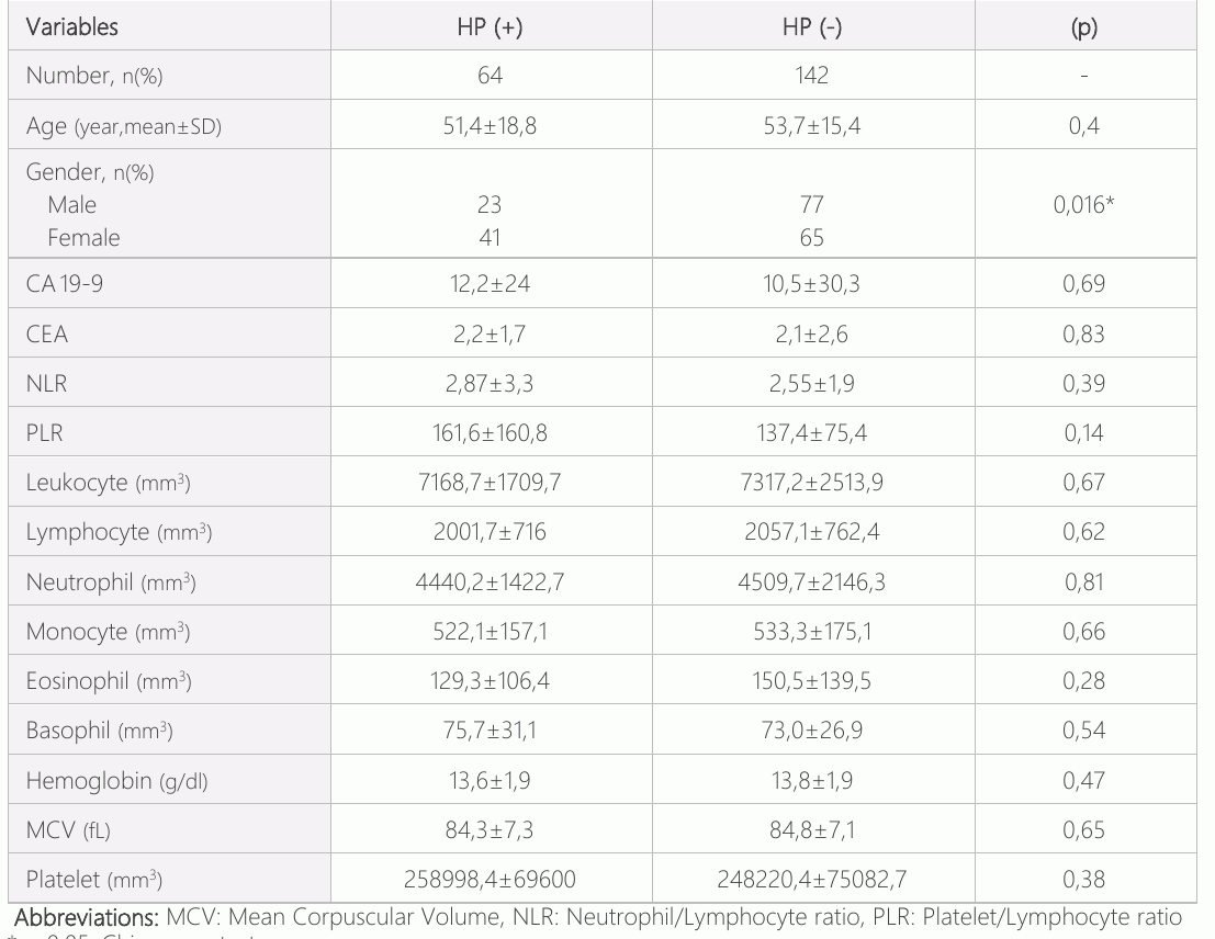
The mean age of 206 patients included in the study was 53±16.5, 64 (31%) were HP (+). The youngest age was 18, while the oldest age was 83. The mean age of HP(+) patients was 51.4± 18.8 years, 41 (64%) women and 23 (36%) men. HP (-) was found in 142 (68.9%) of the patients. The mean age of HP (-) was 53.7±15.4 years, 65 (45.7%) women and 77 (54.2%) men [Table-1]. In both groups, there was no difference in age. (p=0.40). The number of HP (+) individuals was found to be statistically significantly higher in the female gender than in the male gender (p=0.016). In terms of CA19-9, CEA, NLR, PLR, and other hemogram parameters, there was no significant difference between individuals with HP (+) and (-).
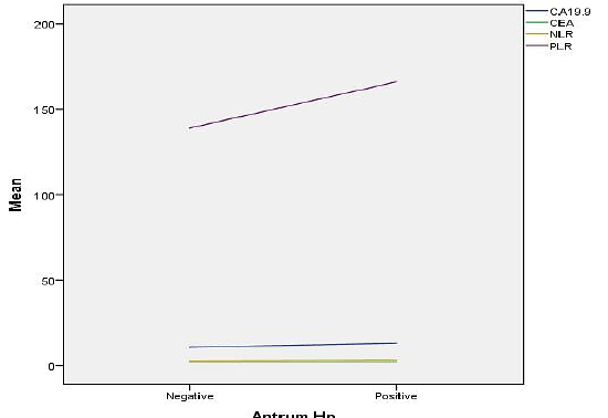
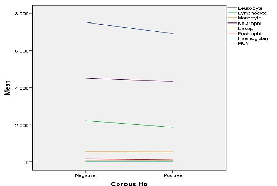
The data were analyzed for the antrum and corpus based on the presence of HP. According to the presence of HP in the gastric antrum and also corpus region, the distribution of serum CA 19-9, CEA, NLR, PLR levels was divided into two groups as HP(+) and HP(-). In terms of serum CA 19-9, CEA, NLR, and PLR levels, there was no significant difference between these groups [Figure-1]. As a result of endoscopic biopsy and histopathological evaluation of gastric corpus region, thy were divided into two groups as HP(+)/(-) and hemogram parameters such as leukocyte, lymphocyte, monocyte, neutrophil, basophil, eosinophil, hemoglobin and MCV. The lymphocyte count in the corpus was found to be significantly higher in those with HP(-) compared to HP(+) (p=0.018, 2228± 744.7 vs. 1868±650.9) [Figure-2].
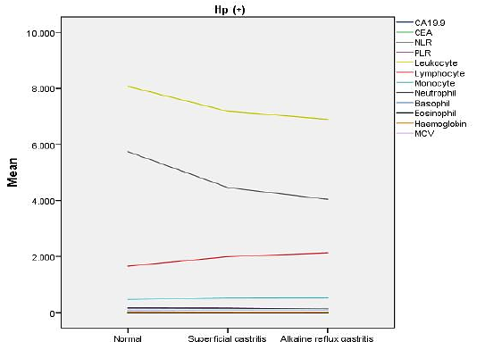
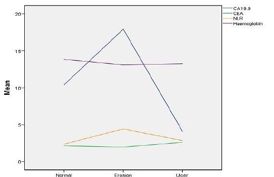
When the patients were evaluated according to their gastritis status as a result of endoscopy, gastric mucosa was found normal in 8 patients, superficial gastritis in 162 patients, and alkaline reflux gastritis in 36 patients. When HP (+) individuals with normal gastric mucosa, those with superficial gastritis and those with alkaline reflux gastritis were analyzed according to serum CEA, CA 19-9, NLR, PLR levels and the other hemogram parameters, no difference was found between individuals with normal gastric mucosa, those with superficial gastritis and alkaline reflux gastritis as in HP(-) [Figure-3]. As a result of their endoscopic evaluation, the patients were divided into three groups those with normal gastric mucosa, those with gastric erosion and those with gastric ulcer, and also were evaluated according to CA 19-9, CEA, NLR levels and the other hemoglobin parameters. The NLR was higher in the gastric erosion than in the normal gastric mucosa (p=0.0001). The levels of serum CA19-9, CEA, and hemoglobin did not differ significantly between the groups (p>0.05) [Figure-4].
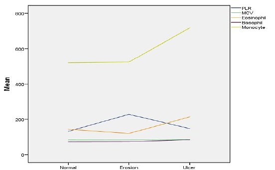
When the patients were divided into three groups as those with normal gastric mucosa, those with gastric erosion and those with gastric ulcer as a result of their upper GIS endoscopic evaluation, they were evaluated according to serum PLR, MCV, eosinophil, basophil and monocytes levels. The PLR level was found to be higher in the gastric erosion group than in the normal gastric mucosa group (p=0.0001). There was no significant difference between the groups in terms of MCV, eosinophil, basophil and monocyte levels. The monocyte count was found to be significantly higher in the gastric ulcer group compared to the normal gastric mucosa and gastric erosion (p=0.002, p=0.008, respectively) [Figure-5].
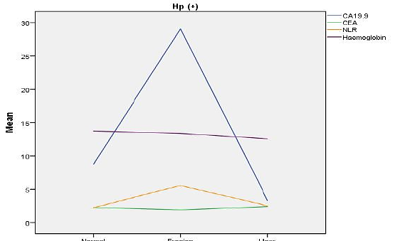
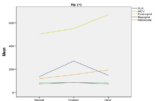
When the individuals with HP (+) were divided into 3 groups as those with normal gastric mucosa, those with gastric erosion and those with gastric ulcer according to the results of upper GI endoscopy, the CA19-9 and NLR levels were higher in the group with gastric erosion compared to the normal gastric mucosa (p= 0.021, p=0.004, respectively). There was no difference between the groups in terms of serum CEA and hemoglobin levels [Figure-6]. When individuals with HP (+) were divided into 3 groups as those with normal gastric mucosa, those with gastric erosion and with gastric ulcer according to the results of upper GI endoscopy, the PLR was higher in the gastric erosion than in the normal gastric mucosa group (p=0.024). There was no difference between the groups in terms of MCV, eosinophil, basophil and monocyte levels [Figure-7].
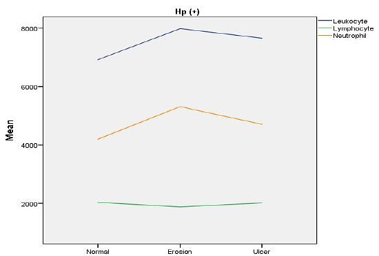
When individuals with HP(+) were divided into 3 groups as those with normal gastric mucosa, those with gastric erosion and those with gastric ulcer according to the results of upper GI endoscopy, the neutrophil count was found to be higher in the gastric erosion group than in the normal gastric mucosa group (p=0.038). In terms of serum leukocyte and lymphocyte levels, there was no difference between the groups [Figure-8].
Discussion
One of the most well-known GIS complaints is dyspepsia. It is more common in men than in women, and its incidence increases with age. It has been observed that the prevalence of dyspepsia varies between 25-50% in different societies (8). Gastritis, duodenal ulcer, gastric ulcer, GERD, NSAID use, and antibiotic use are among the most common causes of organic dyspepsia seen in the clinic (9). HP infection is reported to be more common in dyspepsia patients than in healthy controls (10). Chronic HP infection is most commonly seen in the antrum of the stomach in developed countries, and it is usually associated with duodenal ulcers. In developing countries, HP infection is more localized in the corpus. In this case, HP infection is the initiator of the cascade of chronic gastritis, gastric atrophy, intestinal metaplasia, dysplasia, and invasive carcinoma (11). Although studies are evaluating the relationship between HP and tumor markers and hemogram parameters (6, 12, 13), there is not enough data in the literature for recent studies investigating the relationship between superficial gastritis, alkaline reflux gastritis, gastric ulcer and gastric erosion, and tumor markers and hemogram parameters. The predictability of gastric pathologies diagnosed by some procedures such as endoscopy, with markers that can be seen in serum, has inspired us to work in this direction. Various studies in the literature show that the frequency of HP varies according to years and regions, and is relatively lower in Western societies and the west of Turkey (14–18). Some factors, such as the insufficient level of HP in the environment, the patchy distribution of HP in the gastric mucosa, and the investigation of the presence of HP using histopathology as the only method, may have caused the frequency of HP in our patient group to be lower than the results found in previous studies.
In the present study, we found that there was no significant difference between CA 19-9 and CEA levels in HP (+) and (-) groups. In the study of Xu et al. (12), the CEA level found in those with HP (+) and (-) was significant (p<0.001). An important reason for the differences in the results of the present study is that the results may be more meaningful due to the socio-cultural differences and nutritional habits of this study population and the Chinese individuals, and the sample size in the study is much larger. However, what makes the results of the present study more valuable is the histopathological diagnosis of HP as a method. Xu et al. used only serology for diagnostic purposes. The specificity of histopathology (95-98%) in the diagnosis of HP infection is higher than the specificity of serological tests (86-95%). In terms of CA 19-9 levels, no significant difference was found in terms of HP infection in both studies.
In inflammatory processes and infections, leukocyte, neutrophil, and the platelet count can increase. HP activates mechanisms that both activate and suppress immune response. In HP, leukocytosis, neutrophilia, monocytosis and basophilia are expected as a systemic effect of mucosal inflammation. In the present study, leukocyte and neutrophil counts were found to be statistically insignificantly higher in the HP(-) group (p=0.67, p=0.81)(19). Since the number of PLTs is also an acute phase reactant, it is expected to increase inflammatory processes such as HP infection. It causes an increase in MPV (mean platelet volume), PDW (platelet distribution width), PLR (platelet/lymphocyte ratio) and PLT numbers (20).
In various studies, when HP (+) and (-) patients are compared, it is seen that different results are obtained in terms of hematological parameters. The reason for the results coming out this way is that the bacterial virulence and the response of the host defence system show significant differences from person to person and this is reflected in laboratory values. We could not obtain a meaningful parameter that would guide us in the diagnosis and prognosis of HP infection, especially from the leukocyte formula. The result that we obtained in the study that the NLR and PLR rates were higher in HP (+) patients was in line with the literature stating that these parameters are reliable markers of inflammation. However, the lack of statistical significance of our results does not allow us to make recommendations regarding the use of these parameters in the diagnosis and follow-up of HP infection (20–22, 27, 28).
The HP pathogen exhibits different behavioural modalities in the gastric antrum and corpus. In the gastric antrum, it is mostly associated with ulcers, whereas in the gastric corpus region it is more carcinogenic and has a stronger association with gastric cancer. Therefore, we evaluated endoscopic biopsy results of patients separately for antrum and corpus. In the antrum, there was no difference between the groups with HP(+) and HP(-) and CEA, CA19-9, NLR, PLR and hemogram parameters. The lymphocyte count in the corpus was found to be higher in the HP (-) than in the HP (+) group. (p=0.018). Studies in the literature indicate that the lymphocyte count is both higher and lower in those with HP(+) (19, 22, 23).
There were no differences between CEA, CA 19-9 levels, NLR, PLR, and the other hemogram parameters when the groups with HP (+) and HP (-) were evaluated separately according to the presence of gastritis [Figure-3].
The NLR and PLR values were higher in gastric erosion than in normal gastric mucosa in the present study (respectively p=0.0001, p=0.0001) [Figure-4, 5]. While the NLR and PLR elevations in those with gastric erosion in the HP (+) group remained significant (p=0.04, p=0.024), in the HP(-), although the NLR elevation in those with gastric erosion lost its significance, PLR elevation was higher than the normal gastric mucosa maintained its significance (p=0.011). Patients with gastric ulcers had a higher monocyte count than those with normal gastric mucosa and gastric erosion (respectively, p=0.002, p=0.008) [Figure-5]. Although gastric ulcer is not considered a cause of monocytosis, monocyte levels were higher in patients with gastric ulcer compared with patients with normal gastric mucosa and gastric erosion in the present study. In the HP (+), those with gastric ulcer lost their monocyte uptrend compared to gastric erosion and normal gastric mucosa, whereas in the HP(-); those with gastric ulcers maintained significant monocyte elevation compared to gastric erosion and normal mucosa (p=0.014, p=0.011). Serum monocyte elevation may be a useful parameter in predicting gastric ulcers. There was no study or conclusion on this subject identified in the literature. It has emerged that more extensive research on this subject is required.
CA 19-9 levels were higher in HP (+) patients with gastric erosion versus those with normal gastric mucosa [Figure-6], (p=0.021). There was no difference between CEA levels. In the HP (+) group, CA 19-9 elevation may be an useful parameter in predicting erosion. However, the small number of patients in studies suggesting this relationship, (24, 25) including this present study, indicates that there is a need for larger studies on this subject.
Neutrophil count, NLR, and PLR levels were found to be significantly higher in the HP(+) patients with gastric erosion (p=0.038, p=0.004, p=0.024, respectively) [Figure-6 and Figure-8]. NLR and PLR were found to be significantly higher in patients with gastric erosion in the HP (+) group as compared to patients with normal gastric mucosa in the present study, both were evaluated independently of the presence of HP. According to these results, erosion-related inflammation that developed independently of the presence of HP may have been triggered, and accordingly, NLR and PLR levels, which were accepted as inflammation markers, might be significantly higher than those with normal gastric mucosa (20, 21, 26).
Considering the reliability of the present study, it can be noted that patients with comorbid conditions that may affect the results of the study are excluded from the study and the patient group studied in this way is more homogeneous, the information is based on reliable records, and the presence of HP is revealed histopathological endoscopic biopsy. The study has some limitations; since all the individuals included in the present study were patients who applied to Bolu Abant İzzet Baysal University Faculty of Medicine outpatient clinic with dyspeptic complaints, it may have been insufficient to represent the general population. Our histopathological examination revealed the presence of HP. Its sensitivity for HP positivity is around 80%, largely due to the patchy involvement of HP in the stomach. Therefore, we may have considered some HP(+) as (-). This rate could be reduced by adding a second test such as serology and urea-breath test for diagnosis. The study may have been insufficient to represent larger groups since a total of 206 patients were evaluated because patients were selected according to certain exclusion criteria. Since the present study is an observational retrospective study, it can not provide sufficient information in terms of revealing the reasons for the results evaluating the relationship between HP infection and endoscopic findings and serum tumor marker values, NLR, PLR levels and the other hemogram parameters. Although those with a history of active or previous cancer and those diagnosed with cancer as a result of endoscopy were not included in the study, we can not ignore the possibility that there may be patients with undiagnosed cancer in our sample, and that the laboratory values of these individuals may have affected the results of the present study.
Conclusion
While the origin of most cases of dyspepsia is unknown, we do know that some patients have dyspeptic complaints that can be diagnosed by endoscopic screening. In the present study, we contribute to the literature by investigating the predictability of the results obtained by endoscopic evaluation with serum CA 19-9, CEA levels and the hemogram parameters, and we believe that it will be a source of inspiration for further studies on this subject in a larger and prospective design.
Declarations
- Author contributions (CRediT)
- All authors contributed equally to all 14 CRediT roles (Conceptualization, Data curation, Formal analysis, Funding acquisition, Investigation, Methodology, Project administration, Resources, Software, Supervision, Validation, Visualization, Writing – original draft, Writing – review & editing).
- Funding
-
Funding information was not reported in the published version of this article.
- Conflicts of interest
- All authors declare no conflict of interest.
- Ethics approval
- The present study was approved by the local ethics boards (2019/22) and complies with the provisions of the Declaration of Helsinki, which declares ethical principles to guide medical research. Informed consent was obtained from all patients before the endoscopic procedure.
- Data availability
- A data availability statement was not reported in the published version of this article.
References (32)
18 of 32 references are linked to their DOI.
- Forman D. The prevalence of Helicobacter Pylori infection in gastric cancer. Aliment Pharmacol Ther. 1995;2:71-6 PubMed Google Scholar
- Nedrud JG, Slanchard SS, Czinn SJ. Helicobacter pylori inflammation and immunity. Helicobacter 2002;1: 24-9 DOI Crossref PubMed Google Scholar
- World Health Organization/International Agency for Research on Cancer: IARC Monographs on the Evaluation of Carcinogenic Risks to Humans. Vol 61: Schistosomes, Liver Flukes and Helicobacter pylori. Lyon, International Agency for Research on Cancer, 1994 PubMed Google Scholar
- Perkins G, Slater, E, Sanders, G, Prichard J. "Serum tumor markers". American Family Physician 2003;68(6):1075–1082. PubMed Google Scholar
- Goldstein MJ, Mitchell EP. "Carcinoembryonic antigen in the staging and follow-up of patients with colorectal cancer". Cancer Investigation 2005;23(4):338–51 DOI Crossref PubMed Google Scholar
- Farah R, Hamza H, Khamisy-Farah R. A link between platelet to lymphocyte ratio and Helicobacter pylori infection. J Clin Lab Anal. 2018;32: 1 DOI Crossref PubMed Google Scholar
- Lubrano V, Zym L, Balzan S. Neutrophil to lymphocyte ratio and c reactive protein are independent predictors of severe coronary artery disease: comparison with hydroperoxides and consolidated inflammatory markers. Theor Biol Forum. 2018; 111(1-2):49-56 PubMed Google Scholar
- Knill-Jones RP. Geographical differences in the prevalence of dyspepsia. Scand J Gastroenterology 1991;26:17-24 DOI Crossref PubMed Google Scholar
- Özden A, Şahin B, Yılmaz U et al. Dyspepsia, Helicobacter pylori. Gastroenterology, TGV publication. Ankara 2002:85-72 77 Hematological Parameters Taşçı et al. PubMed Google Scholar
- Armstrong D. Helicobacter pylori infection and dyspepsia. Scand J Gastroenterol Suppl. 1996;215:38 -47 DOI Crossref PubMed Google Scholar
- Smith VC, Genta RM. Role of Helicobacter pylori Gastritis in Gastric Atrophy, Intestinal Metaplasia, and Gastric Neoplasia. Microscopy Research and Technique 2000;48:313-20 DOI Crossref PubMed Google Scholar
- Xu MY, Cao B, Chen Y,et al. Association between Helicobacter pylori infection and tumor markers: an observational retrospective study. BMJ Open. 2018;8(8). DOI Crossref PubMed Google Scholar
- Yıldız R, Cindoruk M, Büyükberber S, et al. CA 19-9, CEA and CA 72-4 levels in erosive gastritis, Gazi Medical Journal, 2009;20(2):55-56 PubMed Google Scholar
- The Eurogast Study Group. Epidemiology of and a risk factor for Helicobacter pylori infection among 3194 asymptomatic subjects in populations. GUT 1993;34(12):1672-6 DOI Crossref PubMed Google Scholar
- Özardalı Hİ, Bitiren M, Nazlıgül Y, Yılmaz N. The frequency of Helicobacter pylori in non-erosive gastritis in Şanlıurfa region. Journal of General Medicine 1998;8(1):149-52 PubMed Google Scholar
- Konakcı N, Gülten M, İbanoğlu MS, et al. The frequency of Helicobacter pylori in patients with chronic active gastritis. Journal of Uludağ University. 2010;36(1):7 -10 PubMed Google Scholar
- Joos A, Nemzeth A, Zsolnay G, et al. Gastric biopsies and Helicobacter pylori. ORV Hetil 1995;136(12):1975 -82 PubMed Google Scholar
- Turan G, Altun E. "The frequency of Helicobacter pylori in endoscopic biopsies with gastritis findings in Balıkesir region". Balıkesir Medical Journal. 2017:17-21 PubMed Google Scholar
- Karttunen TJ, Niemelä S, Kerola T. Blood leukocyte differential in Helicobacter pylori infection. Digest Dis Sci 1996;41,1332-36 DOI Crossref PubMed Google Scholar
- Kaplan M, Ateş İ, Yüksel M, et al. The role of the PLR–NLR combination in the prediction of the presence of Helicobacter pylori and its associated complications. Saudi J Gastroenterol 2018;24:294-300 DOI Crossref PubMed Google Scholar
- Farah R, Farah RK. Association of Neutrophil to Lymphocyte Ratio With Presence and Severity of Gastritis Due to Helicobacter pylori Infection. Journal of Clinical Laboratory Analysis, 2014;28:219–23 DOI Crossref PubMed Google Scholar
- Zeren S, Bayhan Z, Koçak FE, et al. Diagnostic value of platelet/lymphocyte ratio and neutrophil/lymphocyte ratio in investigations for helicobacter pylori gastritis. Int J Clin Exp Med 2016;9(2):5102-6 PubMed Google Scholar
- Güçlü M, Agan AF. Association of Severity of Helicobacter pylori Infection with Peripheral Blood Neutrophil to Lymphocyte Ratio and Mean Platelet Volume. Euroasian J Hepatogastroenterol. 2017;7(1):11–16 DOI Crossref PubMed Google Scholar
- Paul A, Bunn JR, Cohen MI, et al. Simultaneous Gastric and Plasma Immunoreactive Plasma Carcinoembryonic Antigen in 108 Patients Undergoing Gastroscopy Gastroenterology, 1979;76:734-741 DOI Crossref PubMed Google Scholar
- Ventrucci M, Pozzato P, Cipolla A, Uomo G. Persistent Elevation of Serum CA 19-9 With No Evidence of Malignant Disease. Dig Liver Dis. 2009;41(5):357-63 DOI Crossref PubMed Google Scholar
- Jafarzadeh A, Akbarpoor V, Nemati M, Rezayati MT. Total Leukocyte Counts and Neutrophil-Lymphocyte Count Ratios Among Helicobacter Pylori-Infected Patients With Peptic Ulcers: Independent of Bacterial CagA Status. Southeast Asian J Trop Med Public Health. 2013;44(1):82-8 PubMed Google Scholar
- Gasparyan AY, Ayvazyan L, Mukonova U, et al. The Platelet-to-Lymphocyte Ratio as an Inflammatory Marker in Rheumatic Diseases. Ann Lab Med. 2019; 39(4):345-57 DOI Crossref PubMed Google Scholar
- Pan L, Du J, Li T, Liao H. Platelet-to-lymphocyte Ratio and Neutrophil-To-Lymphocyte Ratio Associated With Disease Activity in Patients With Takayasu's Arteritis: Case-Control Study. BMJ, 2017;7(4). DOI Crossref PubMed Google Scholar
- The EUROGAST Study Group. Gut 1993;34:1672-6 PubMed Google Scholar
- Malaty HM, Engstrad L, Pedersen NL, Graham DY. Helicobacter pylori infection. Genetic and environmental influences. A study of twins. Ann Intern Med 1994;120:982-6 DOI Crossref PubMed Google Scholar
- Helicobacter pylori. Biologic Agents: A Review of Human Carcinogens, vol 100B. Lyon, International Agency for Research on Cancer. 2012;385-435 PubMed Google Scholar
- Di Napoli A, Petrino R, Boero M, et alınarak. Quantitive assessment of histologic changes in chronic gastritis after eradication of Helicobacter pylori. J Clin Pathol 1992;45:796-8 DOI Crossref PubMed Google Scholar
Respond to this article
Comments on a published article are welcome as a Letter to the Editor.
Get an e-mail when new articles in your fields are published
Questions to the authors
Questions are moderated: an editor approves each question, the authors answer, and an editor approves the answer before both appear here.
No answered questions yet.
Ask the authors a question
Discussion
Comments are moderated: they appear after the writer confirms the e-mail address and an editor approves them. Each comment carries a conflict-of-interest statement.
No comments yet.
Write a comment
Read the full text
Open the PDF here
Loading the PDF…
If the PDF does not show here, use the download button above.
How to cite this article
Tasci B, Gurler M. Retrospective Evaluation of the Role of CA19-9, CEA and Hematological Parameters in the Prediction of Helicobacter Pylori, Gastritis Etiology and Endoscopic Lesions. Ulutas Med J. 2022;8(1):64-72. doi:10.5455/umj.20220118095245
Tasci, B., & Gurler, M. (2022). Retrospective Evaluation of the Role of CA19-9, CEA and Hematological Parameters in the Prediction of Helicobacter Pylori, Gastritis Etiology and Endoscopic Lesions. The Ulutas Medical Journal, 8(1), 64–72. https://doi.org/10.5455/umj.20220118095245
Tasci, B. and Gurler, M. (2022) 'Retrospective Evaluation of the Role of CA19-9, CEA and Hematological Parameters in the Prediction of Helicobacter Pylori, Gastritis Etiology and Endoscopic Lesions', The Ulutas Medical Journal, 8(1), pp. 64–72. doi: 10.5455/umj.20220118095245.