The Ulutas Medical Journal · ISSN 2149-0430 (印刷版), 2149-388X (在线版) · https://www.ulutasmedicaljournal.com/?mno=80098
原创论著 · 开放获取
将CT和Choledochal Stones的超声波学诊断值与ERCP结果的比较:1152案件的证据
标题、摘要和全文为机器翻译。 英文原文
- Department of General Surgery, Mehmet Akif İnan Training, and Research Hospital, Şanlıurfa, Turkey ROR
- Department of Radiology, Harran University, Faculty of Medicine, Şanlıurfa, Turkey ROR
- Department of General Surgery, Afyon Kocatepe, University, Faculty of Medicine, Şanlıurfa, Turkey ROR
- 开放获取
- CC BY-NC 4.0
- ORCID
- 资助
- 伦理批准
- 数据可用性
摘要
导言:内分泌复古乔木板造影法(ERCP)、磁共振乔木板造影法(MRCP)、计算成形法(CT)和超声波造影法(US)被广泛用于诊断乔木石。 这项研究旨在确定美国、CT和MRCP在检测常见的胆管结石时的敏感性和诊断性贡献,并将其与ERCP进行比较。 材料和方法: 这项研究包括1 152名被诊断为胆红宝石的ERCP患者。 最初,每个病人都接受了USG、CT和MRCP。 ERCP在2-7天后进行. 将结果与ERCP结果进行比较。 计算出灵敏度,特异性,正预测值(PPV),负预测值(NPV)和F1分数. 结果:与ERCP相比,诊断胆管胆炎最有效的方法是MRCP(F分数:0.84),CT(F分数:0.73),MR+BT(F分数:0.75),MR+BT+USG(F分数:0.55)和美国(F分数:0.43). 结论:ERCP是处理普通桶管中的石头的黄金标准。 然而,由于这种方法具有侵入性,因此由于可能出现并发症,用于诊断目的的用途有所减少. 在诊断常见的胆红宝石时,无论采用其他成像方法,单是MRCP就足够了.
关键词
全文
全文为机器翻译;以英文原文为准。 英文原文
导言
Choledocholithiasis仍然是导致手术的常见医学问题之一. 内分泌逆转性Cholangiopancreatography(ERCP)是一种可用于诊断和治疗胆汁胆炎的方法,但作为胰腺炎呈现的并发症可能以8%至12%的速度出现. (1, 2)由于其侵入性和并发症,用于诊断目的的使用逐渐减少 (3)然而,在强烈怀疑胆红宝石的病人中,应优先使用ERCP;否则,使用超声波学(美国)、计算成像法(CT)或Mr Cholangio胰腺学(MRCP)更可取。 (4)。 。 。
腹部美国(英語:Abdomal US)是诊断胆固醇胆炎的一种方法. 然而,Wilcox et al. (5) 和邱 al等. (6) 报告的两项研究表明,定期肝功能测试的病人中存在具有不同技术和百分比的胆管结石,美国的调查结果。 在紧急情况下,CT更倾向于评价腹痛. 在这种情况下,图画的诊断价值有争议,文献中称不应优先使用图画. (7)今天,MRCP和内视USG是常规的放射性方法,以避免诸如ERCP等侵入性程序。 特别是,MRCP现在被认为是检测胆红宝石最合适的非侵入性方法,敏感度为93%,特异性为96%. (8)。 。 。
由于胆管结石导致的阻塞性黄油,胆炎,败血症,多器官衰竭,凝血,肾衰竭等严重并发症可能出现. (9)为此原因,正确和及早诊断疑似双管障碍病例对于确定适当的治疗方案至关重要。 (9)一些研究表明,美国、CT和MRCP在对常见的胆管结石造成的胆管阻塞进行差别诊断方面的有效性。 (9)在这项研究中,我们的目标是确定美国、CT和MRCP在初步诊断胆球菌病后接受ERCP治疗的病人的诊断中的效率和优越性。
材料和方法
学习设计
2019年6月至2022年4月,在Şanlıurfa Mehmet Akif İnan训练研究医院总外科门诊中,共有1152例确诊为胆囊结石的病例被纳入研究范围.
道德声明
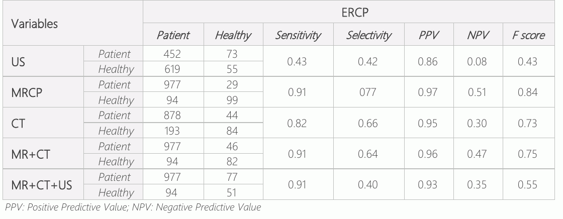
伦理委员会批准该研究的是哈兰大学临床研究伦理委员会(HRü/22.15.03)。 所有病人在入院时都接受了反差增强的腹部CT,在入院1-2天后,整个腹部都接受了美国和MRCP。 放射科医生报告进行了放射检查。 成像研究揭示了胰腺结膜管,并发现了病理的局部化和病因. 这些病人平均在2至7天的时间里接受了ERCP。 计算了F1分数 (10)5. F1分被作为评价本研究结果的基础(表1)。 F1 分值显示敏感度和特异性值的谐波平均值 (11)。 。 。
结果
1 152名病人的平均年龄为58.5±20.2岁,他们因胆汁石而接受ERCP治疗。 性别分布为754名妇女(65.4%)和398名男子(34.6%)。
讨论情况
在这项研究中,恢复了MRCP在探测胆红宝石方面的效力。 然而,美国诊断价值不足,它常常被用来评估常见的管道。 此外,诊断胆红石时使用的结合试验(MR+CT和MR+CT+US)并没有提高效果.
美国腹腔是一种非侵入性、廉价、实用的检查,几乎可以在任何地方使用。 (3). Wilcox等人著. (5) 和邱 al等. (6) 报告的两项研究表明,在进行例行肝功能测试的病人中和美国存在具有不同技术和百分比的胆红宝石。 因此,腹部的美国应该成为第一个选择,但如果怀疑胆固醇胆炎,除了例行的血清学和美国的评估之外,还需要进行进一步的放射检查。 (7)在我们的研究中,发现美国的诊断价值很低。 虽然足以检测到石头[正预测值(PPV):0.86]的患者,但将没有石头的患者排除在外[负预测值(NPV):0.08]却远远不够. 在对Kalthantaner等人的研究中,MRCP对胆固醇胆炎的敏感度在0.50-1和0.83-之间的特异性之间。 页:1 (12). MRCP的诊断值在双胞胎障碍物中相对较高,特别是胆固醇胆碱化,对ERCP的入侵性检查可以降低诊断值. (12)在另一项研究中,经ERCP确认的患者胆固醇胆碱化的MRCP灵敏度为100%,特异性为86%,精度为96%. (13)在另一项研究中,MRCP在确定常见管子的损伤时的敏感性为85%,特异性75%,PPV89.5%,NPV66.7%,精度82%. (14) 在排除那些经ERCP无法确定的病人时,报告了胆固醇胆碱化的MRCP敏感度(66.6%)、特异性(82.8%)、PPV(86.9%)和NPV(59.1%)。 在考虑5毫米和更大的石头时,MRCP的灵敏度已提高到91.9%。 (15). 在我们的研究中,MRCP的灵敏度为0.91,选择性077,PPV0.97,NPV0.51,F得分0.84. 这些结果与以前的研究是一致的。
在MRCP中检测胆红宝石可能存在错误. 生理变化,如Oddi的螺旋体机能失调,在病理上看似石块或腐烂的常见胆管瘤,以及各种技术文物,如良性严格等,都可能发生. 因此,人们知道,在MRCP中无法不时发现小石头 (16, 17)常规的MRCP,没有胆碱化石的实验室检测是主要限制,因为它无法负担得起 (18)多数治疗方案的其他缺点是,在非兽医机构无法治疗,而且缺乏治疗效果。 因此,在怀疑胆管石时,应优先考虑可用于治疗和诊断的技术。 重度肥胖、体内有金属异体和幽闭恐惧症的患者也很难实施MRCP。 众所周知,小于5毫米的石块和近缘水肿在探测胆红宝石方面是不利的。 (19)尽管存在许多缺点,但它是一种优于USG的技术,因为它是非侵入性的,提供了具有专有软件的更高空间和三维图像 (7)然而,由于这是一种非侵入性技术,在将患者转诊到其他侵入性手术之前,应当始终倾向于采用这种技术。 CT通常在紧急情况下优先评价腹痛. 在这种情况下,图画的诊断价值有争议,文献中称不应优先使用图画. (7). 由于许多胆石中缺乏钙会导致密度与周围的bile相似,因此CT的能见度和灵敏度有限. (7) . CT扫描增加了患者的X光照射,费用高于美国 (7). CT的诊断力高于美国,而MRCP在常见的胆结石的诊断力较低. (20, 21)在一些研究中,CT胆固醇学与MRCP具有相似的敏感性和特异性. (21, 22)新的多探测器CT显示敏感度为78%,特异性为96% (23). 另一方面,Kim et al. (24) 报告称,2013年17%的病例中,64名检测员CT由于尺寸 < 5毫米、黑色/棕色以及患者年龄,没有显示胆红石。 辐射照射增加,诊断功率降低,使CT比MRCP低效. (7)。 。 。
据报道,MRCP比USG和CT在检测常见的胆红宝石时更敏感. MRCP在检测外肝小管结石方面与ERCP具有相似的敏感性和特异性,据报道在诊断内肝小管结石方面优于ERCP. (25)研究表明,MRCP和美国-CT的结合可以更精确地显示双体障碍的程度,从而能够在导线因闭塞障碍而无法推进而ERCP不诊断的情况下进行诊断。 (26)虽然MRCP具有高度的敏感性和特殊性,但当我们进行综合试验应用(MR-CT和MR-CT-US组合)以提高敏感性和选择性时,其敏感性并没有积极变化。 另一方面,我们观察到,选择性逐渐减少,假阳性随试验每次新增加而增加,这是平行试验应用中预期的.
结论
虽然麻醉,临床,和实验室的发现在诊断常见的胆红素石时往往给出一个想法,但早期的成像对于诊断和避免不必要的ERCP程序非常重要. MRCP是一种非侵入性,容易获取的方法,可以与ERCP相比,以区别胆固醇胆碱化. 应确定最快和最敏感的方法,以避免不必要的成像。 根据临床和实验室发现选择成像方法对正确诊断至关重要。
声明
- 作者贡献(CRediT)
- 所有作者对全部 14 项 CRediT 角色贡献均等(Conceptualization, Data curation, Formal analysis, Funding acquisition, Investigation, Methodology, Project administration, Resources, Software, Supervision, Validation, Visualization, Writing – original draft, Writing – review & editing)。
- 资助
-
No financial support was provided from any institution or organization within the scope of the research.
- 利益冲突
- The authors declare no conflict of interest for the present study.
- 伦理批准
- The ethics committee approval of the study was obtained from Harran University Clinical Research Ethics Committee (HRÜ/22.15.03).
- 数据可用性
- A data availability statement was not reported in the published version of this article.
参考文献 (26)
26 条参考文献中有 20 条已链接 DOI。
- Williams EJ, Taylor S, Fairclough P, Hamlyn A, Logan RF, Martin D, et al. Risk factors for complication following ERCP; results of a large -scale, prospective multicenter study. Endoscopy. 2007;39(9):793-801 DOI Crossref PubMed Google Scholar
- Freeman ML, Nelson DB, Sherman S, Haber GB, Herman ME, Dorsher PJ, et al. Complications of endoscopic biliary sphincterotomy. N Engl J Med. 1996;335(13):909-18 DOI Crossref PubMed Google Scholar
- Committee ASoP, Maple JT, Ben-Menachem T, Anderson MA, Appalaneni V, Banerjee S, et al. The role of endoscopy in the evaluation of suspected choledocholithiasis. Gastrointest Endosc. 2010;71(1):1-9 DOI Crossref PubMed Google Scholar
- Karakan T, Cindoruk M, Alagozlu H, Ergun M, Dumlu S, Unal S. EUS versus endoscopic retrograde cholangiography for patients with intermediate probability of bile duct stones: a prospective randomized trial. Gastrointest Endosc. 2009; 69(2):244-52 DOI Crossref PubMed Google Scholar
- Wilcox CM, Kim H, Trevino J, Ramesh J, Monkemuller K, Varadarajulu S. Prevalence of normal liver tests in patients with choledocholithiasis undergoing endoscopic retrograde cholangiopancreatography. Digestion. 2014;89(3):232-8 12 77 MRCP, CT and US in Choledochal Stones Ugurlu et al. DOI Crossref PubMed Google Scholar
- Qiu Y, Yang Z, Li Z, Zhang W, Xue D. Is preoperative MRCP necessary for patients with gallstones? An analysis of the factors related to missed diagnosis of choledocholithiasis by preoperative ultrasound. BMC Gastroenterol. 2015;15:158 DOI Crossref PubMed Google Scholar
- Cianci P, Restini E. Management of cholelithiasis with choledocholithiasis: Endoscopic and surgical approaches. World J Gastroenterol. 2021;27(28):4536-54 DOI Crossref PubMed Google Scholar
- Lammert F, Gurusamy K, Ko CW, Miquel JF, Mendez-Sanchez N, Portincasa P, et al. Gallstones. Nat Rev Dis Primers. 2016; 2:16024 PubMed Google Scholar
- Gutt C, Schlafer S, Lammert F. The Treatment of Gallstone Disease. Dtsch Arztebl Int. 2020;117(9):148-58 DOI Crossref PubMed Google Scholar
- Cohen JF, Korevaar DA, Altman DG, Bruns DE, Gatsonis CA, Hooft L, et al. STARD 2015 guidelines for reporting diagnostic accuracy studies: explanation and elaboration. BMJ Open. 2016;6(11):e012799 DOI Crossref PubMed Google Scholar
- Sasaki YJTtm. The truth of the F-measure. 2007;1(5):1-5 PubMed Google Scholar
- Kaltenthaler EC, Walters SJ, Chilcott J, Blakeborough A, Vergel YB, Thomas S. MRCP compared to diagnostic ERCP for diagnosis when biliary obstruction is suspected: a systematic review. BMC Med Imaging. 2006;6:9 DOI Crossref PubMed Google Scholar
- Gautier G, Pilleul F, Crombe-Ternamian A, Gruner L, Ponchon T, Barth X, et al. Contribution of magnetic resonance cholangiopancreatography to the management of patients with suspected common bile duct stones. Gastroenterol Clin Biol. 2004;28(2):129-34 DOI Crossref PubMed Google Scholar
- Bostanci E, Yol S, Ozogul Y, Kayaalp C, Atalay F, Akoglu MJTJoS. Comparison of magnetic resonance cholangio pancreatography with endoscopic retrograde cholangio pancreatography in patients with suspected common bile duct stone. 2003;19(3):157-62 PubMed Google Scholar
- Baykan AR, Buran T, Gerçeker E, Yüceyar H, Tarhan S, Kasap E, et al. Hepatobiliyer Hastalıklarda Tanıya Yönelik Görüntüleme Yöntemlerinden Endoskopik Retrograd Kolanjio pankreatografi ve Manyetik Rezonans Kolanjio pankreatografinin Etkinliğinin Karşılaştırılması.2021;8(1):116-21 DOI Crossref PubMed Google Scholar
- Dusunceli E, Erden A, Erden I. [Anatomic variations of the bile ducts: MRCP findings]. Tani Girisim Radyol. 2004;10(4):296- PubMed Google Scholar
- Srinivasa S, Sammour T, McEntee B, Davis N. Selective use of magnetic resonance cholangiopancreatography in clinical practice may miss choledocholithiasis in gallstone pancreatitis. Can J Surg. 2010;53(6):403-7 PubMed Google Scholar
- Epelboym I, Winner M, Allendorf JD. MRCP is not a cost-effective strategy in the management of silent common bile duct stones. J Gastrointest Surg. 2013;17(5):863-71 DOI Crossref PubMed Google Scholar
- Polistina FA, Frego M, Bisello M, Manzi E, Vardanega A, Perin B. Accuracy of magnetic resonance cholangiography compared to operative endoscopy in detecting biliary stones, a single center experience and review of literature. World J Radiol. 2015;7(4):70-8 DOI Crossref PubMed Google Scholar
- Mitchell SE, Clark RA. A comparison of computed tomography and sonography in choledocholithiasis. AJR Am J Roentgenol. 1984;142(4):729-33 DOI Crossref PubMed Google Scholar
- Soto JA, Alvarez O, Munera F, Velez SM, Valencia J, Ramirez N. Diagnosing bile duct stones: comparison of unenhanced helical CT, oral contrast-enhanced CT cholangiography, and MR cholangiography. AJR Am J Roentgenol. 2000;175(4): 1127-34 DOI Crossref PubMed Google Scholar
- Kondo S, Isayama H, Akahane M, Toda N, Sasahira N, Nakai Y, et al. Detection of common bile duct stones: comparison between endoscopic ultrasonography, magnetic resonance cholangiography, and helical-computed-tomographic cholangiography. Eur J Radiol. 2005;54(2):271-5 DOI Crossref PubMed Google Scholar
- Anderson SW, Rho E, Soto JA. Detection of biliary duct narrowing and choledocholithiasis: accuracy of portal venous phase multidetector CT. Radiology. 2008;247(2):418-27 DOI Crossref PubMed Google Scholar
- Kim CW, Chang JH, Lim YS, Kim TH, Lee IS, Han SW. Common bile duct stones on multidetector computed tomography: attenuation patterns and detectability. World J Gastroenterol. 2013;19(11):1788-96 DOI Crossref PubMed Google Scholar
- Kim TK, Kim BS, Kim JH, Ha HK, Kim PN, Kim AY, et al. Diagnosis of intrahepatic stones: superiority of MR cholangiopancreatography over endoscopic retrograde cholangiopancreatography. AJR Am J Roentgenol. 2002; 179(2):429-34 DOI Crossref PubMed Google Scholar
- Hacim NA, Akbas A, Meric S, Karabay O, Ulgen Y, Tokocin M, et al. Predictive value of ultrasonography and magnetic resonance cholangiopancreatography in the diagnosis of biliary obstruction. Ann Ital Chir. 2020;91:277-82 PubMed Google Scholar
阅读全文
在此打开 PDF
正在加载 PDF…
若 PDF 未在此显示,请使用上方的下载按钮。
如何引用本文
Ugurlu ET, Dere O, Yilmaz S. Comparison of the Diagnostic Value of MRCP, CT, and Ultrasonography in Choledochal Stones with the Results of ERCP: Evidence from 1152 Case. Ulutas Med J. 2023;9(1):8-12. doi:10.5455/umj.20221205103820
Ugurlu, E. T., Dere, O., & Yilmaz, S. (2023). Comparison of the Diagnostic Value of MRCP, CT, and Ultrasonography in Choledochal Stones with the Results of ERCP: Evidence from 1152 Case. The Ulutas Medical Journal, 9(1), 8–12. https://doi.org/10.5455/umj.20221205103820
Ugurlu, E.T., Dere, O. and Yilmaz, S. (2023) 'Comparison of the Diagnostic Value of MRCP, CT, and Ultrasonography in Choledochal Stones with the Results of ERCP: Evidence from 1152 Case', The Ulutas Medical Journal, 9(1), pp. 8–12. doi: 10.5455/umj.20221205103820.